

Functional dyspepsia and the gut microbiota
By Prof. Lucas Wauters
Gastroenterology and Hepatology, University Hospitals Leuven, Leuven, Belgium
Lay public section
Find here your dedicated section

About this article
Created
09 November 2026
Updated
09 November 2026
Functional dyspepsia (FD) refers to chronic epigastric symptoms, with postprandial distress syndrome and epigastric pain syndrome as the major subgroups. It is part of the disorders of gut-brain interaction, with visceral signaling (gut-brain) via duodenal hyperpermeability and immune activation, potentially triggered by an altered microbiome composition or dysbiosis. Interestingly, first-line therapy with proton pump inhibitors (PPI) may improve both symptoms and duodenal alterations in the short term but adversely influence the duodenal environment in the long-term. In addition, other treatments, including prokinetics and neuromodulators, have limited efficacy or raise safety issues. Therefore, alternative treatments have been studied, including the spore-forming Bacillus coagulans MY01 and Bacillus subtilis MY02. Besides the clear efficacy and safety in a randomized, double-blind, placebo-controlled trial, beneficial immune and microbial changes were found and more pronounced in FD patients on PPIs, including a reduction in small intestinal bacterial overgrowth. These and other studies have provided insights into potential underlying mechanisms of FD, including the role of gut microbiota.
Definition and diagnosis
Dyspepsia refers to chronic upper abdominal symptoms originating from the gastroduodenal region with a significant impact on patients’ quality of life. According to the Rome criteria, functional dyspepsia (FD) is characterized by bothersome epigastric symptoms that remain unexplained after routine investigations¹⁻³. Disorders of gut-brain interaction (DGBI), including FD, are related to any combination of dysmotility, visceral hypersensitivity, altered luminal or central factors. In contrast, organic dyspepsia may result from erosive esophagitis, peptic ulcer disease or cancer in a minority of patients¹⁻³.
Functional dyspepsia is characterized by bothersome epigastric symptoms that remain unexplained after routine investigations.
Image
Two subgroups of FD were proposed by the Rome III consensus and reiterated in the Rome IV version:
- postprandial distress syndrome (PDS) with postprandial fullness or early satiation,
- epigastric pain syndrome (EPS) with epigastric pain or burning¹.
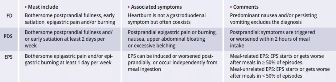
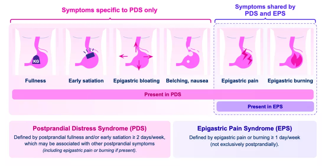
The recent Rome V version specifies that symptoms must be severe enough to affect usual activities with a minimal frequency of 1 (EPS) or 2 (PDS) days per week², which is lower than the previous Rome IV criteria for PDS (3 days per week) (table 1). As PDS is characterized by meal-related symptoms (occurring < 2 hours after meal ingestion), postprandial epigastric pain in the presence of PDS symptoms should also be characterized as PDS and not EPS according to Rome V². In the absence of PDS, epigastric pain symptoms could be further subdivided into meal-related or meal-unrelated EPS, which requires further studies (figure 1). Nausea (especially postprandially) and vomiting can coexist in PDS and EPS, but predominant nausea or recurrent vomiting is more common in chronic nausea and vomiting syndrome or gastroparesis².
Using the Rome IV criteria, PDS was consistently more prevalent (61% vs. 18% for EPS and 21% for overlap) with a similar distribution across the USA, UK and Canada⁴. In addition, FD often coexisted with gastroesophageal reflux (GER) and/or irritable bowel syndrome (IBS). Of note, bowel disorders should be considered when epigastric symptoms are consistently relieved by bowel movements¹⁻³.
While the exclusion of structural, systemic, or metabolic diseases is necessary for confirmation of FD, the yield of routine endoscopy is low in (young) patients with dyspeptic symptoms and no alarm or risk factors (see below). The prevalence of dyspepsia was higher in women, smokers, users of nonsteroidal anti-inflammatory drugs (NSAIDs) and after Helicobacter pylori (H. pylori) infection¹⁻³. Thus, a non-invasive “test & treat” strategy (i.e., urea breath test or H. pylori fecal antigen detection) is common, with endoscopy for alarm factors (e.g., age > 55 years old, or lower in certain regions of the world, evidence of gastrointestinal bleeding or iron-deficiency anemia, dysphagia, unintentional weight loss and family history of gastric or esophageal cancer)¹⁻³. Nevertheless, the Rome criteria for FD do not exclude the presence of microscopic pathology in the gastroduodenal region or the stomach and proximal small intestine.
Image
Duodenum and dysbiosis
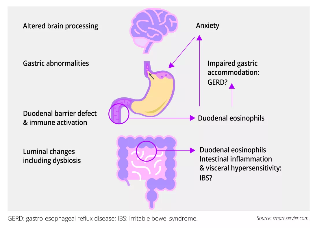
Abnormalities of gastric function in FD include impaired accommodation or expansion of the stomach, hypersensitivity to distention and delayed emptying, but these changes correlate poorly or not at all with symptoms¹⁻². Both visceral sensory signaling (gut to brain) and central modulation (brain to gut) can be abnormal in FD, as up to 50% of subjects identify stress as a trigger for their symptoms, and anxiety and depression are common. Recent research focused on mucosal and luminal alterations in the proximal small intestine or duodenum⁵. Interestingly, duodenal mucosal eosinophilia was linked to impaired gastric accommodation, reflux symptoms and anxiety, which may influence gastric function⁵. Overlapping IBS symptoms in FD may also be explained by more extensive intestinal inflammation with visceral hypersensitivity (figure 2).
Image
Current and novel therapies
Despite the recognition of meal-related symptoms, no specific dietary factors have been convincingly linked with the pathophysiology of FD. In the absence of controlled dietary intervention studies, standard dietetic advice includes frequent small size meals with avoidance of high‐fat food items, alcohol or coffee and smoking cessation¹⁻³. Acid-suppressive therapy with PPI is recommended as first-line therapy in H. pylori-negative patients or after eradication if H. pylori-positive³. Indeed, PPIs are more effective than placebo in the reduction of global symptoms of FD with a number needed to treat (NNT) of 11 in a Cochrane meta-analysis⁹. This includes a standard once-daily dose of PPI during 4 to 8 weeks, with discontinuation of therapy in case of insufficient improvement. While PPIs are beneficial for overlapping reflux symptoms, we have also shown beneficial effects of short-term PPI-therapy on duodenal permeability and immune activation in FD patients⁷.
Recent research focused on mucosal and luminal alterations in the proximal small intestine or duodenum.
Duodenal mucosal changes in FD patients include a barrier defect and immune activation, with increased numbers and degranulation of eosinophils and mast cells. Even if a mucosal T-helper (Th) 2-signal was not commonly found, increased interleukin (IL)5/13-production after stimulation of peripheral blood mononuclear cells (PBMC) from FD patients suggested a shift from Th1- to Th2-type inflammation⁵. In addition, increased IL1β-production of cultured PBMC pointed to a Th17-response, and production of GM-CSF from Th17 cells may also drive mucosal eosinophil recruitment in FD (see below). Moreover, CD4+ T cells co-expressing integrin α4β7 and CCR9, indicative of small intestinal mucosal migration, were upregulated in FD and correlated with gastric emptying. This also suggests a primary role for duodenal mucosal changes, but the luminal or other triggers are unknown⁵.
Duodenal luminal changes include an altered microbial composition or “dysbiosis”.
We found a lower abundance of Neisseria and Porphyromonas and microbial diversity in duodenal brush samples of FD patients compared to healthy controls, which inversely correlated with symptoms and duodenal eosinophils⁶. In contrast, microbial changes were associated with the effects of proton pump inhibitors (PPI) on duodenal bile salt concentrations in healthy controls, which persisted after long-term PPI-therapy in FD patients⁶⁻⁷. The presence of intestinal-like bacteria in the gastric fluid of FD patients also suggested small intestinal bacterial overgrowth (SIBO) in a subset, even if a standardized diagnosis of SIBO is not available yet and the majority of studies have used non-invasive breath tests in FD with different substrates (glucose or lactulose), doses or type of exhaled gases (hydrogen and/or methane)⁸.
We and others have hypothesized that the duodenal barrier defect may lead to mucosal immune activation through antigen presentation with a predominant Th2- or Th17-response and recruitment and degranulation of eosinophils, triggering visceral hypersensitivity and dysmotility. Possible causes include the duodenal microbiome, which may also be a therapeutic target⁵’⁸.
A significant reduction in dyspeptic symptoms was also found with prokinetics, with a NNT of 12 when cisapride (withdrawn from the market due to cardiac adverse events) was excluded in a systematic review and meta-analysis¹⁰. However, QT-prolongation (domperidone) or extrapyramidal side effects (metoclopramide) limit their chronic use and other prokinetics such as itopride or acotiamide are not widely available. In addition, amitriptyline (tricyclic antidepressant) but not escitalopram (selective serotonin-reuptake inhibitor) was effective for ulcer-like FD, with threefold higher odds of reporting adequate relief of symptoms compared to placebo¹¹. Another systematic review and network meta-analysis confirmed the superior efficacy of antidepressants or neuromodulators for symptom improvement, and that symptom resolution was most common with standard-dose PPIs¹².
However, inappropriate and prolonged use of PPIs is frequently reported, and may increase the risk of dysbiosis. Probiotics are live microorganisms that exert a health benefit on the host, and spore-forming probiotic strains may outperform traditional supplements because of gastric-acid resistant endospores with improved survival in the small intestine. We showed that Bacillus coagulans MY01 and subtilis MY02 spore-forming probiotics (daily dose of 5·10⁹ colony-forming units or CFU) were effective and safe in FD patients as mono- or add-on therapy to long-term intake of PPI¹³. Importantly, PDS (primary outcome), EPS and individual symptoms significantly decreased with spore-forming probiotics compared to placebo. We extended the clinical efficacy during an open-label phase (total 16 weeks), with symptom-improvement in the original placebo-group.
Inappropriate and prolonged use of PPIs is frequently reported and may increase the risk of dysbiosis.
Interestingly, longer-term probiotic treatment decreased systemic Th17-signaling, including Th17 cells and IL-17A, which were associated with clinical efficacy. Additional decreases were found in Th2, gut-homing and GM-CSF+ T cells in FD patients on-PPI, suggesting that the probiotic effects may be more pronounced in case of PPI-related dysbiosis. Moreover, a reduction in SIBO was found in FD patients on-PPI after 8 weeks. Finally, despite the absence of major shifts in relative or quantitative fecal microbiota community composition, a proportional increase in the beneficial Faecalibacterium was also associated with probiotic efficacy. Another RCT found a higher clinical efficacy for the probiotic Bifidobacterium animalis subsp. lactis BL-99 in a high (5·10¹⁰ CFU/day) compared to low dose (1·10¹⁰ CFU/day) or positive-drug control (rabeprazole) and placebo after 8 weeks¹⁴. Interestingly, this effect was sustained 2 weeks after stopping therapy, including the persistence of the probiotic strains in stool samples. Moreover, an increase in short-chain fatty acid (SCFA)-producing microbiota such as Faecalibacterium and Roseburia was also found with the probiotic, with increased levels of SCFA in stool and blood samples, pointing to underlying microbial mechanisms.
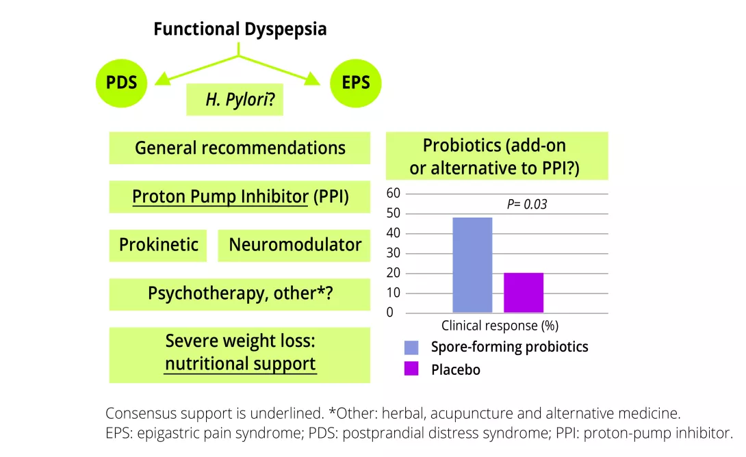
While the use of probiotics was not yet included in the European consensus on FD³, agreement or consensus support was only reached for PPI and nutritional support in case of severe weight loss (figure 3). Based on the novel evidence, probiotics (particularly spore-forming) could be considered as add-on or alternative to (long-term) PPI-therapy.
Image
Conclusion
Emerging data increasingly point towards the duodenum as a key integrator in FD, including the microbiome of the small intestine¹⁵. While first-line or PPI-therapy may be effective in the short term, long-term therapy is frequently reported with an increased risk of dysbiosis. Alternative therapies include probiotics, with not only beneficial clinical but also immunological and microbial effects of spore-forming probiotics. Although these could be considered as monotherapy, changes in immune activation were more pronounced in FD patients on-PPI, suggesting additional effects on the observed chronic alterations with PPI-therapy, which is especially useful in case PPI-withdrawal is not feasible.
Sources: