


Magazine
Artificial sweeteners, gut microbiota and metabolic health: an interaction requiring close examination
By Pr. Karine Clément
Sorbonne University, Inserm, Unité de recherche nutrition et obésités ; approches systémiques [Nutrition and Obesity Research Unit (systemic approaches)], Paris, France; Nutrition Department, Assistance-Publique - Hôpitaux de Paris, Pitié-Salpêtrière Hospital, Paris, France

Summary
Off
Migrated content
Désactivé
Updated content
Désactivé
Urinary and Urethral Microbiota #18
By Pr. Satu Pekkala
Academy of Finland Research Fellow, Faculty of Sport and Health Sciences, University of Jyväskylä, Finland

Summary
Off
Migrated content
Désactivé
Updated content
Désactivé
Vaginal Microbiota #18
By Pr. Satu Pekkala
Academy of Finland Research Fellow, Faculty of Sport and Health Sciences, University of Jyväskylä, Finland

Summary
Off
Migrated content
Désactivé
Updated content
Désactivé
Gut Microbiota #18
By Pr. Satu Pekkala
Academy of Finland Research Fellow, Faculty of Sport and Health Sciences, University of Jyväskylä, Finland

Summary
Off
Migrated content
Désactivé
Updated content
Désactivé
Highlights of Gut Microbiota for Health - World Summit 2023
By Dr. Nicolas Benech
Gastroenterology and hepatology, Microbiota Study Group, Hospices Civils de Lyon, Lyon, France

Summary
Off
Migrated content
Désactivé
Updated content
Désactivé
Rural environment reduces allergic inflammation by modulating the gut microbiota
COMMENTED ARTICLE - Children’s section
By Pr.Emmanuel Mas
Gastroenterology and Nutrition Department, Children’s Hospital, Toulouse, France

Comment on the article by Yang et al. (Gut Microbes [1])
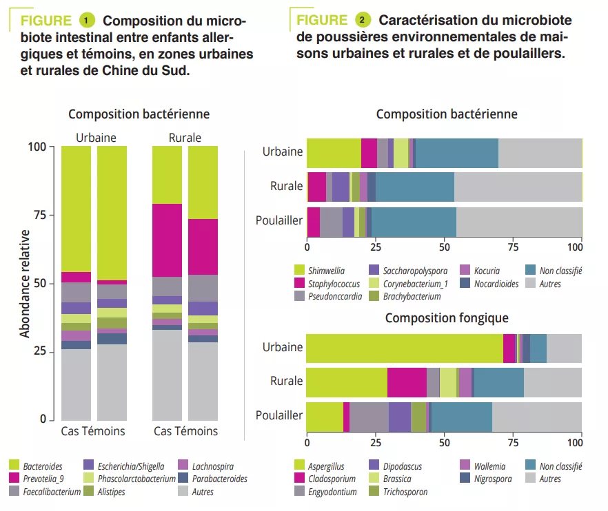
Rural environments and microbiota are linked to a reduction in the prevalence of allergies. However, the mechanism underlying this reduction is unclear. The authors assessed gut bacterial and fungal composition in urban and rural children in Southern China (EuroPrevall-INCO cohort). The bacterial and fungal composition of airborne dusts from homes in the city and countryside (including mattress dust) as well as dust from henhouses (rural environment) were analysed by 16S rRNA sequencing. Mice were repeatedly exposed to intranasal dust extracts and evaluated for their effects on ovalbumin (OVA)-induced allergic airway inflammation. It was found that children in rural areas had fewer allergies and unique gut microbiota with fewer Bacteroides and more Prevotella. Dusts from rural environments contained a higher level of endotoxins and diversity of bacteria and fungi, whereas indoor urban dusts were enriched with Aspergillus and contained a higher number of potentially pathogenic bacteria. Intranasal administration of rural dusts before OVA sensitisation reduced respiratory eosinophils and blood IgE level in mice and also led to a recovery of gut bacterial diversity and Ruminiclostridium in the mouse model. Faecal microbiota transplant restored the protective effect by reducing OVA-induced lung eosinophils in recipient mice. These results support a cause-effect relationship between exposure to dust microbiota and allergy susceptibility in children and mice. Specifically, rural environmental exposure modulated the gut microbiota, which was essential in reducing allergy in children.
What do we already know about this subject?
The prevalence of allergic diseases has increased dramatically. It has been shown that children living in the countryside are less prone to asthma than those living in cities. Indoors, dust is the main source of bacteria and fungi. Its composition reflects the outdoor environment and is influenced by external activities (e.g., agricultural), building materials and animals.
The establishment of gut microbiota during the first 1,000 days of life shapes the subsequent development of allergic diseases. Some well-known factors influence the composition of the gut microbiota of infants including antibiotics, delivery method and diet. Any resulting dysbiosis is thought to increase the subsequent likelihood of developing allergic diseases. In contrast, breastfeeding and vaginal delivery protect against the subsequent development of allergic diseases. The intestinal microbiota of these types of infants is characterised by a predominance of bifidobacteria, especially Bifidobacterium breve. Decreased contact with nature was seen to favour intestinal dysbiosis with a dysregulation of Th1/Th2 immune balance in favour of Th2, which is the adaptive immune response involved in allergic diseases (Cukrowska Nutrients).
The mechanisms by which gut dysbiosis in early life influences the development of allergies and asthma are little understood. Farm dust and bacterial lipopolysaccharide are known to induce endotoxin tolerance, thus reducing allergic asthma.
What are the main insights from this study?
The authors compared urban and rural environmental exposures in China in a human and mouse study. The EuroPrevall-INCO human cohort included 5,139 urban and 5,542 rural school-age children. The prevalence of food allergies and especially asthma, rhinitis, and eczema were higher in urban children (p < 0.001).
A case-control study included 225 children: 151 urban and 74 rural children. The gut microbiota of all children was analysed via 16S rRNA sequencing and metabolic pathways were assessed via shotgun sequencing. Clinical data and allergen sensitizations were collected. The Prevotella-to-Bacteroides ratio was significantly higher in rural children (p < 0.001). This difference was due to Prevotella_9 accounting for 25% of amplified variants in rural children and <5% in urban children (figure 1). However, no significant difference was observed in gut microbiota composition between cases and controls in both urban and rural participants. The analysis of metabolic pathways identified 14 different pathways between urban/rural participants and nine between controls/ cases. Among these, the L-lactate producing pathway was strongly associated with allergy and pathways involved in sugar degradation and lipopolysaccharide synthesis were abundant in the microbiota of control children.
To mimic the microbial exposure of children in urban and rural environments, mattress dusts were collected from ten urban and ten rural families and dusts from five henhouses from rural families (on the assumption that these may contribute to the microbial environment in rural families). Enterobacteriaceae and Rhizobiaceae were predominant only in urban house dusts (figure 2). A significantly higher a diversity and endotoxin content was observed in rural house dusts compared to urban house dusts and henhouse dusts. Finally, urban house dusts had a significantly higher proportion of potentially pathogenic bacteria. In addition, Aspergillaceae dominated in urban house dusts, whereas Trichocomaceae (genus Penicillium) was more abundant in rural house dusts (hence the higher diversity) and henhouse dusts (figure 2).
Image
To test the impact that exposure to environmental dusts may have on allergic disease by altering the gut microbiota, the researchers exposed mice to dust by intranasal exposure (OVA-induced allergic model). Prior exposure to house dust in rural areas attenuated allergic inflammation (eosinophilic infiltration of airways, in bronchoalveolar lavage (BAL), increased sIgE). Mice exposed to rural dusts showed the lowest increase in the proportion of potentially pathogenic bacteria. The abundance in the gut of Bacteroidales increased and while Clostroidales (including species belonging to both Lachnospiraceae and Ruminococcaceae families) decreased in control mice exposed to PBS as well as those exposed to urban dusts. Finally, the relative abundance in the gut microbiota of Bacteroides and Ruminiclostridium was correlated to eosinophils in BAL (r = 0.59 and p = 0.001 and r = – 0.45 and p = 0.05 respectively).
What are the consequences in practice?
Early modulation of the gut microbiota, targeting the beneficial effect of rural house dusts could prevent the development of allergic diseases.
CONCLUSION
This study reported differences in the composition of dust microbiota between urban and rural areas in China. These modulate differently the gut microbiota and its immune response in allergic diseases.
Summary
Off
Migrated content
Désactivé
Updated content
Désactivé
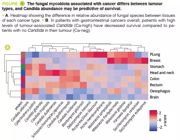
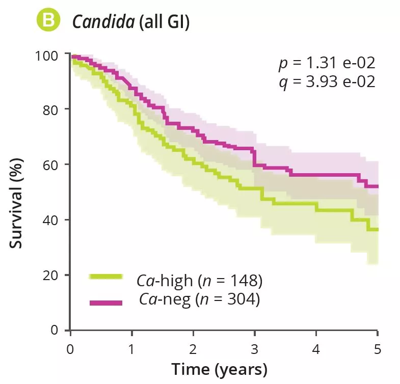
A pan-cancer mycobiome analysis reveals fungal involvement in gastrointestinal and lung tumours
COMMENTED ARTICLE - ADULTS’ SECTION
By Pr. Harry Sokol
Gastroenterology and Nutrition Department, Saint-Antoine Hospital, Paris, France
Comment on the article by Dohlman et al. (Cell 2022 [1])
Fungal microorganisms (mycobiota) comprise a small but immunoreactive component of the human microbiome, yet little is known about their role in human cancers. Pan-cancer analysis of multiple body sites revealed tumour-associated mycobiomes at up to 1 fungal cell per 104 tumour cells. In lung cancer, Blastomyces was associated with tumour tissues. In stomach cancers, high rates of Candida were linked to the expression of proinflammatory immune pathways, while in colon cancers Candida was predictive of metastatic disease and attenuated cellular adhesions. Across multiple GI sites, several Candida species were enriched in tumour samples and tumour-associated Candida DNA was predictive of decreased survival. The presence of Candida in human GI tumours was confirmed by external ITS sequencing of tumour samples and by culture-dependent analysis in an independent cohort. These data implicate the mycobiota in the pathogenesis of GI cancers and suggest that tumour-associated fungal DNA may serve as diagnostic or prognostic biomarkers.
What do we already know about this subject?
Cancer is one of the leading causes of death worldwide. The tumorigenesis, progression and treatment-response of cancer are influenced by various interactions between the immune system of the host and bacteria in the microbiota. However, the role of fungi (mycobiota) in these processes remains largely unexplored. Fungi and bacteria co-colonise the gastrointestinal tract, skin epithelium, airways and reproductive organs of mammals, forming a complex ecosystem of microbe-microbe and host-microbe interactions with significant implications for human health. While fungal infections account for over 1.5 million deaths worldwide each year, they only account for 0.1% of microbial DNA in the gut, suggesting a disproportionate influence of species from this kingdom on the overall gut microbiome and host immunity. Whether referring to viruses, bacteria or fungi, an ever growing body of scientific evidence suggests a link between the human microbiome and cancer and its outcomes. Several cases showing the association between bacterial species and cancer development/progression have been observed in recent years. Helicobacter pylori is responsible for approximately 75% of the risk attributable to gastric cancer, while genotoxic Escherichia coli, Bacteroides fragilis, Streptococcus bovis/gallolyticus and Fusobacterium nucleatum have been implicated in colorectal carcinogenesis [2]. The common feature of these bacteria is their ability to trigger chronic inflammation, a feature considered to contribute to their tumorigenic capacity. Recent reports have also identified intracellular bacteria in many types of tumour [3].
The mycobiome plays a key role in the activation of innate immunity in the gut. Mycotoxins and bioactive amines have been associated with carcinogenesis. Recent experimental studies support fungal involvement in cancer in some contexts [4]. Sequencing data from tumour banks have revealed the presence of microbial sequences, although the fungal component has not yet been explored.
What are the main insights from this study?
By analysing several types of cancer using “The Cancer Genome Atlas” (TCGA), the authors extracted tumour-associated mycobiome profiles with a species-level resolution. After eliminating contamination and false-positive signals, the authors reported that fungal compositions varied according to the type of cancer, and that some fungi were tumour-type specific, in both gastrointestinal and non-gastrointestinal locations (figure 1A). Overall, up to one fungal cell per 104 human tumour cells were observed, a rate consistent with the finding that fungi make up 0.1-1% of the microbiome, while bacteria are estimated to make up less than 1% of tumour cells [2, 3]. Abundant numbers of several species of Candida, Saccharomyces cerevisiae and Cyberlindnera jadinii have been found in gastrointestinal tumours, while Blastomyces and Malassezia species are abundant in lung and breast tumours, respectively. The authors then showed that several Candida species are alive and transcriptionally active in the tumour. Finally, the abundance of some fungi within the tumour could predict host tumour gene expression, disease status and survival (figure 1B), although these findings still need to be confirmed. Overall, these results suggest an involvement of fungi, especially Candida, in the pathogenesis of gastrointestinal cancers but also highlight their potential as a therapeutic target and prognostic tool.
Image
Image
What are the consequences in practice?
Alongside bacteria, this study reported the presence of fungi in many gastrointestinal and non-gastrointestinal tumours, with some degree of specificity across tumour types and a potential for predicting severity. These results suggest that fungi play a role in the cancer process and its severity. They could also pave the way for the development of new biomarkers or new cancer treatments targeting the fungal component.
CONCLUSION
An analysis of multiple gastrointestinal and non-gastrointestinal tumours identified tumour-associated fungi, especially Candida enrichment in gastrointestinal cancers. Fungi may also play a role in carcinogenesis. Tumourassociated fungal DNA could serve as a prognostic marker in this context and fungi could represent a new therapeutic target in cancer.

"Interesting developments. Thank you for caring about humanity." -sturehp (From Biocodex Microbiota Institute on X)
"Tumour-associated fungal DNA may serve as diagnostic or prognostic biomarkers. Very interesting. I am curious to see what comes out of this." -Just me. (From Biocodex Microbiota Institute on X)
Sources
1. Dohlman AB, Klug J, Mesko M, et al. A pan-cancer mycobiome analysis reveals fungal involvement in gastrointestinal and lung tumors. Cell 2022 ; 185 : 3807-22.e12.
2. Sepich-Poore GD, Zitvogel L, Straussman R, et al. The microbiome and human cancer. Science 2021 ; 371 : eabc4552.
3. Nejman D, Livyatan I, Fuks G, et al. The human tumor microbiome is composed of tumor type-specific intracellular bacteria. Science 2020 ; 368 : 973-80.
4. Alam A, Levanduski E, Denz P, et al. Fungal mycobiome drives IL-33 secretion and type 2 immunity in pancreatic cancer. Cancer Cell 2022 ; 40 : 153-67.e11.
Summary
Off

Migrated content
Désactivé
Updated content
Désactivé
Oral microbiota and chronic conditions
By Dr. Jay Patel
Usher Institute of Population Health Sciences and Informatics, University of Edinburgh, United Kingdom
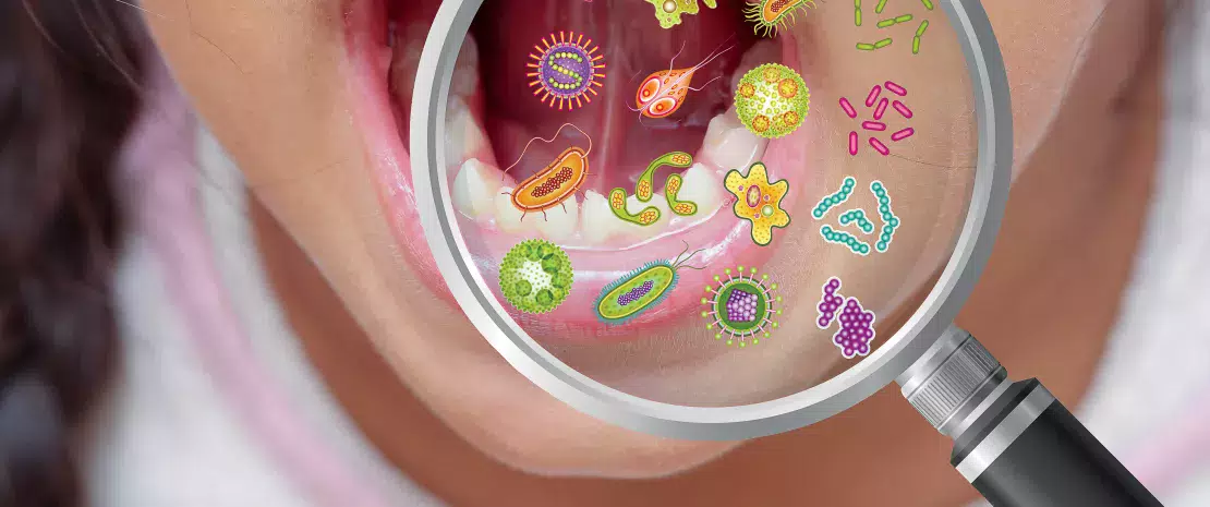
Although the co-evolutionary role of the human microbiome as a determinant of human health is increasingly recognised in modern medicine, the oral microbiome remains a largely siloed factor contributing to general health and well-being. In health, the oral microbiome maintains a careful symbiotic equilibrium with the host, with harmful bacteria at clinically inconsequential levels. However, external environmental pressures readily turn the oral microbiome dysbiotic, where an improper proportional and diversity of microbes colonise the mouth. These environmental pressures are often highly modifiable risk factors. An expanding body of evidence suggests that the relevance of this disturbance is not merely confined to local disease activity but has a disseminated risk profile for other major chronic diseases of the body, with a high global burden of disease including diabetes, atherosclerotic cardiovascular disease, and rheumatoid arthritis.
In health, the oral microbiome represents a carefully balanced, diverse community protecting the mouth from disease. Modern lifestyle choices can readily upset this balance, rendering the community less protective and increasingly harmful.
Mechanism
The morphology, warmth and moisture of the mouth affords the oral microbiota a highly diverse habitat for colonisation and growth. From birth, children acquire a simple oral microbiome, and with age, the eruption of teeth, and the additive role of external factors, this community becomes increasingly complex. Both host-derived and microbially-derived factors maintain the homeostatic equilibrium of the oral microbiome required for health.
Poor oral hygiene can be a profound ecological pressure that steers complex microbial communities in the mouth into dysbiosis [1].
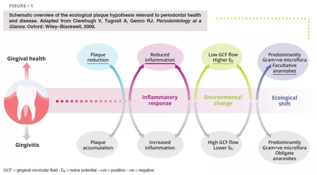
Ecological shifts in a dysbiotic ecosystem favour the colonisation and proliferation of pathogenic oral bacteria (figure 1). When these species increase in quantity, the risk of oral disease significantly increases. Periodontal disease is a chronic, non-resolving, inflammatory process leading to tissue breakdown of the tooth-supporting apparatus, and can lead to tooth loss, if untreated. Routine activities including chewing, flossing, and toothbrushing can induce bacteraemia, which facilitate haematogenous dissemination of oral bacteria and inflammatory mediators, inducing systemic inflammation in some patients [2]. Patients with periodontal disease––the sixth most prevalent condition affecting humankind globally [3]––show micro-ulcerated sulcular epithelia and damaged periodontal tissues, and thus seem more susceptible to bacteraemia. Therefore, the inflammatory state from periodontal disease metastasises to other sites of the body, which can occur at clinically-relevant levels. Good oral hygiene is therefore essential for controlling the total bacterial load in the mouth, maintaining or re-establishing the oral symbiotic equilibrium, and preventing the dissemination of oral bacteria to other sites in the body.
The characteristics of the oral microbiome are not only confined to oral pathological changes but can influence systemic health, and in cases, this influence is measurable in both positive and negative directions.
Image
Diabetes
The strongest evidence supporting a bidirectional role between oral and systemic health exists for the dose-dependent relationship between the severity of periodontitis and complications arising from diabetes.
Type II diabetes is a metabolic disorder characterised by an insufficiency of insulin production and the subsequent inability for the body to metabolise glucose, leading to elevated levels of blood glucose (chronic hyperglycaemia). Severe periodontitis strongly influences glycated haemoglobin (HbA1c) and fasting blood glucose levels in people with and without diabetes [4]. Periodontitis is thus recognised as the sixth major complication of diabetes, as the risk for periodontitis is raised by 2-3 times for people with the condition [5].
19–33% Compared to individuals with periodontal health, patients with severe periodontitis are at a elevated risk of developing diabetes [6].
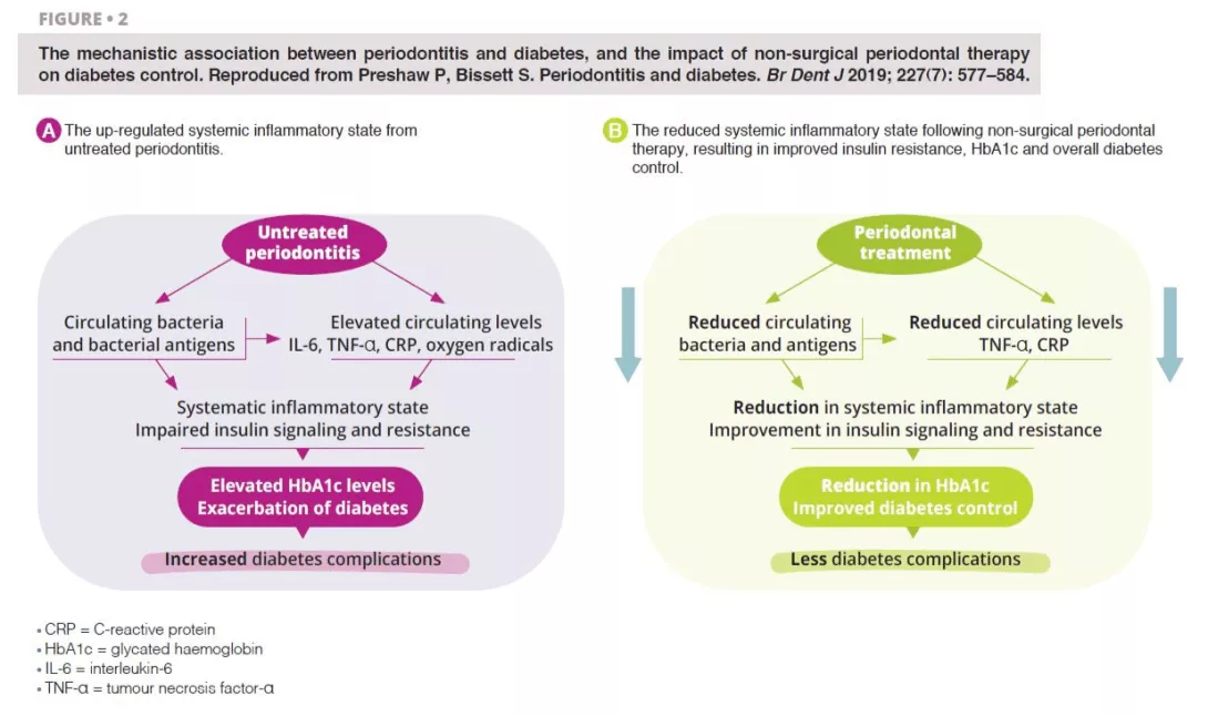
Untreated severe periodontitis is associated with an increase in the circulating levels of bacteria and bacterial antigens, pro-inflammatory mediators and cytokines, and increased levels of interleukin 6, tumour necrosis factor alpha, C-reactive protein, and oxygen free radicals. This combined effect fosters the conditions for systematic inflammation, impairing insulin signalling and resistance [6]. Clinically, this is recognised through increased HbA1C and the progression of diabetes, with greater risk of diabetic complications. Periodontal treatment reduces the oral bacterial load, and therefore lowers the circulating levels of inflammatory mediators, thereby reducing the degree of the systematic inflammatory state (figure 2). Hence, the dental management of periodontitis can lead to a clinically-relevant improvement in glycemic control, where patients with diabetes experience HbA1c reductions of 0.3–0.4% up to four months after treatment.
Image
Atherosclerotic cardiovascular disease
Atherosclerosis describes the accumulation of fats, cholesterol, and blood cells that form hardened plaque deposits within the artery walls, occluding blood flow through the vessels, increasing the risk of cardiovascular complications.
Oral bacteria are contributory infectious agents in the pathogenesis of atherosclerosis, through the invasion of cardiovascular host cells, namely endothelial cells [7].
Chronic periodontal disease can lead to endothelial dysfunction, through an elevated systematic inflammatory state, which can be shown through increased levels of IL-6, fibrinogens, and periodontopathic bacterial products, such as outer membrane vesicles and gingipains [8]. Much of the atherosclerotic pathology appears to be attributable to Porphyromonas gingivalis. However, Aggregatibacter actinomycetemcomitans, Prevotella intermedia, Tannerella forsythia and Fusobacterium nucleatum have each been studied in relation to this association. The primary microbial implications are endothelial dysfunction and the promotion of atherosclerosis in cardiovascular cells. P. gingivalis has the ability to attach to endothelial target cells, and external factors mediate its cellular entry, where it induces pro-coagulant effects. The results of a parallel-group, single-blind, randomised, controlled trial found that although intensive periodontal therapy led to systemic inflammation and endothelial dysfunction in the immediate term, 6 months after treatment, clinical and biochemical improvements in endothelial function were noted [9]. This study added to the theory that periodontal control may modulate atherosclerotic cardiovascular processes.
Rheumatoid arthritis
Rheumatoid arthritis is a chronic, autoimmune inflammatory condition affecting the synovial fluid of joints in a symmetrical pattern, and if untreated, other organs. Porphyromonas gingivalis is implicated in the pathophysiology of rheumatoid arthritis, where the bacteria produce enzymes with the capacity of citrullinating proteins, increasing the probability of reductions in the host immune tolerance and promoting the release of autoantibodies characteristic to the condition [10].
82% Cross-sectional data from the United States showed an 82% increase in rheumatoid arthritis associated with periodontitis, identified through gain in periodontal attachment loss [12].
Several studies have shown that periodontitis caused by dysbiotic oral biofilms, can trigger rheumatoid arthritis with systemic inflammation and increased bone erosion. A bidirectional relationship has been postulated between the inflammatory conditions, but further evidence is needed to verify this [11]. Clinicians involved in the rheumatological care of arthritis patients should be aware of the role of periodontitis as a factor modulating the efficacy of biologic disease- modifying anti-rheumatic therapies, since the maintenance of systemic inflammation could affect treatment response.
Non-surgical periodontal therapy appears to improve the biochemical expression of rheumatoid arthritis, but its role in improving clinical outcomes remains to be fully understood.
In patients with dysbiotic oral biofilms, where the proportions of periodontopathic bacteria capable of citrullinating proteins are higher than in health, preventive and curative treatment to stabilise the oral microbiome and periodontal inflammation would be prudent to include as a core aspect of the rheumatological care plan.
Prevention
Scientific advances in the understanding of the oral microbiome demonstrate its contribution to both oral and general health and well-being. The ecological plaque hypothesis is the currently accepted theory desiring microbiological changes in the mouth, where shifts in the ecology of the oral microbiome result in disharmony, which foster key harmful pathogens to increase in number [13]. The dissemination of oral bacteria to systemic bodily sites is substantially reduced by enhancing control of the oral microbial load. Daily mechanical removal of plaque, through a systematic and comprehensive toothbrushing and interdental cleaning technique, reduces the volume of this load, and prevents the colonisation of pathogenic species. Good plaque control also prevents the risk of developing periodontal diseases, characterised by micro-ulceration of the gingival architecture, thereby producing channels for the leakage of bacteria and inflammatory mediators.
Supplemented with professional interventions by dental practitioners (commonly oral hygiene instruction, risk factor control, and mechanical plaque removal), periodontal disease processes can be stabilised and if mild, reversed.
Where the microbial balance has been disturbed in disease, the symbiotic equilibrium of the oral microbiome can be re-established and stabilised through relatively simple personal and professional interventions.
Conclusion
Research exploring the association between changes in the oral microbiome and systemic chronic conditions continues to expand.There are multiple plausible reasons to justify the bidirectionality of these putative connections. Dysbiosis in the oral microbiome, the primary driver for local and general disease onset and progression is mediated by highly modifiable risk factors, reinforcing the value of prevention, and the need for health systems to re-orient their mode of care delivery to accommodate the delivery of preventive oral health care.
Sources
1. Kilian M, Chapple IL, Hannig M, et al.. The oral microbiome - an update for oral healthcare professionals. Br Dent J 2016; 221: 657–66.
2. Patel J, Sampson V. The role of oral bacteria in COVID-19. Lancet Microbe 2020; 1: e105.
3. GBD 2019 Diseases and Injuries Collaborators. Global burden of 369 diseases and injuries in 204 countries and territories, 1990-2019: a systematic analysis for the Global Burden of Disease Study 2019. Lancet 2020; 396: 1204–22.
4. Preshaw PM, Alba AL, Herrera D, et al. Periodontitis and diabetes: a two-way relationship. Diabetologia 2012; 55: 21–31.
5. Teeuw WJ, Kosho MX, Poland DC, Gerdes VE, Loos BG. Periodontitis as a possible early sign of diabetes mellitus. BMJ Open Diabetes Res Care 2017; 5: e000326.
6. Preshaw P, Bissett S. Periodontitis and diabetes. Br Dent J 2019; 227: 577–84.
7. Tonetti MS, Van Dyke TE; working group 1 of the joint EFP/AAP workshop. Periodontitis and atherosclerotic cardiovascular disease: consensus report of the Joint EFP/AAP Workshop on Periodontitis and Systemic Diseases. J Periodontol 2013; 84(Suppl 4): S24–S29.
8. Reyes L, Herrera D, Kozarov E, Roldán S, Progulske-Fox A. Periodontal bacterial invasion and infection: contribution to atherosclerotic pathology. J Clin Periodontol 2013; 40 (Suppl 14): S30-S50.
9. Tonetti MS, D’Aiuto F, Nibali L, et al. Treatment of periodontitis and endothelial function. N Engl J Med 2007; 356: 911–20.
10. Quirke AM, Lugli EB, Wegner N, et al. Heightened immune response to autocitrullinated Porphyromonas gingivalis peptidylarginine deiminase: a potential mechanism for breaching immunologic tolerance in rheumatoid arthritis. Ann Rheum Dis 2014; 73: 263–9.
11. González-Febles J, Sanz M. Periodontitis and rheumatoid arthritis: What have we learned about their connection and their treatment? Periodontol 2000 2021; 87: 181–203.
12. de Pablo P, Dietrich T, McAlindon TE. Association of periodontal disease and tooth loss with rheumatoid arthritis in the US population. J Rheumatol 2008; 35: 70–6.
13. Marsh PD. Microbial ecology of dental plaque and its significance in health and disease. Adv Dent Res 1994; 8: 263–71.
Summary
Off
Migrated content
Désactivé
Updated content
Désactivé
Influence of vaginal intercourse on male urogenital microbiota
The male urethra, which has been little described until now, is also thought to harbor microbiota. Vaginal intercourse could reshape its composition, making it a reservoir for the bacteria responsible for vaginosis in women.
Little is known about male urethral microbiota, since sampling is painful and often reserved for men with sexually transmitted infections (STIs). However, there is increasing evidence to suggest that microorganisms colonize this mucosa, even in healthy individuals. An American study including 110 men with no urethral symptoms, infections or inflammation has finally uncovered them.
A “core” microbiota...
Most of the individuals were heterosexual. A swab was inserted 2 cm into each man’s urethra to analyze the microbiota (shotgun sequencing). In total, 117 different species of bacteria were detected. Most of the samples contained lactic acid bacteria (e.g., Streptococcus mitis) and corynebacteria, which could represent the “core” microbiota responsible for the health of the urethra. But that’s not all. Scientists have also identified bacteria in some men that are associated with bacterial vaginosis in women, in particular Gardnerella vaginalis. As a consequence, the male genital tract could be colonized by bacteria that are potentially pathogenic for women, even though on the whole its microbiota differs greatly from that of the vagina.
10% of the microbiota present in the male urethra is influenced by sexual intercourse, in particular vaginal penetration.
And two types of urethral microbiota
Two types of urethral microbiota (or urethrotypes, UT) were identified: type 1 microbiota (UT1) which is less abundant and diversified, mainly dominated by S. mitis, and type 2 microbiota (UT2) which is more abundant and diversified, dominated by G. vaginalis and composed of nine bacteria associated with vaginal disorders (bacterial vaginosis, vaginitis, etc.) and capable of forming biofilms with G. vaginalis. Given the bacteria’s degree of affinity for oxygen, researchers believe that these two microbiomes are located in different niches: UT1 is located close to the urinary meatus and UT2 slightly deeper.
In addition, UT2 is associated with vaginal intercourse and certain bacteria related to bacterial vaginosis are still detectable 60 days after sexual intercourse and, to a lesser extent, after a year or even over a lifetime. Vaginal intercourse in the last two months alone explains 4.26% of the variance in the composition of male urethral microbiota. And sexual practices as a whole (oral, vaginal, anal) account for around 10% of this variance.
Finally, no other variable, whether anal or oral intercourse, age, ethnicity or history of STI, was associated with either urethrotype.
Is bacterial vaginosis an STI?
The male urogenital microbiota could therefore be linked to sexual behavior and the male urethra could, in some men, harbor a wide range of agents that are potentially pathogenic for the female vaginal flora. Could they represent a reservoir for sexually transmitted microorganisms and risk contaminating women during unprotected sex? If so, shouldn't bacterial vaginosis be considered an STI? Currently it is not considered as such but this hypothesis (which is not new) will be explored in further research involving couples this time.
Recommended by our community
"Yes. Experts who contribute to the Embedded/Chronic UTIs support group on FB say the same about what used to be known as ‘Interstitial’ Cystitis." - Hazel Bowden (From Biocodex Microbiota Institute on X)
 Idiopathic urethritis in men: new infectious etiologies?
 Using recombinant endolysins to treat bacterial vaginosis
Summary
Off
Migrated content
Désactivé
Updated content
Désactivé