The microbiota of the male urethra has been little studied and yet it has the potential to disrupt the vaginal bacterial ecosystem. How? Read on to find out more.
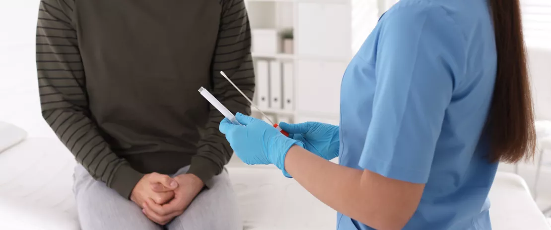
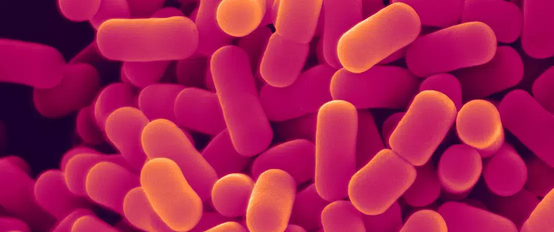
The male urethra, i.e., the canal that runs through the penis and carries urine and sperm, is, like many other parts of the body, home to a whole microscopic world. This microbiota is generally only talked about when it is invaded by pathogenic bacteria that cause painful inflammation, such as the “clap” caused by gonococcal infection. Poorly understood and little studied (understandably, few men volunteer to be swabbed...), the microbiota of the male urethra has just revealed some of its secrets, thanks to the courage of 110 healthy Americans who agreed to have a swab taken.
Definition
The urethra, i.e., the small canal that connects the bladder to the tip of the penis, measures around 15 cm in men, compared with 3.5 cm in women.
The samples show that most of these men harbor a relatively simple community of (sidenote:
Microorganisms
Living organisms that are too small to be seen with the naked eye. They include bacteria, viruses, fungi, archaea and protozoa, and are commonly referred to as “microbes”.
What is microbiology? Microbiology Society.), including oxygen-loving bacteria which probably live at the tip of the penis close to the open air. Their relatively constant presence suggests that they form a sort of core community, ensuring the health of the penile urethra.
However, some men have a more complex set of microorganisms, including bacteria known to disrupt women’s vaginal bacterial ecosystem and cause all sorts of disorders (vaginosis, etc.).
These bacteria could live a little deeper in the urethra of the penis, far from any source of oxygen. More importantly, only men who reported having had vaginal sex are carriers of these pathogenic bacteria. It's only a short step from there to imagining that they brought them back from a “close encounter” with a vagina...
Is a man's urethra a reservoir of harmful bacteria for women?
Based on the findings of the researchers who examined the reported sexual practices of the 110 participants, sexual behavior (i.e., vaginal, oral or anal intercourse and combinations thereof) over the last two months explained around 10% of the total variation in the composition of the penile urethra microbiota, of which 4.26% can be explained by vaginal intercourse alone. Men in good health appear to be able to be colonized for extended periods by bacteria that they could pass on to their subsequent female partners, with these women running the risk of getting bacterial vaginosis. This is a public health issue that the team is continuing to investigate, with plans to study couples to find out more about potential transmissions.
A team of Irish researchers has recently discovered that the gut microbiota of people suffering from social phobia has specific characteristics that differ from those of healthy individuals 1. A first!
What about the microbiota of people with social phobia? Does it also display characteristics suggesting it plays a role in the disease via communication with the brain?
Until now, a lack of data has made it difficult to answer this question. However, a study by a team of Irish researchers from University College Cork (UCC) suggests that this may indeed be the case.
The scientists enrolled 31 people diagnosed with social phobia, together with 18 controls not suffering from the condition. They collected the participants’ stools to analyze and compare the composition of their microbiota. What did the analyses show?
13%
The percentage of the population in Europe and the United States affected by social phobia.
Compared to the control group, the microbiota of the “social phobia” group contained more Anaeromassilibacillus. Several studies have shown this bacterial genus to be implicated in autism and depression, two disorders that share physiological processes with social phobia.
Their microbiota also contained more Gordonibacter, a bacterial genus that can produce the metabolite urolithin from the breakdown of polyphenols, which studies show has an impact on mental health.
The social phobia group also had lower levels of Parasutterella excrementihominis. Several studies have shown this bacterium to be less abundant in people suffering from autism spectrum disorders, but also in those with a high (sidenote:
Indice de masse corporelle
Rapport du poids en kg sur le carré de la taille en m2
) or sugar intake, which is common in people suffering from phobias, including those in the study.
Another notable difference was that their microbiota showed a significant enrichment in a metabolic pathway for aspartate degradation via a specific protein. According to the authors’ analysis, this protein is similar to another involved in the metabolism of tryptophan, one of the messengers involved in the functioning of the gut-brain axis.
When the fear of being judged becomes an illness
Social phobia, also known as social anxiety disorder, is a persistent and intense fear of:
Being judged by others;
Feeling humiliated, ridiculous, or embarrassed in the presence of others.
The fear is intense and pervasive and is accompanied by particularly disturbing physical symptoms: trembling, palpitations, excessive sweating, nausea, and at times panic attacks.
Social phobics may be unable to do things as simple as going to a restaurant, asking for directions, taking an exam, or attending a meeting. They are also at greater risk of alcoholism and depression.
This illness differs from stage fright or shyness in the intense psychological suffering it causes.
Treatment relies mainly on medication (antidepressants, beta-blockers) and cognitive behavioral therapies (CBT). 2
Towards new treatments and biomarkers
According to the researchers, this small study is highly important and will lay the foundations for larger-scale studies to confirm the involvement of the gut-brain axis and specific bacteria in social anxiety disorder.
Long term objective
The ultimate aim is to identify biomarkers and develop new treatments for social phobia, a common and particularly disabling disorder, for which treatments are not yet very effective.
News, accrediting training, infographics, expert’s video, thematic folder… Let’s deep dive to the Biocodex Microbiota Institute’s materials dedicated to microbiota gut-brain axis. Tools and contents adapted to your practice to improve your knowledge and be(come) easily a gut-brain axis expert!
The health benefits of prunes in postmenopausal women appear to be linked to their positive effects on the gut microbiota, according to a study conducted by American researchers 1.
Delicious; rich in fiber and antioxidants; good for bowel function, the heart, the blood vessels, and bone health; excellent allies for weight loss... In terms of health benefits, prunes get two thumbs up!
Prunes: Their mode of action is still a mystery…
The inclusion of these dried fruits in the diet of postmenopausal women is no exception to the rule. The health benefits of prunes for postmenopausal women, particularly in improving bone health, are well documented, but there are many unknowns surrounding how they work, particularly when it comes to the role of the gut microbiota.
What exactly are the effects of these dried fruits on the gut bacteria during menopause? A team of American researchers has tried to answer this question.
They recruited 143 postmenopausal women aged 55 to 75 and randomly assigned them to 3 different groups:
one in which they had to eat 4 to 6 prunes per day (50 g),
another in which they had to eat 10 to 12 prunes per day (100 g)
and a third in which they did not eat any prunes (control group).
Before and after the experiment, the scientists collected stool samples from the volunteers to analyze and compare the evolution of their microbiota.
Osteoporosis: 3 prunes a day may keep the orthopedic surgeon away!
Osteoporosis after menopause affects one in three women. Could prunes be an ally in the fight against this widespread problem? A review 2 of studies conducted on this subject suggests that they could. According to its authors, eating prunes regularly could:
prevent and reduce bone loss,
improve bone mineral density and bone biomarkers,
have anti-inflammatory effects (which is useful, given that inflammation is a risk factor for osteoporosis!),
suppress the production of cytokines (the body’s pro-inflammatory messengers),
increase antioxidant enzymes (which combat the oxidative effects of inflammation).
More beneficial gut bacteria
They also collected blood and urine samples to measure inflammatory markers (a risk factor for many diseases) and “phenolic metabolites.”
Phenolic metabolites are beneficial compounds derived from the breakdown of antioxidants (polyphenols). The level of urinary phenolic metabolites reflects the level of activity of the microbiota bacteria that break down polyphenols.
After 12 months of the experiment, the results indicate that women in the “prune” groups show significant changes in their microbiota compared to those in the control group. These changes are different depending on the dose (50 or 100 g).
In particular, the researchers’ analyses show an enrichment in bacteria of the Lachnospiraceae family, already known for their capacity to maintain the intestinal barrier.
Anti-inflammatory effects mediated by the microbiota
It appears from the study that Lachnospiraceae are able to metabolize prune polyphenols and ferment their fibers to produce (sidenote:
Short chain fatty acids (SCFA)
Short chain fatty acids (SCFA) are a source of energy (fuel) for an individual’s cells. They interact with the immune system and are involved in communication between the intestine and the brain.
Silva YP, Bernardi A, Frozza RL. The Role of Short-Chain Fatty Acids From Gut Microbiota in Gut-Brain Communication. Front Endocrinol (Lausanne). 2020;11:25.) that have anti-inflammatory properties. Calculations show that the presence of some of these bacteria is negatively correlated with inflammatory markers and positively correlated with phenolic metabolites.
According to the researchers, by providing fiber and polyphenols, prunes exert a selection pressure that favors good bacteria in the long term, which could explain their health benefits.
A commensal species of the gut microbiota producing trans fatty acids and saturated fatty acids is believed to aggravate obesity induced by a high-fat diet. Its metabolites are thought to harm the host’s lipid metabolism and intestinal barrier, according to a study published in Cell Metabolism.
As the prevalence of obesity and associated metabolic disorders increases worldwide, dietary changes are not enough for many patients. Understanding the impact of other environmental factors is crucial to developing alternative treatment strategies. Numerous studies point to a link between dysbiosis of the gut microbiota and the development of obesity, which could at least partly explain the variations from one individual to another in terms of susceptibility to metabolic diseases. However, the molecular mechanisms and the causal link between the bacteria of the gut microbiota, especially their metabolites, and the development of obesity are not fully understood.
High-fat diet and Fusimonas intestini: A synergistic effect on weight gain
It is well accepted that a diet high in fat, especially saturated fat, increases the risk of obesity and its metabolic comorbidities. But we do not know to what extent certain metabolites (such as long-chain fatty acids) produced by the bacteria of the gut microbiota influence the pathogenesis of these conditions. A Japanese team looked at the Lachnospiraceae, a bacterial family in the gut microbiota linked with obesity and type 2 diabetes in previous studies. The team showed that one of the bacterial family’s commensal species, Fusimonas intestini, is significantly more present in cases of obesity and hyperglycemia, in both mice and humans.
To identify a potential causal link between this species and obesity, the researchers compared mice whose gut microbiota was colonized by Escherichia coli and F. intestini or by E. coli alone, fed a normal or high-fat diet. They found a significant increase in body weight and body fat only in those mice fed the high-fat diet and colonized with F. intestini, even in very small amounts. In addition, these mice had elevated levels of plasma cholesterol and of expression of pro-inflammatory TNF-α, lipopolysaccharide-binding proteins, and genes encoding for leptin. By colonizing gnotobiotic mice with F. intestini and 9 species representative of the human microbiota, the researchers found this fat gain. These results suggest that high dietary fat intake and F. intestini act synergistically to change the host metabolism.
Altered lipid metabolism and intestinal impermeability
The researchers found that F. intestini produced an abundance of various long-chain fatty acids. On the high-fat diet, the gut microbiota colonized by this bacteria contained twice as much elaidate, a trans fatty acid that is known to increase the risk of cardiovascular disease, obesity and insulin resistance. It also had more saturated fatty acids such as palmitate, stearate and margarate. According to the researchers, the high-fat diet leads to an overexpression of microbial genes involved in lipid production, in particular FadR (Fatty acid metabolism regulator), which regulates fatty acid metabolism. Their blood and tissue analyses suggest that metabolites of F. intestini degrade the intestinal barrier, leading to endotoxemia that promotes the development of obesity.
This study highlights one of the molecular mechanisms linking gut microbiota and obesity through overproduction of lipid metabolites. According to its authors, improving our knowledge of the metabolism of bacteria in the gut microbiota could pave the way for new treatment options for obesity.
Is obesity the consequence of over-eating and a high-fat diet? It’s not that simple. The composition and metabolism of the bacteria in our gut microbiota are also believed to play a role in obesity. A recent study published in the journal Cell Metabolism reveals that in cases of a high-fat diet, the species Fusimonas intestini seems to promote weight gain by producing harmful fatty acids.
Obesity and its accompanying metabolic disorders, such as type 2 diabetes, are taking an increasing toll on public health worldwide. For many sufferers, just correcting the diet is not a good enough solution. Over the past ten years or so, various studies have highlighted the role of the gut microbiota in obesity. It may explain, at least in part, individual differences in vulnerability to obesity and the effects of diets. However, not all the mechanisms involved have been elucidated yet. Researchers are therefore exploring the specific composition and functioning of the microorganisms of the gut microbiota that can influence obesity.
Microbiota bacteria that “make fat”
We all know that a diet that is too rich in fats, especially saturated fats, increases the risk of obesity. But the bacteria of the gut microbiota also produce fatty acids. To what extent and in what way could their metabolism contribute to the development of the condition? To answer this question, a Japanese team from the research institute RIKEN looked at the Lachnospiraceae, a bacterial family in the gut microbiota that has already been linked with obesity and type 2 diabetes in previous studies. The team showed that one of the bacterial family’s species, Fusimonas intestini, is significantly more present in the gut microbiota in individuals with obesity and high blood sugar, in both mice and humans.
To determine whether this bacterium could be a cause of obesity, the researchers compared mice whose gut microbiota was colonized by Fusimonas intestini or not, fed normally or fed a high-fat diet. They found that the “fatty” diet increased body fat gain in the presence of Fusimonas intestini bacteria even in very small amounts.
Hindered metabolic genes and leakage through the intestinal barrier
The researchers found that Fusimonas intestini produced an abundance of various “long-chain” fatty acids. Only under the effect of the high-fat diet, the gut microbiota colonized by this bacteria contained twice as much elaidate, a (sidenote:
Trans fatty acid
Trans fatty acids (TFAs) are not synthesized in the human body but are generally consumed in our meals. They come from ruminants (via meat and dairy products) or are of industrial origin. TFAs, especially those of industrial origin, are believed to contribute to cardiovascular disease, obesity and diabetes. Sarnyai F, Kereszturi É, Szirmai K, Mátyási J, Al-Hag JI, Csizmadia T, Lőw P, Szelényi P, Tamási V, Tibori K, Zámbó V, Tóth B, Csala M. Different Metabolism and Toxicity of TRANS Fatty Acids, Elaidate and Vaccenate Compared to Cis-Oleate in HepG2 Cells. Int J Mol Sci. 2022 Jun 30;23(13):7298.) known to increase the risk of developing cardiovascular disease, obesity and insulin resistance. Furthermore, the intestinal flora had more saturated fatty acids such as palmitate, which is also implicated in these diseases. The high-fat diet is thought to modify the expression of microbial genes that regulate fatty acid metabolism, thus increasing lipid production. But that’s not all: the metabolites of Fusimonas intestini appear to compromise the integrity of the intestinal barrier, making it more permeable and allowing the passage of harmful molecules. This leads to a phenomenon called (sidenote:
Endotoxemia
Endotoxemia is a condition characterized by the presence of endotoxins in the blood. Endotoxins are components of the cell walls of certain bacteria. They are released when the bacteria die or multiply. When the gastrointestinal barrier is compromised, endotoxins enter the bloodstream and cause inflammation. André P, Laugerette F, Féart C. Metabolic Endotoxemia: A Potential Underlying Mechanism of the Relationship between Dietary Fat Intake and Risk for Cognitive Impairments in Humans? Nutrients. 2019 Aug 13;11(8):1887.), known to cause inflammation in the body and implicated in the development of obesity and type 2 diabetes.
In short, Fusimonas intestini and dietary fat seem to act together to cause fat gain! From a scientific point of view, this study sheds light on one of the mechanisms linking the gut microbiota and obesity. According to its authors, improving our understanding of lipid metabolism in the bacteria of the gut microbiota could also lead to new treatments for people suffering from obesity.
After three years of Covid-19, there is accumulating evidence that gut microbiota but also oral, nasal and lung are significantly altered in patients with COVID-19. How does it work? Is there a link between the virus, immunity and the microbiota?
Irina Spacova & Sarah Lebeer (respectively Senior postdoctoral researcher and Professor at Antwerp University in Belgium) review the latest major findings.
Which role may play the microbiota in Covid-19 infection?
Prof. Irina Spacova and Prof. Sarah Lebeer: COVID-19 does not have the same effect on everyone: some of us remain asymptomatic, while others suffer for months or even years from residual symptoms such as fatigue and muscle weakness. In addition to sociodemographic factors such as age, recent studies suggest that individual differences in our microbiota play an important role in determining COVID-19 outcomes. Indeed, our bodies are inhabited by diverse microbial communities in the gastrointestinal tract and the airways where the SARS-CoV-2 infection takes place. Many of the (sidenote:
Microorganisms
Living organisms that are too small to be seen with the naked eye. They include bacteria, viruses, fungi, archaea and protozoa, and are commonly referred to as “microbes”.
What is microbiology? Microbiology Society.) within the microbiota play a protective gatekeeper function against invading (sidenote:
Pathogen
A pathogen is a microorganism that causes, or may cause, disease.
Pirofski LA, Casadevall A. Q and A: What is a pathogen? A question that begs the point. BMC Biol. 2012 Jan 31;10:6.).
Important
Some microbiota members are themselves (sidenote:
Opportunistic infection
An infection caused by a microorganism that is normally non-pathogenic, but which becomes so when the host microbiota loses its balance (through factors such as a weakened immune system, disease, age, certain medication, etc.).
) that can cause bacterial or fungal superinfections and additional inflammation when the barrier and immunological defenses are disrupted. Thus, a balanced microbiota is key for respiratory and gastrointestinal health, especially during viral infection.
Does the virus impact the gut, oral, nasal and lung microbiota in the same way?
I. S. & S. L.: COVID-19 is linked to microbiota disruptions (sometimes also named dysbiosis) of the gut, oral, nasal and lung microbiota, with many studies reporting less diverse microbial communities in infected patients at these major sites of infection and multiplication of the virus. However, not all studies observe the same alterations in microbiota diversity.
We summarize the main overall findings in what follows:
The nasal passage, the mouth and especially the throat (the ENT microbiota) are two key sites of SARS-CoV-2 infection and multiplication. Patients with confirmed COVID-19 have generally a lower microbial diversity in nasopharyngeal swabs. The microbial community richness also seems to decrease with increasing disease severity 1. An increased abundance of a specific bacterium, for example bacterial pathogens such as Pseudomonas aeruginosa is also found in the nasal microbiota of hospitalized patients with COVID-19 2. This indicates that SARS-CoV-2-induced inflammation could promote the growth of opportunistic pathogens in the nose, resulting in superinfection. In the mouth, the oral microbiota also seems to be less diverse associated with the severity of the COVID-19 symptoms. Finally, opportunistic fungal pathogens Candida and Aspergillus, as well as bacteria associated with poor oral hygiene and periodontitis, are more abundant in COVID-19 patients 3.
Severe COVID-19 can result in acute respiratory distress syndrome (ARDS) associated with widespread inflammation in the lungs (pulmonary microbiota), often requiring prolonged mechanical ventilation in hospital settings. There appears to be a significant association between severe COVID-19 requiring mechanical ventilation and a lower microbial community diversity compared to a healthy lung samples 4. Furthermore, lung samples from these patients are frequently dominated by single bacterial genera that contain potential pathogens such as Staphylococcus and Enterococcus.
In the gastrointestinal tract (gut microbiota), COVID-19 is associated with symptoms such as diarrhea and loss of appetite. Therefore, it is not surprising that it has been linked with an intestinal dysbiosis. Notably, Candida and Aspergillus (opportunistic fungal pathogens) appear to also increase in the fecal microbiota of COVID-19 patients 5 and potentially beneficial bacteria such as Faecalibacterium prausnitzii to decrease 6. A striking research finding was that the composition of gut microbiota at admission might be predictive of long-term complications of COVID-19. At admission compared to long-term COVID-19 at 6 months, a total of 13 bacteria species, including Bifidobacterium longum were negatively correlated with long COVID-19: that means that the more these bacteria are present in your gut, the less risk you will have to develop long term COVID-19, indicating a putative protective role of these species in the recovery from the infection 6. Other species such as Atopobium parvulum were positively correlated with symptoms: the more we found this bacteria in your gut, the more you will suffer severe infection. These differences open up possibilities for better monitoring and predicting of long COVID-19 symptoms.
What is the link between the virus, immunity and the microbiota?
I. S. & S. L.: It is still not well understood whether these observed microbiota changes are cause or consequence of the disease. To better understand this, it is also important to take the immune system into account. Effective immune responses need to be generated upon SARS-CoV-2 infection to clear the virus and prevent future reinfections.
Even before the COVID-19 infection takes place, the resident microbiota can serve a protective function by training our immune system, enhancing the barrier function 7 or even directly inhibiting adherence or infectivity of the virus 8. Conversely, a disrupted gut microbiota can increase susceptibility to viral disease through disrupting the intestinal mucosal barrier function, impaired antiviral responses and increase in pathogen colonization and adhesion 9.
A small-scale study administering oral probiotic mix of bacteria in patients infected with SARS-CoV-2 reported a decreased risk for respiratory failure and quicker resolution of diarrhea 13.
While endometriosis affects 10% of women of childbearing age, it remains poorly understood and difficult to treat. A study on mice advances our understanding of the disease, highlighting the involvement of the gut microbiota and its metabolites in the progression of endometriosis.
Despite decades of research on endometriosis, the means of providing relief are limited for patients who experience recurring issues despite hormone therapy or surgical removal of lesions. The factors contributing to the development of the disease remain poorly understood. Retrograde menstruation towards the peritoneal cavity, which affects 90% of women, is not eliminated by immune cells in 10% of them. The endometrial tissue is then believed to proliferate under the influence of inflammatory cytokines and growth factors, leading to the formation of ectopic lesions. In addition, a growing number of studies on patients highlights the role of the gut microbiota: could the gut dysbiosis observed and the role of gut metabolites be involved in the pathophysiology and progression of endometriosis?
The gut microbiota: A factor in the progression of endometriosis lesions
To explore the role of the gut microbiota in the progression of endometriosis, researchers have developed a new mouse model of endometriosis in which (sidenote:
These mice have less bacteria in their gut microbiota compared to the control mice.
).The first finding: the depletion of the gut microbiota does not have a harmful effect on the general uterine morphology. However, the growth of endometriosis lesions is reduced in the DM mice compared to endometriosis mouse models whose gut microbiota has not been depleted. Fecal microbiota transplantation (FMT) from these control mice to the DM mice resulted in a resumption of endometriosis lesion growth, whereas another experiment with FMT from healthy mice (without endometriosis, with non-depleted gut microbiota) did not result in this resumption of growth. This confirms that the gut microbiota is essential for the growth of endometriosis lesions.
10%
Endometriosis affects 10% of women of childbearing age.
A bacterial metabolite promotes the survival of endometrial cells
Another finding is that the uterine microbiota is not necessary for lesion growth. Researchers also suppose that the gut microbiota has an impact on the growth of endometriosis lesions through modulation of the peritoneal immune cells. Finally, they identify an intestinal metabolic signature specific to endometriosis. One of the metabolites, quinic acid, promotes the survival of endometrial epithelial cells in vitro, and the growth of lesions in vivo.
These results suggest that the gut microbiota and its metabolome contribute to the growth of endometriosis lesions in mice, possibly through the modulation of certain immune cell populations. These results could be useful for the development of tools for the early diagnosis of the disease and for assessing its progression.
Can certain probiotic bacteria, or even pieces of their cell walls (postbiotics), mitigate the effects of malnutrition? A study conducted with a strain of Lactiplantibacillus plantarum raises hope.
Food is not the only thing that influences growth… The gut microbiota also plays an essential role. A research team had previously shown in a mouse model that the gut microbiota may buffer the deleterious effects of chronic malnutrition by limiting the body’s resistance to growth hormone (GH) and elevating circulating levels of growth factor IGF-1. The result is less stunting of growth. The researchers focused on a particular strain of Lactiplantibacillus plantarum because of its ability to promote growth in a model of malnourished fruit flies (Drosophila). This time, the team sought to decipher the underlying mechanisms 1.
45%
Around 45% of deaths among children under 5 years of age are linked to undernutrition. These mostly occur in low- and middle-income countries.
Positive effect of Lactiplantibacillus plantarum on chronic malnutrition
For this new experiment, the researchers took newly weaned mice and subjected them to a reduced-protein diet, where daily protein intake was 75% lower than in a standard diet. The mice were separated into two groups, one receiving the bacteria and the other a placebo for 56 days. At the end of the experiment, the mice receiving the bacteria were larger and heavier than the placebo group, even if they did not fully make up for the growth delay caused by malnutrition. The researchers also observed metabolic and hormonal changes in the mice receiving the bacteria, including improved circulating levels and activity of IGF-1 and insulin. These changes were mediated by components of the bacterial cell wall, particularly muramyl dipeptide.
149 million children
In 2020, 149 million children under 5 were estimated to be stunted (too short for age).
Chronic malnutrition reduces the number of gut stem cells, leading to a reduction in epithelial cells, which also have shorter villi, resulting in a lower absorption of nutrients. Based on their findings, the authors suggest that after the ingestion of Lactiplantibacillus plantarum certain bacterial compounds, including muramyl dipeptide, are released from the bacterial cell wall. The NOD2 receptor in the intestinal wall detects muramyl dipeptide and is stimulated by it, leading to a cascade of cellular signaling that increases intestinal cell proliferation and allows for the maturation of the epithelium, thus improving the absorption of nutrients. This in turn stimulates the activity of the nutrient-sensitive GH/IGF-1/insulin axis, improving postnatal growth among mice.
Three ways to fight malnutrition
These results suggest that, coupled with renutrition, certain strategies may mitigate the stunting caused by malnutrition:
The bacterium Lactiplantibacillus plantarum seems to have a superpower: it reduces the effects of malnutrition on growth (weight and size) in young mice. A ray of hope for malnourished children?
First, take mice that have just been weaned after 21 days of breastfeeding. Then, give some of them Lactiplantibacillus plantarum, a bacterium known to boost the growth of malnourished fruit flies, and a placebo to the rest. Lastly, observe the growth of the mice, which have been intentionally undernourished for the needs of the study. The mice that did not receive the bacteria develop into very stunted young adults after 56 days, 10% lower in weight and 3%-4% smaller in size than the other mice in the experiment. On the other hand, in the mice that did receive Lactiplantibacillus plantarum, the effects of malnutrition were largely mitigated, even though they developed less than mice fed a normal diet.
45%
Around 45% of deaths among children under 5 years of age are linked to undernutrition. These mostly occur in low- and middle-income countries.
Lactiplantibacillus plantarum, a bacterium with superpowers
Still missing from this experiment is the reason for this difference. This study 1, makes it clear that diet is not the only key factor in the growth of baby mice: bacteria in the gut microbiota also play an essential role. What mechanisms allow this super bacterium to make such a difference in malnourished mice? According to the scientists, once the bacterium is ingested, a compound in its cell wall binds to a specific gut receptor, promoting the maturation of the mouse’s digestive tract. A more mature digestive tract lets young mice benefit more from their food by absorbing nutrients more effectively, partly compensating for malnutrition, and reducing resistance to growth hormone. And that’s how it’s done!