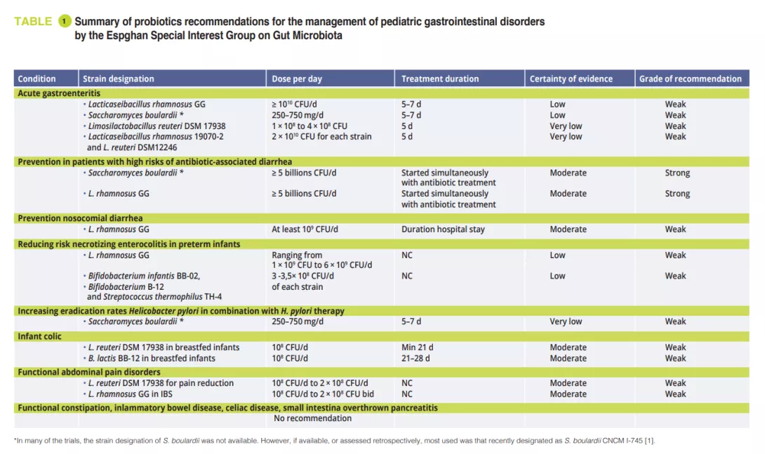
Gut Microbiota #19

By Pr. Satu Pekkala
Academy of Finland Research Fellow, Faculty of Sport and Health Sciences, University of Jyväskylä, Finland

Microbial metabolism of 5-ASA in inflammatory bowel disease

Mehta RS, Mayers JR, Zhang Y, et al. Gut microbial metabolism of 5-ASA diminishes its clinical efficacy in inflammatory bowel disease. Nat Med 2023; 29: 700-9.

Inflammatory bowel disease (IBD) is treated with 5-aminosalicylic acid (5-ASA). However, more than half of the patients do not respond to the treatment. Previous studies have suggested that this is partly due to that 5-ASA can be metabolized by gut bacteria into N-acetyl 5-ASA that is clinically ineffective. In this elegant study Mehta et al. aimed at identifying the gut microbial enzymes that generate N-acetyl 5-ASA. The human stool samples were analyzed using multi-omics. The untargeted metabolomic analyses of the samples pre-5-ASA and post-5-ASA administration revealed potential microbial mediators of the anti-inflammatory effects of 5-ASA. These included a decrease in 2-aminoadipate, a bacterial metabolite that has been linked to greater oxidative stress. In addition, 5-ASA seemed to alter nicotinate metabolism, which may also explain some anti-inflammatory effects. The authors further sought to identify the microbial enzymes that potentially metabolize 5-ASA. By combining metatranscriptomics and metabolomics, they identified three Acetyl-CoA C-acetyltransferases (Acyl-CoA NAT) that associated with the N-acetyl 5-ASA levels in 5-ASA users. In addition, some thiolases were identified as potential candidate enzymes. The candidate enzymes were then heterologously expressed in Escherichia coli and their biochemical activity was measured. Firmicutes CAG:176 thiolase and Faecalibacterium prausnitzii acyl-CoA NAT were able to acetylate 5-ASA using acetyl-CoA. Ultimately, a metagenomic analysis of the stool samples revealed that the gut microbial 5-ASA-inactivating acetyltransferases associated with greater risk of treatment failure in 5-ASA users. Altogether, the findings of this study can help in advancing the possibility of microbiome-based personalized treatment of IBD.

Image

Helicobacter pylori and colorectal cancer

Ralser A, Dietl A, Jarosch S, et al. Helicobacter pylori promotes colorectal carcinogenesis by deregulating intestinal immunity and inducing a mucus-degrading microbiota signature. Gut 2023; 72: 1258-70.

Helicobacter pylori infection can lead to gastric cancer and increase the risk for colorectal cancer (CRC). However, mechanistic data on the latter is lacking. In this paper, Ralser and co-workers identify in a rodent model the underlying mechanisms of how H. pylori infection contributes to CRC. When the authors infected Apc mice that are excellent animal models bearing multiple intestinal neoplasia with H. pylori, an increased tumor burden in the small intestine and colon was observed. It is known that the T-cell immune response of the host contributes to gastric carcinogenesis, and therefore, the authors studied these responses in the intestines. They found a reduction in regulatory T cells and pro-inflammatory T cells as well as an increase in IL-17A, which is shown to be one of the main players in the immune response to H. pylori. The infected mice were characterized by higher abundance of so-called pro-inflammatory gut microbes, and mucus degrading bacteria, such as Akkermansia. By studying transcriptomic profiles of the intestinal epithelial cells, they found that H. pylori induced the activation of NF-κB and STAT3 pathways. Activation of these pathways has been previously shown also in CRC patients. Interestingly, germ-free mice infected with H. pylori scarcely showed activation of STAT3 signaling suggesting that H. pylori induced carcinogenesis in the small intestine is partly dependent on the gut microbiome. Ultimately, the authors show that H. pylori induced colorectal carcinogenesis can be prevented by eradication of the bacterium with antibiotics. The authors conclude that implementation of H. pylori status into preventive measures of CRC should be considered.

Image

Viral diversity in the healthy infant gut

Shah SA, Deng L, Thorsen J, et al. Expanding known viral diversity in the healthy infant gut. Nat Microbiol 2023; 8: 986-98.

In infancy, the gut microbiome contributes to the maturation of the immune system to protect against chronic disease later in life. While it is known that bacteriophages (i.e., bacteria infecting viruses) can control the growth of bacterial populations, the gut viromes have not been extensively studied. By using metagenome sequencing, this study analyzed 647 viromes of a Danish cohort of 1-year-old infants. The first striking finding was that infant gut vOTUs were largely absent from gut virus databases. This suggests that the infant gut harbors specialized viruses distinct from the adult gut. The most predominant viral clades of the infants were largely undescribed. However, vertebrate-infecting ssDNA anelloviruses (Anelloviridae) and bacterial ssDNA microviruses (Petitvirales) were among the most abundant. In addition, virulent caudoviral families Skunaviridae, Salasmaviridae, β-crassviridae and Flandersviridae were also well represented in the infant viromes. Overall, temperate viruses were less prevalent than the virulent ones despite being found in more children. Family-level abundance was not significantly linked to phage lifestyle as determined by integrase as an indicator of a temperate lifestyle. However, temperate caudoviral families were genetically more diverse than the virulent families. Prediction analysis of the bacterial hosts of the viromes showed that Bacteroides, Faecalibacterium and Bifidobacterium were the three most prominent host genera in the infants’ gut. Of these, Bacteroides-infecting families were more often virulent and host specific. While no clear conclusions were provided, the study increases the knowledge on phage taxonomy and aids in the development of future infant gut virome research.

Image
Summary
Off
Sidebar
Off
Migrated content
Désactivé
Updated content
Désactivé
Hide image
Off
Press review Gut microbiota

Highlights from the 55th Espghan

By Dr. Tania Mahler
MD, Pediatric Gastroenterology & Nutrition - Chronic Pain & Functional Gastrointestinal Disorders - Qualifications in mindfulness and medical hypnosis for functional GI disorders. Clinical adjunct at Queen Fabiola Children’s University Hospital, Belgium

At the ESPGHAN (European Society of Pediatric Gastroenterology Hepatology and Nutrition) 2023 meeting held in Vienna, 4,300 participants from all over the world gathered. Following the challenging period of the Covid-19 pandemic, this event offered a revitalizing experience, as attendees were able to engage in live presentations and have faceto-face discussions This in-person interaction proved to be significantly more enjoyable and enriching than virtual alternatives. Various research groups focused their work on the gut microbiome and presented compelling data in the field of pediatrics.

The microbiome’s impact on health and disease is widely recognized, so it is evident that also pediatric clinicians and researchers try to get a better understanding of how we can manipulate the microbiome and how by using the signature of the microbiome we can detect disease in an early stage. This review aims to shed light on several key topics that have been extensively discussed.

Recommendations for the use of probiotics in selected pediatric gastrointestinal disorders

In February 2023, the Espghan special interest group on gut microbiota published recommendations for the use of probiotics for the management of selected pediatric gastrointestinal disorders based on systematic reviews and/or meta-analyses using the modified Delphi-process [1]. At the meeting of the special interest group on gut microbiota and modifications Prof. Szajewska showed us the results of this work. Only a few specific probiotic strains proof some utility in certain conditions. In the original paper [1] you find a clear overview of the actual recommendations.

FMT in adolescents suffering from refractory IBS

Dr De Bruijn from the group of Amsterdam UMC reported in the plenary session of the highest scoring abstract their study on the efficacy of FMT in adolescents with refractory IBS in a randomized double-blind placebo controlled trial [2]. Her talk was fascinating but also stirred a lot of reactions in the audience when she showed a slide from a patient receiving syringes with fecal material. To our knowledge only one other pediatric study has evaluated FMT to relieve abdominal bloating, another feature of disorders of the gut-brain interaction and which is often present in IBS [3].

The chronic pain in IBS can have an enormous impact on the functioning of children and adults leading to absenteeism at school and work and a poor quality of life. The origin of the disease is multifactorial and can best be explained by the biopsychosocial model. One of the key role factors is dysbiosis of the gut microbiota. In adults’ different studies have been published over the positive effect seen with FMT [4].

In pediatrics non-pharmacological treatments as education, hypnosis, mindfulness are more effective than pharmacological therapy [5]. Still in approximately 25% of patients’ symptoms persist. Pre-, pro-, and synbiotics are tested with various outcomes to correct the dysbiosis in IBS. Changing to FODMAP diet can also influence the gastrointestinal flora [5]. But in specific groups of patients FMT could be, if safe, the ultimate treatment to effectively restore a healthy gastrointestinal microbiome. In the study of De Bruijn et al., 32 patients with refractory IBS between 16 and 21 years old were recruited and randomized. One group received allogeneic (healthy donor) and the other group autologous (own) fecal infusions by nasogastric tube at baseline and 6 weeks later. Clinical efficacy was defined as the proportion of patients with a reduction of more than 50 points in the IBS Severity-Scoring-System (IBS-SSS). Patients were evaluated 12 weeks and 6 months after TMF. Both groups had similar IBSSSS at baseline. After the first evaluation there was no statistical difference, but at 6 months of follow-up there was improvement in 60% of the patients that received allogeneic TMF versus 25% in the autologous group (p = .048). Secondary outcome included health-related quality of life (QoL). Total QoL score at baseline did not differ between groups but became significantly better after allogeneic TMF. No adverse events were recorded. Allogeneic TMF seems an exciting way to treat refractory IBS in youth, but further studies are needed.

Image

Microbiota and IBD

In the gastroenterology session on IBD a Czech group presented a study that had as aim to evaluate if the changes in microbiota in CD was due to the anti-TNFα treatment or was the result of decreased mucosal inflammatory activity [6]? Therefore, they compared children on anti-TNFα treatment with active Crohn’s disease (CD) and juvenile idiopathic arthritis (JIA). Their results showed that mucosal healing in CD was essential to obtain changes in the bacteriome. The antiTNFα treatment in JIA had no impact on the bacteriome of these patient group. Schwerd et al. followed 20 newly diagnosed pediatric CD patients treated first with exclusive enteral nutrition (EEN) with stool sampling [7]. Fifteen out of twenty patients went in remission. They demonstrated clear temporal and individual intestinal microbial and metabolite changes with reduced abundance of Lachnospiraceae and enriched unsaturated long chain fatty acids. Ex vivo fermentation with an EEN-like media and subsequent transfer in gnotobiotic mouse models showed a protective effect in contrast to the fiber-rich media and to those colonized directly with patient’s baseline microbiota. Based on those results they concluded that EEN-modulated patient microbiomes are regulating intestinal inflammation. They also elaborated on the possibility of using a low-fiber diet for long-term remission. A multicenter study in the UK (children and adults) studied the possibility to use a Crohn’s Disease TReatment-with-EATing (CD-TREAT) solid food diet to create a more palatable diet that could influence gut inflammation by changing the gut bacteria [8]. The diet is personalized for each patient but excludes specific dietary components such as gluten, lactose, alcohol. The 55% of patients adhering to this regime had a significant lower fecal calprotectin and had microbial and metabolic changes in the same line as patients under successful EEN. This was not seen in those not respecting the diet. Based on those findings, it could be interesting to use autologous feces of successfully treated EEN CD patients for FMT. The group of Schwerd analyzed this possibility using autologous capsule FMT. They concluded that this approach was unsuitable since there was still a to high pathogen burden and a to low microbiota diversity [9].

The Cologne group had a very interesting poster on the follow-up of 2 cases of very early IBD refractory to steroids and antiTNFα treatment. The first patient has ulcerative colitis and is now 3 years in total remission with weekly administered enema of donor stool preparation. The second patient with CD is only in partial remission after one year of follow-up [10].

Combined efforts of scientific researchers and clinicians will further unravel the mystery of the gut microbiota and will eventually bring new ways to treat and to prevent diseases.

Sources

1. Szajewska H, Berni Canani R, Domellöf M, et al. Probiotics for the Management of Pediatric Gastrointestinal Disorders: Position Paper of the ESPGHAN Special Interest Group on Gut Microbiota and Modifications. J Pediatr Gastroenterol Nutr 2023; 76: 232-47
2. De Bruijn C, Zeevenhoven J, Vlieger A, et al. Efficacy of fecal microbiota transplantation in adolescents with refractory irritable bowel: a randomized, double-blind, placebo-controlled trial. J Pediatr Gastroenterol Nutr 2023; 76(S1 Suppl 1): 1-1407
3. Wang YZ, Xiao FF, Xiao YM, et al. Fecal microbiota transplantation relieves abdominal bloating in children with functional gastrointestinal disorders via modulating the gut microbiome and metabolome. J Dig Dis 2022; 23: 482-92
4. El-Salhy M, Winkel R, Casen C, et al. Efficacy of Fecal Microbiota Transplantation for Patients with Irritable Bowel Syndrome at 3 Years After Transplantation. Gastroenterology 2022; 163: 982-94.e14
5. Mahler T, Hoffman I, Smets F, et al. The Belgian consensus on irritable bowel syndrome: the paediatric gastroenterologist view. Acta Gastroenterol Belg 2022; 85: 384-6
6. Hurych J, Mascellani Bergo A, Lerchova T, et al. The faecal microbiome and metabolome changes in Crohn’s disease are associated with decreased mucosal inflammatory activity. J Pediatr Gastroenterol Nutr 2023; 76 (S1 Suppl 1): 1-1407
7. Schwerd S, Häcker D, Siebert K, et al. Exclusive enteral nutrition initiates protective functions in the gut microbiota and metabolome to induce remission in pediatric Crohn’s disease. J Pediatr Gastroenterol Nutr 2023; 76 (S1 Suppl 1): 1-1407
8. Macdonald J, Wilson D, Henderson P, Din S, e Chantges in faecal microbiome and metabolome are more pronounced in Crohn’s disease patients who adhered to the CD-TREAT diet and responded by calprotectin. J Pediatr Gastroenterol Nutr 2023; 76 (S1 Suppl 1): 1-1407
9. Hölz H, Heetmeyer J, Tsakmaklis A, et al . Autologous fecal microbiota transfer in pediatric Crohn ́s disease patients under treatment with exclusive enteral nutrition harbors major challenges - a feasibility test. J Pediatr Gastroenterol Nutr 2023; 76 (S1 Suppl 1): 1-1407
10. Fritz T, Huenseler C, Broekaert I. Safety and efficacy of long-term faecal microbiota transfer in very early onset inflammatory bowel disease. J Pediatr Gastroenterol Nutr 2023; 76 (S1 Suppl 1): 1-1407

Summary
Off
Sidebar
Off
Migrated content
Désactivé
Updated content
Désactivé
Hide image
Off
Congress review Gut microbiota

Cholestasis impairs gut microbiota development and bile salt hydrolase activity in preterm neonates

COMMENTED ARTICLE - Children’s section

By Pr. Emmanuel Mas
Gastroenterology and Nutrition Department, Children’s Hospital, Toulouse, France

Commentary on the original article by Lynch LE et al. Gut Microbes [1]

Cholestasis refers to impaired bile flow from the liver to the intestine. In neonates, cholestasis causes poor growth and may progress to liver failure and death. Normal bile flow requires an intact liver-gut-microbiome axis, whereby liver-derived primary bile acids are transformed into secondary bile acids. Microbial bile salt hydrolase (BSH) enzymes are responsible for the first step, deconjugating glycine- and taurine-conjugated primary bile acids. Cholestatic neonates often are treated with the potent choleretic bile acid ursodeoxycholic acid (UDCA), although interactions between UDCA, gut microbes, and other bile acids are poorly understood. This study was conducted on 124 stool samples collected from 24 preterm infants to find novel associations linking isomeric bile acids and BSH activity to neonatal growth trajectories. These data highlight deconjugation of bile acids as a key microbial function that is acquired in early neonatal development and is impaired by cholestasis.

What do we already know about this subject?

Preterm infants born before 37 weeks of amenorrhoea (WA) are at a greater risk of developing cholestasis. Cholestasis (i.e., impaired bile flow) is more likely in the presence of various risk factors such as prematurity, low birth weight and parenteral nutrition. In the absence of other causes, this is referred to as transient neonatal cholestasis. To improve cholestasis, ursodeoxycholic acid (UDCA) is often administered.

Bile acids are necessary for the absorption of lipids and fat-soluble vitamins. Reduced quantities of bile acids in the gut is observed in cholestasis, as are changes in the proportions of the different bile acids. Primary bile acids are produced from cholesterol and conjugated in the liver. Their interactions with the intestinal microbiota play an important role leading to the formation of secondary bile acids through the intermediary of a microbial enzyme, bile salt hydrolase (BSH).

Bile acids and intestinal microbiota have an impact on the growth and development of preterm infants. The authors looked at the impact of cholestasis on gut microbiota development and bile acid deconjugation in very premature neonates.

What are the main insights from this study?

The authors included 24 preterm neonates, 12 with cholestasis and 12 controls, born at 27.2 ± 1.8 WA, with a mean birth weight of 946 ± 249.6 g. Mean peak conjugated bilirubin was 7.0 mg/dL. No differences existed between the two groups in terms of intrauterine environment, delivery method and antibiotic use over time.

Image

Stools were collected from birth to six weeks. Their sequencing (shotgun method) showed that in controls alpha-diversity increased during the first months of life. In terms of phyla, Proteobacteria and Firmicutes were the most abundant. In terms of genera, Staphylococcus was the most prominent at birth, its abundance then decreased while Klebsiella gradually increased in abundance (figure 1). Clostridium perfringens increased the most in relative abundance over time, based on postmenstrual age (PMA, which is the sum of postnatal age and WA at birth) (p = 0.01). According to the metagenomic analysis, the metabolic pathway most enriched in mature stools (32-40 weeks PMA) compared with less mature stools (25-28 weeks PMA) was secondary bile acid biosynthesis.

In the control group, the principal component analysis showed that the main factor influencing the composition of the intestinal microbiota was PMA, whereas it had no effect in cholestatic premature infants. Secondary bile acid biosynthesis was the most enriched pathway in stools from the control group relative to the cholestatic group at 32-40 weeks PMA (p = 0.04). Similarly, the authors observed a 55% reduction in the relative abundance of the BSH gene (p = 0.04) and Clostridium perfringens (p = 0.0008) in the cholestatic neonates (figure 2).

Image

Faecal bile acid profile measured by mass spectrometry showed that the proportion of unconjugated bile acids increased from 4% at 25–28 weeks PMA to 98% by 32– 40 weeks PMA in the control group but was only 46% in cholestatic infants. However, it should be noted that some isomers may have a predictive value as they increased before the onset of cholestasis. UDCA used on five of the 12 premature neonates was found in their faeces at a concentration 522-fold higher compared to the seven other untreated neonates. UDCA administration increased the relative abundance of Firmicutes and decreased Proteobacteria (p < 0.05), with an enrichment in Clostridium perfringens at a species level.

Finally, premature neonates with faecal BSH gene abundance > 0.005% at 32–40 weeks PMA exhibited a 1.2-fold increased length and weight rate compared to those with an abundance < 0.005%. Equally, neonates with a faecal cholic acid composition of > 30% demonstrated increased length (14%), weight (18%), and head circumference (15.8%) (p < 0.05).

What are the consequences in practice?

This study offers an insight into the pathophysiological mechanisms that disrupt the liver-gut-microbiome axis during cholestasis. This opens the way to the possibility of correcting the enterohepatic cycle using (BSH-carrying) probiotics or other medicinal products.

CONCLUSION

In extremely preterm neonates, cholestasis disrupts the development of the intestinal microbiota, by reducing the acquisition of Clostridium perfringens and the capacity to synthesise secondary bile acids. In contrast, an increase in certain bile acids, linked to BSH activity, is associated with improved neonatal growth.

Summary
Off
Sidebar
Off
Migrated content
Désactivé
Updated content
Désactivé
Hide image
Off
Commented article Preterm birth

Microbiota-derived 3-IAA (indole-3-acetic acid) influences chemotherapy efficacy in pancreatic cancer

COMMENTED ARTICLE - ADULTS’ SECTION

By Pr. Harry Sokol
Gastroenterology and Nutrition Department, Saint-Antoine Hospital, Paris, France

Commentary on the article by Tintelnot et al. Nature 2023 [1]

Pancreatic ductal adenocarcinoma (PDAC) is expected to become the second most deadly cancer by 2040, owing to the high incidence of metastatic disease and limited responses to treatment. Less than half of all patients respond to the primary treatment for PDAC, and genetic alterations alone cannot explain sufficiently the different responses. Diet is an environmental factor that can influence the response to therapies, but its role in PDAC is unclear. Here, using shotgun metagenomic sequencing and metabolomic screening, the authors showed that the microbiota-derived tryptophan metabolite indole-3-acetic acid (3-IAA) is enriched in patients who respond to treatment. Faecal microbiota transplantation, dietary manipulation of tryptophan and oral 3-IAA administration increase the efficacy of chemotherapy in humanised mouse models of PDAC. Using a combination of loss- and gain-of-function experiments, the authors showed that the efficacy of 3-IAA and chemotherapy requires the presence of neutrophil-derived myeloperoxidase (MPO). MPO oxidises 3-IAA which, in combination with chemotherapy induces a downregulation of the reactive oxygen species (ROS)-degrading enzymes glutathione peroxidase 3 and glutathione peroxidase 7. This all results in the accumulation of ROS and the downregulation of autophagy in cancer cells, which compromises their metabolic fitness and, ultimately, their proliferation. In humans, the authors observed a significant correlation between 3-IAA levels and the efficacy of therapy in two independent PDAC cohorts. In summary, the authors identified a microbiota-derived metabolite that has clinical implications in the treatment of PDAC, and provided a motivation for considering nutritional interventions when treating cancer patients.

What do we already know about this subject?

Polychemotherapy, either with 5-fluorouracil (5-FU), irinotecan and oxaliplatin in combination with folinic acid (FOLFIRINOX), or with gemcitabine and nabpaclitaxel (GnP), is considered the standard of care for patients suffering from metastatic PDAC (mPDAC). However, less than half of all patients are responsive to the therapy, and patients who do not respond (NR, non-responder patients) suffer pain and die within a few weeks. Genetic alterations in PDAC poorly explain the differences between patients who respond to therapy (R, responder patients) and NR patients, which leaves environmental factors, including the intestinal microbiota, as the potential mediators of chemotherapy efficacy. Thus, there is an urgent need to identify environmental factors that might explain the differences between R and NR patients to develop new concepts for future therapies. The intestinal microbiota has been shown to induce a response to immunotherapy in patients with melanoma, and can be modulated by the diet 2, 3 . In rare long-term survivor patients with localised PDAC, bacteria can pass from the intestine to the tumour and thus control anti-tumour immune activation. However, most patients suffering from aggressive immunotherapy-resistant mPDAC are treated with polychemotherapy, and it is presently unclear whether and how the microbiota or dietary habits affect the efficacy 4 .

Image
Key points
  • The intestinal microbiota is different in PDAC patients who are responders and non-responders to chemotherapy
  • The tryptophan metabolite 3-IAA, produced by the microbiota, is enriched in responder patients
  • 3-IAA increases the efficacy of chemotherapy by boosting ROS production by neutrophils and decreasing autophagy in cancer cells

What are the main insights from this study?

Analysing the microbiota of 30 mPDAC patients revealed differences between R and NR patients. Transferring the microbiota of R patients to mice with pancreatic tumours reduced the size of the tumours after chemotherapy. The tryptophan metabolite 3-IAA was enriched in R patients and mice with R microbiota, potentially contributing to the response to chemotherapy. The administration of 3-IAA increased the efficacy of chemotherapy in mice (figure 1). The analysis of immune cells in mice showed an increase in CD8+ T cells and a reduction in neutrophils in mice with a microbiota associated with a good response to chemotherapy. 3-IAA affected the MPO of neutrophils, thereby reducing their survival. The combination of 3-IAA and chemotherapy reduced the number of neutrophils and inhibited tumour growth, with MPO playing a crucial role. It is suggested that MPO induces the ROS production leading to cell death during chemotherapy. In-vitro experiments have shown that 3-IAA increases ROS levels. This effect was confirmed in vivo and the inhibition of ROS by N-acetylcysteine abolished the efficacy of FIRINOX in mice with high 3-IAA levels. The authors then showed that the effect of 3-IAA was linked to downregulation of autophagy. Finally, high serum concentrations of 3-IAA were correlated to a reduction in neutrophil counts and improved survival in the two cohorts of human patients.

What are the consequences in practice?

The intestinal microbiota has an effect on chemotherapy responses. The mechanisms involved in this study demonstrates the role of microbial metabolites, particularly tryptophan metabolites. Among these, 3-IAA is not only a predictive marker of response to chemotherapy in PDAC, but could also offer an adjuvant therapeutic drug.

CONCLUSION

The intestinal microbiota has an effect on chemotherapy responses in PDAC. One of its metabolites, 3-IAA, is a predictor of a good response to chemotherapy and enhances its effect by inducing an accumulation of ROS and reducing autophagy in cancer cells.

Image
Summary
Off
Sidebar
Off
Migrated content
Désactivé
Updated content
Désactivé
Hide image
Off
Commented article Pancreatic cancer

Gut microbiome and antimicrobial resistance

By Dr. Joseph Nesme

Assistant professor, Section of Microbiology, Department of Biology, University of Copenhagen, Denmark

Antibiotic resistance is ancient and prevalent and the environmental resistome is a reservoir of resistance genes fueling the antimicrobial resistance silent pandemic. Horizontal gene transfer, population dynamics and co-selection accelerate emergence and dissemination of the resistome and by year 2050, more than 10 million death per year will be due to antibiotic resistance. The early colonization of the gut microbiome is prone to dysbiosis that can lead to increased abundance and diversity of the gut resistome of infants. Adopting a One Health approach, considering the interconnectedness of human health, animal health, and the environment, is essential for addressing the complex challenges posed by AR.

Antibiotic resistance in the environment: A pre-existing challenge

Antibiotic resistance (AR) is an ancient and prevalent phenomenon in the environment existing long before the introduction of antibiotic molecules as therapeutic. The environment serves as a vast reservoir of antibiotic resistance genes with diverse microbial communities harboring resistance mechanisms. AR has been found in various environmental settings, including soil, water, plants, animals, and even in 30,000 years old Arctic permafrost 1, 2 . The ecological role of antibiotic molecules and associated resistance in non-clinical settings remains unclear but highlights the fact that a readily available pool of genes predates clinical antibiotic usage and explain rapid emergence in pathogens. The current antibiotic crisis is an evolutionary phenomenon and mitigation strategies needs to account for microbial ecology. It is the rapid acquisition of resistance by pathogens that were previously sensitive leading to therapy failures that is problematic, especially when very few novel antimicrobials are expected to reach the market 3 .

Mechanisms driving the emergence and dissemination of the resistome

The resistome refers to the complete set of genes that encode for antibiotic resistance (AR)-related proteins or those proteins that could potentially evolve into powerful AR agents 4 (figure 1). This includes recognized AR genes in pathogenic bacteria (the problematic ones), AR genes from antibiotic-producing organisms, such as Streptomyces spp. producing streptomycin antibiotic and associated resistance gene[5], cryptic AR genes (i.e., genes that could provide resistance in a different genetic setting; e.g., upregulated efflux pumps or downregulated porins), and precursor AR genes that code proteins with a minimal level of affinity or resistance to antibiotic compounds.

It is noteworthy that a considerable portion of these gene subsets overlap due sequences homology, indicating they likely have a shared evolutionary background.

Image

The notion of resistome is distinct from “functional and clinically relevant AR” important. Indeed, resistome genes can transition between the different states described above by horizontal gene transfer (HGT), point mutations, or recombination which are leading to new hosts or genetic context where clinically relevant AR phenotype can be expressed. A resistance gene is therefore not problematic per se as this is host and genetic context dependent, but all resistome genes are potential threat with different associated public health risk outcomes. The discovery of a resistance gene towards a clinically relevant molecule, located on a mobile element and hosted by a human pathogen is a critical risk but the same gene, or close homologues, found in a non-pathogenic soil bacterium and not associated with mobile genetic element is a very low risk ARG. Ranking risk of antibiotic resistance in resistome studies is therefore of paramount.

Horizontal gene transfer (HGT) is a key mechanism responsible for the rapid spread of AR genes among bacteria, even across distant lineages. For example, Bacteroides spp., a predominant group in the human gut microbiota, possess macrolide resistance gene ermB identical to those found in several Clostridium perfringens, Streptococcus pneumoniae and Enterococcus faecalis isolates from various geographical origin, indicating genetic connection between Bacteroides and some Gram-positive bacteria that are not prevalent in the human gut . Genetic elements, such as plasmids, facilitate the transfer of resistance genes between different microbial species . HGT enables the dissemination of genes across diverse environments and bacterial populations, contributing to the overall prevalence and diversity of AR. Co-selection is another significant factor in the spread of AR. The use of non-antibiotic compounds, such as heavy metals and biocides, can co-select for AR genes by exerting selective pressures on microbial populations, either by co-resistance (different resistance determinants present on the same genetic element) and cross-resistance (the same genetic determinant responsible for resistance to antibiotics and metals) 8 . Exposure to naturally occurring antimicrobial compounds such as those produced by competing microorganisms or any co-selective compounds can drive the selection of resistant strains 9 . The presence of antibiotics in the environment, either from natural sources or human activities, further contributes to the selection pressure for resistance. Additionally, the use of antibiotics in agriculture and veterinary practices can lead to the contamination of the environment, promoting the emergence and spread of antibiotic environmental resistance genes overtime 10 .

Understanding barriers and trends to the transfers of genetic vectors harboring resistance genes is crucial to devise eco-evolutionary informed mitigations strategies to limit the dissemination of AR in the early life and in general in clinical settings.

Dysbiosis and the gut resistome in infants: A delicate balance

The environmental reservoir diversity of AR genes and its potential to be transfered poses a threat to the early life human gut microbiome. Strategies such as improved wastewater treatment, responsible use of antibiotics in agriculture and veterinary medicine, and reducing environmental contamination with antibiotic residues and antibiotic resistant bacteria can help mitigate the spread of resistance 11 . Additionally, monitoring and surveillance of environmental reservoirs can provide valuable insights into the emergence and persistence of AR and inform public health interventions.

Adopting a One Health approach, considering the interconnectedness of human health, animal health, and the environment, is essential for addressing the complex challenges posed by AR.

Image

Our gut is rapidly colonized after birth by microorganisms acquired from their mothers and their surrounding environment. It is during the first years of life that changes are drastic and characterized by a low resilience compared to the more stable healthy adult’s gut microbiome. Newborns and infants are therefore more prone to disruptions in microbial communities, known as dysbiosis. During that period, many factors can influence and perturbate gut maturation and potentially lead to long-term consequence on health 12 . Mice studies showed that during this critical developmental window, rather than a direct effect of antibiotic molecules, it is the alteration of the gut microbiota composition that is triggering metabolic consequences, such as obesity 13 .

Unraveling the antibiotic resistome in infants gut: Insights from a large cohort study

While antibiotic resistance is problematic at all age of life, the establishment of the gut microbiome at early age represent a window of opportunity to limit the buildup of an AR genes reservoir in the gut. It is important therefore to identify the various factors increasing or reducing the abundance of AR genes that could spread to infectious pathogens and cause antibiotic therapy failure throughout life. To study the global human gut resistome researchers rely on holistic approaches interrogating both species presence and genomes functional potential, including the antibiotic resistome. Researchers rely on environmental DNA extraction from proxy samples (e.g., stool samples for the gut) followed by untargeted high-throughput sequencing (shotgun metagenomics). Approximately 80% of human gut bacterial species detected by molecular tools are unculturable, especially the specialized anaerobes inhabiting the gut. It is likely that many microorganisms are organized in multi-species cell aggregates with metabolic co-dependencies making pure strain isolation delicate if not impossible. Using computational methods, it is however possible to reconstruct quasi-complete genomes from metagenomes and associate encoded genes with specific species or even strains of bacteria. In a recent study, researchers analyzed fecal samples from 662 infants from a cohort which followed children from birth to age 7 14 . The objectives of the study were to establish a cohort scale overview of the resistome at 1 year of age and identify perinatal and environmental factors associated with ARG abundance and diversity. The researchers used shotgun metagenomic sequencing of samples obtained at 1 year of age from the 662 children to identify ARGs and bacterial taxa present in the samples (figure 2). Making use of their large dataset, the authors were able to reconstruct Metagenomes Associated Genomes (MAGs), allowing them to confidently annotate the taxonomy of recovered genomes and their AR genes content.

A first result observed was that all children had at least one type of multiple drug ARG in their gut, indicating that even in absence of antibiotic treatment, there is a resident resistome associated with the gut microbiome. Indeed, many multidrug resistance genes were identified as efflux-pumps. These proteins are normal component of every bacterial cell but some can confer AR and are very easily co-selected by cross-resistance, for example towards heavy metals or biocides, potentially explaining their high abundance in the gut, but also in all environments 8, 15 . Another striking result was the clear separation of the cohort in two groups based on their resistome profile. The first group was characterized by a higher ARGs diversity and relative abundance with Escherichia coli as the main contributor of ARGs, as shown in (figure 2). This agrees with previous observations that Enterobacteriaceae are abundant early in life but should decrease rapidly when Bacteroidetes population starts colonizing the gut. Alteration of this maturation in some children can be associated to a combination of several factors, such as antibiotic usage, mode of delivery, rural or urban household that seems to delay the reduction of the population of Escherichia coli and lead to an increased resistome at 1 year of age. This is also confirmed by the observation that the higher abundance of ARGs is associated with a lower gut microbiome maturity score, based on ratios of specific age-related taxa 16 .

Antibiotic treatment during pregnancy and the first year of life of course had significant impact on the abundance of the infant gut resistome. A clear temporal trend could be observed between the time (more than 6 months or less) since the last antibiotic course taken by a child and the abundance of ARGs in its gut.

This indicates some level of resilience at an early age which could be potentially improved with targeted intervention, e.g., with pro- or prebiotics, and remains to be tested. At the cohort scale, the largest differences in ARG abundance were however found for resistance genes towards antibiotic that were not prescribed to the children, underlying that perinatal and environmental factors besides antibiotic therapy are also driving the gut resistome. Another observation from that study that connects the surrounding environment, and associated resistome, and the gut resistome was that children whose households was in urban areas had a significantly higher load of ARGs than children living in rural areas. There is a myriad of potential confounding factors that could explain this but it strengthens the fact that the environment contribution to the development of the early life microbiome is extremely important.

It can be hypothesized that urban living is associated with less contact with the outdoor and a decreased microbiome diversity, or that the type of lodging found in rural (house) or urban (appartement) environment are associated have consequences on the indoor microbiome as seen in bed dust.

Conclusion

Antibiotic resistance is ancient and prevalent and the environmental resistome is a reservoir of potential clinical resistance genes. Mechanisms such as horizontal gene transfer, intricate population dynamics, and co-selection effects, have been identified as significant accelerators in the evolution and spread of the antibiotic resistome making previously sensitive strains of pathogens now resistant to antibiotic therapy. The human gut microbiome and its early developmental phase is prone to dysbiosis and potential invasion, selection, and co-selection of bacteria harboring resistance genes, which can have long term health consequences. It is unclear to what extent these are related to future failed antibiotic therapies. High temporal resolution, long term multi-omics studies are needed to identify first longitudinal patterns of gut microbiome development and second, most probable routes of AR genes transfer by HGT. Together, these results will provide insights to devise preventive strategies for minimizing the development of AR already early in life and limit transfer to problematic pathogens. This effort is of the utmost importance to reduce the burden of the worldwide AMR pandemic and improve global public health by preserving the efficacy of the few antibiotic available to fight infectious diseases.

Sources

1. Nesme J, Cécillon S, Delmont TO, et al. Large-scale metagenomic-based study of antibiotic resistance in the environment. Curr Biol 2014; 24: 1096-100.
2. D’Costa VM, King CE, Kalan L, et al. Antibiotic resistance is ancient. Nature 2011; 477: 457-61.
3. Renwick MJ, Brogan DM, Mossialos E. A systematic review and critical assessment of incentive strategies for discovery and development of novel antibiotics. J Antibiot 2016; 69: 73-88.
4. Wright GD. The antibiotic resistome: the nexus of chemical and genetic diversity. Nat Rev Microbiol 2007; 5: 175-86.
5. Benveniste R, Davies J. Aminoglycoside antibiotic-inactivating enzymes in actinomycetes similar to those present in clinical isolates of antibiotic-resistant bacteria. Proc Natl Acad Sci USA 1973; 70: 2276-80.
6. Shoemaker NB, Vlamakis H, Hayes K, et al. Evidence for extensive resistance gene transfer among Bacteroides spp. and among Bacteroides and other genera in the human colon. Appl Environ Microbiol 2001; 67: 561-8.
7. Martínez JL, Coque TM, Baquero F. Prioritizing risks of antibiotic resistance genes in all metagenomes. Nat Rev Microbiol 2015; 13: 396.
8. Baker-Austin C, Wright MS, Stepanauskas R. Co-selection of antibiotic and metal resistance. Trends Microbiol 2006; 14: 176-82.
9. Wencewicz TA. Crossroads of Antibiotic Resistance and Biosynthesis. J Mol Biol 2019; 431: 3370-99.
10. Knapp CW, Dolfing J, Ehlert PA, et al. Evidence of increasing antibiotic resistance gene abundances in archived soils since 1940. Environ Sci Technol 2010; 44: 580-7.
11. Wolters B, Hauschild K, Blau K, et al. Biosolids for safe land application: does wastewater treatment plant size matters when considering antibiotics, pollutants, microbiome, mobile genetic elements and associated resistance genes? Environ Microbiol 2022; 24: 1573-89.
12. Dogra S, Sakwinska O, Soh SE, et al. Rate of establishing the gut microbiota in infancy has consequences for future health. Gut Microbes 2015; 6: 321-5.
13. Cox LM, Yamanishi S, Sohn J, et al. Altering the intestinal microbiota during a critical developmental window has lasting metabolic consequences. Cell 2014; 158: 705-21.
14. Li X, Stokholm J, Brejnrod A, et al. The infant gut resistome associates with E. coli, environmental exposures, gut microbiome maturity, and asthma-associated bacterial composition. Cell Host Microbe 2021; 29: 975-87.e4.
15. Mulder I, Siemens J, Sentek V, et al. Quaternary ammonium compounds in soil: implications for antibiotic resistance development. Rev Environ Sci Bio/Technology 2018; 17: 159-85.
16. Subramanian S, Huq S, Yatsunenko T, et al. Persistent gut microbiota immaturity in malnourished Bangladeshi children. Nature 2014; 510: 417-21.

Summary
Off
Sidebar
Off
Migrated content
Désactivé
Updated content
Désactivé
Hide image
Off
Article Gut microbiota Off

Is the microbiota key to the early diagnosis of Alzheimer’s disease?

Even before patients show any clinical signs of Alzheimer’s disease, imbalances in their gut microbiota may betray its onset. Is it thus possible to detect the disease earlier?

The gut microbiota
Photo: Le microbiote, la clé du diagnostic précoce de la Maladie d'Alzheimer ?

Alzheimer’s disease is progressive and silent. During the so-called pre-clinical phase, cognitive state appears normal. However, in-depth tests have already shown the progressive accumulation of two proteins in the brain, β-amyloid (Aβ) and tau proteins, which cause brain damage and a slow degeneration of neurons, beginning in the memory center and then spreading to the rest of the brain.

An interval of at least 10 years exists between the first deposition of Aβ plaques in the brain and the first clinical signs of impairment.

After this silent phase, the first symptoms of (sidenote: Dementia Brain disorders that affect memory, thinking, behavior, and emotions. Changes in mood and behavior sometimes precede memory problems. Symptoms worsen over time. Most sufferers eventually require assistance in their day-to-day life. Sources: OMS and Alzheimer’s Disease International ) appear. This is the clinical stage of Alzheimer’s disease, marked by mood and personality changes, memory lapses, forgetting certain words to the point of being difficult to understand, disorientation in space and time, leaving things in inappropriate places (keys in the fridge), etc.

More than 55 million people worldwide suffer from dementia.

60% to 70% of dementia cases are caused by Alzheimer’s disease.

Gut dysbiosis in pre-clinical Alzheimer’s disease

What role does the gut microbiota play in all this? We already know that in the clinical stage of the disease, patients have an unbalanced gut microbiota. According to a US study published in 2023, this imbalance also exists in the pre-clinical stage and is all the more pronounced the more β-amyloid proteins have accumulated. This imbalance in the gut microbial ecosystem (or dysbiosis) is not linked to diet: future Alzheimer’s patients who do not yet show any signs of dementia have a diet comparable to that of healthy patients who are not slowly succumbing to the disease.

75% of those surveyed were unaware that diseases such as Parkinson’s, Alzheimer’s, and autism may be linked to microbiota.

Can this help predict the subsequent clinical form of the disease?

The team identified gut bacteria that are generally over- or under-represented in the pre-clinical stage. These bacteria allowed them to use (sidenote: Machine Learning Automatic learning whereby artificial intelligence solves a task based on metagenomic and metabolomic data collected, in this case the identification of discriminating bacterial species. Wazid M, Das AK, Chamola V, et al. Uniting cyber security and machine learning: Advantages, challenges and future research. ICT Express, 2022; 8(3), 313-321. ) to improve their predictive models for Alzheimer’s disease. Admittedly, the gain is small when the initial model incorporates β-amyloid proteins, which are the main pre-clinical signature of Alzheimer’s disease. However, the latter requires lumbar punctures and brain neuroimaging, complex and uncommon procedures. When models are based solely on readily available data (age, sex, hypertension, family history, etc.), the addition of bacterial data from a stool sample improves model (sidenote: Sensitivity The sensitivity of a medical test measures its ability to correctly detect those suffering from a disease (identification of as many sufferers as possible). A sensitivity of close to 100% means the test has little chance of missing cases of a disease, i.e. few false negatives (few undetected true sufferers). Bertrand D, Fluss J, Billard C. Efficacité, sensibilité, spécificité : comparaison de différents tests de lecture. L’Année psychologique, 2010 ; 110, 299-320. ) by 6.8% and (sidenote: Specificity Specificity is the probability that the test will be negative knowing that the subject is healthy. It therefore measures a test’s ability to detect healthy individuals. The closer the specificity is to 1, the fewer the false positives. Bertrand D, Fluss J, Billard C. Efficacité, sensibilité, spécificité : comparaison de différents tests de lecture. L’Année psychologique, 2010 ; 110, 299-320. ) by 27.1%.

This makes it easier to pre-identify at-risk patients, who can then be prescribed more in-depth examinations. If bacteria are indeed the cause of these changes, which has yet to be confirmed, these results also suggest the possibility of modifying the gut microbiota to limit the progression of Alzheimer’s disease.

Alzheimer’s: how the gut makes us lose our head

Learn more
Summary
Off
Sidebar
On
Migrated content
Désactivé
Updated content
Désactivé
Hide image
Off
News

Gut microbiota: an early indicator of Alzheimer’s disease?

Before even the slightest clinical sign of Alzheimer’s disease appears, future sufferers see a change in their gut microbiota. These changes may help identify patients at the pre-clinical stage.

Previous studies have highlighted dysbiosis in the gut microbiota of patients showing symptoms of Alzheimer’s disease. But what about before the first symptoms appear? A team from the University of Washington School of Medicine in the US examined this question by analyzing the microbiota of 164 people aged between 68 and 94 who had no cognitive impairment, but 49 of whom showed (sidenote: Biomarkers Pathogenic β-amyloid (Aβ) and tau proteins measured via positron emission tomography (PET) or by levels in cerebrospinal fluid (CSF), markers of neurodegeneration (temporo-parietal hypometabolism, hippocampal atrophy, etc.) identified via CSF and magnetic resonance imaging (MRI). ) . The results were unequivocal: the gut microbial taxonomic profiles of the 49 “pre-patients” differed from those of the 115 controls.

55 million More than 55 million people worldwide suffer from dementia.

60% to 70% 60% to 70% of dementia cases are caused by Alzheimer’s disease.

A gut microbiota specific to pre-clinical stages

This dysbiosis correlates with markers of pre-clinical stages of the disease, notably the deposition of β-amyloid plaques in the brain. However, it is not linked to biomarkers of neurodegeneration (temporo-parietal hypometabolism, hippocampal atrophy, etc.). Thus, the gut microbiota seems to change from a very early, asymptomatic stage of the disease.

More specifically, the content of certain bacteria either increases or decreases, including Dorea formicigenerans, which has pro-inflammatory properties, Oscillibacter sp. 57_2, which may be associated with reduced epithelial integrity, anti-inflammatory Faecalibacterium prausnitzii, and to a lesser extent, Coprococcus catus, Anaerostipes hadrus, Methanosphaera stadtmanae, and Ruminococcus lactaris. Some of these gut bacteria may therefore be involved in the causal chain, although further experiments are required to confirm any such causal link and rule out a simple co-occurrence.

10 years An interval of at least 10 years exists between the first deposition of Aβ plaques in the brain and the first clinical signs of impairment.

Simplifying and improving identification of at-risk patients

In any case, this bacterial signature may help improve disease prediction. This is based on a test carried out on a sub-group of 65 patients, where the addition of these bacterial taxa improved the accuracy of predictive models. This improvement is slight (sensitivity +1.5%, specificity +5.0%) when the initial model includes the β-amyloid protein, the main pre-clinical signature of the disease. However, the latter requires complex tests. When models are based solely on readily available data (demographics, clinical covariates, and genetics), the addition of taxonomic characteristics, which require only a stool sample, improves sensitivity by 6.8% and specificity by 27.1%. This makes it easier to pre-identify at-risk patients, who can then be prescribed more in-depth examinations (lumbar puncture and neuroimaging). Lastly, the study could open the door to interventions on the microbiota aimed at limiting the progression of Alzheimer’s disease towards clinical stages.

Summary
Off
Sidebar
On
Migrated content
Désactivé
Updated content
Désactivé
Hide image
Off
News Neurology Gastroenterology

Stroke: gut microbiota directly implicated

A Mendelian randomization study 1 has confirmed the causal effects of gut microbiota on ischemic stroke: certain bacteria have been identified as being able to increase or reduce this risk, opening up the prospect of prevention with probiotics.

Observational studies have one drawback: they cannot distinguish the chicken from the egg, and when it comes to microbiota, they cannot show whether dysbiosis is the cause or consequence of an illness. The solution? Mendelian randomization, named after the famous Austrian botanist Gregor Mendel, who laid the foundations of genetics with a few pea plants.


Mendelian randomization

Mendelian randomization is a statistical and genetic approach used in epidemiological research to assess cause-and-effect relationships between an exposure (e.g. a risk factor) and an outcome (e.g. a disease). It is based on the natural genetic variations in people, inherited at random from their parents. Use of this method can make it possible to establish (or refute) a causal link between an exposure (e.g. gut microbiota) and the genetic variants associated with a disease — ischemic stroke (or cerebrovascular accident) — and, more precisely, 3 subtypes: large artery stroke (LAS), small vessel stroke (SVS) and cardioembolic stroke (CES), based on data from the European (sidenote: European Megastroke Consortium: 40,585 stroke cases (including 4,373 LAS, 5,386 SVS and 7,193 CES) and 406,111 controls of European origin.   ) Consortium 2.

2nd Stroke was the 2nd leading cause of death and the 3rd leading cause of disability worldwide in 2016.

70 to 80% 70 to 80% of strokes are ischemic, i.e. caused by the blockage of vessels carrying blood to the brain.

Identification of a handful of gut bacteria

To do this, the Chinese team carried out a Mendelian randomization analysis based on 194 bacterial traits of the European participants in the MiBioGen 3 consortium (18,340 individuals from 24 population cohorts). 

The results from these cohorts show that gut microbiota is not linked to ischemic stroke subtypes. However:

  • 4 bacteria increase the risk of LAS and 5 reduce it;
  • 3 bacteria raise the risk of SVS and 6 decrease it;
  • 4 bacteria amplify the risk of CES and 5 restrict it.

These results suggest a causal effect of the abundance of certain bacteria on the risk of stroke subtypes. In particular, Intestinimonas protects against the risk of LAS and SVS, and the Lachnospiraceae NK4A136 group against SVS and CES. According to the authors, these 2 bacteria could potentially be probiotics capable of mitigating the risk of ischemic stroke through metabolic regulation, if longitudinal studies and clinical trials go on to support their findings.

Recommended by our community

"Thank you for sharing"  -@longphan368 (From Biocodex Microbiota Institute on X)

Summary
Off
Sidebar
On
Migrated content
Désactivé
Updated content
Désactivé
Hide image
Off
News Neurology Gastroenterology

Digital Accessibility

Biocodex Microbiota Institute is committed

Committed to providing the best possible experience for all its users, Biocodex Microbiota Institute is making its website accessible to people with disabilities. We are firmly convinced that the web should be available and accessible to everyone, and we are committed to providing a website that is accessible to as many people as possible, regardless of circumstances and abilities.

In an ongoing effort to optimize, aligned with the group's general policy in favor of diversity, we have partnered with AccessiWay.
Visit AccessiWay's website

With this collaboration, we aim to be part of this civic and supportive approach
for the long term. During this digital transition and with the support of AccessiWay, we commit to
improving the digital accessibility of our website in the coming years.

As of September 22, 2023, the website www.biocodexmicrobiotainstitute.com has a compliance rate of 70.77% according to RGAA 4.1 and is Partially Compliant.

Accessibility statement

View the accessibility statement

What is Digital Accessibility?

Digital accessibility means that people with disabilities can use the Web. More specifically, they can perceive, understand, navigate, and interact with the Web,
and they can contribute to the Web. Digital accessibility also benefits others, especially the elderly, whose abilities may change with age. Web accessibility encompasses all disabilities that affect access, whether they are visual, cognitive, or motor.


An accessible website, for example, allows for:

  • Navigation with voice synthesis and/or a braille display (particularly used by blind and visually impaired people);
  • Customization of the site display according to individual needs (such as enlarging text, changing colors, etc.);
  • Navigation without using a mouse (using the keyboard only, via a touch screen, by voice, or any other adapted device).

For this to be possible, the site must comply with current standards during its. creation and updates.

Navigation Assistance by AccessWidget


AccessWidget enhances site accessibility for users suffering from a wide range of disabilities (blind and visually impaired, people with cognitive disorders, motor
impairments, epilepsy, etc.), in compliance with WCAG 2.2 guidelines, as well as European and French regulations (RGAA 4.1). For this, AccessiWay combines digital accessibility consulting services with an AI-based application that runs in the background to constantly optimize its level of accessibility.


Notable improvements include:

Screen Reader and Keyboard Navigation:

Implementation of WAI-ARIA (Accessible Rich Internet Applications) attributes, along with various behavioral modifications, to ensure that blind users visiting
the site with screen readers can read, understand, and take advantage of all the site's features.

Keyboard Navigation Optimization:

The background process also adjusts the website's HTML code and adds various behaviors through JavaScript to make the website keyboard-friendly. This allows users to navigate the site using the Tab and Shift+Tab keys, use dropdown menus with arrow keys, close them with the Esc key, and activate buttons and links using the Enter key.

Activatable Profiles :

Safety Profile for People with Epilepsy: This profile allows people with epilepsy to safely use the website by eliminating the risk of epileptic seizurescaused by flashing animations and risky color combinations.

Profile for Visually Impaired People: This profile adapts the website to beaccessible for the majority of visual impairments, such as deteriorating eyesight,
tunnel vision, cataracts, glaucoma, etc.

Profile Adapted for Cognitive Impairments: This profile provides various assistive features to help users suffering from cognitive disabilities like autism, dyslexia, strokes, and others, to more easily focus on essential elements.

Profile Adapted for Hyperactive People: This profile significantly reduces distractions and noise, aiding people with ADHD and other neurodevelopmental
disorders to navigate, read, and concentrate on essential elements more easily.

Profile Adapted for Blind People (Screen Reader): This profile adapts the website to be compatible with screen readers like JAWS, NVDA, VoiceOver, and TalkBack.

Profile Adapted for Motor Disabilities (Keyboard Navigation): This profile allows people with limited mobility to navigate the website using the keyboard and the Tab, Shift+Tab, and Enter keys. Users can also use shortcuts like "M" (menus), "H" (headings), "F" (forms), "B" (buttons), and "G" (graphics) to access
specific elements.

Additional Adjustments to User Interface, Design, and Readability:

Font Adjustments: Users can increase and decrease font size, change the font family (type), adjust spacing, alignment, and line height, etc.

Color Adjustments: Users can select different contrast color profiles such as light, dark, inverted, and monochrome. Additionally, users can switch color schemes for titles, texts, and backgrounds, with more than seven different coloration options.

Animations: Users with epilepsy can stop all ongoing animations by clicking a button. Animations controlled by the interface include videos, GIFs, and flashing
CSS transitions.

Content Highlighting: Users can opt to highlight important elements like links and titles. They can also choose to highlight only the targeted or hovered-over
elements.

Audio Mute: Users with hearing aids may experience headaches or other issues due to autoplaying audio. This option allows users to instantly mute the entire
website.

Cognitive Disorders: AccessiWay utilizes a search engine linked to Wikipedia and Wiktionary, allowing individuals with cognitive impairments to decipher the
meaning of sentences, acronyms, slang, etc.

Additional Features: AccessiWay offers users the option to modify the cursor's color and size, use a print mode, enable a virtual keyboard, and many other
functions.

Feedback and Contact


If you are unable to access content or a service, send a message to contact@biocodexmicrobiotainstitute.com to be directed to an accessible alternative or to obtain the content in another form.


Recourse Options


Use this procedure in the following case:
You have reported an accessibility issue to the website manager that prevents you from accessing content or one of the services on the portal, and you have not received a satisfactory response.

  • Write a message to the Rights Defender: https://formulaire.defenseurdesdroits.fr/
  • Contact the regional delegate of the Rights Defender: https://www.defenseurdesdroits.fr/saisir/delegues
  • Send a letter by mail (free, no postage required)

Rights Defender
Free reply 71120
75342 Paris CEDEX 07

Summary
On
Sidebar
Off
Migrated content
Désactivé
Updated content
Désactivé
Hide image
Off
Article

Accessibility statement

Biocodex Microbiota Institute is committed

Biocodex Microbiota Institute is committed to making its internet, intranet, extranet sites, and software accessible (as well as its mobile applications) in accordance with Article 47 of Law No. 2005-102 of February 11, 2005. We value inclusion and diversity, and we ensure that our website: https://www.biocodexmicrobiotainstitute.com is accessible to everyone, including people with disabilities.

In this digital accessibility initiative, we are proud to collaborate with Accessiway.

We are firmly committed to a web for all. With a long-term and responsible vision, we aim to fully meet regulatory requirements, including the General Accessibility Improvement Framework (RGAA) 4.1.

To this end, Biocodex Microbiota Institute implements the following strategy and actions:

  • Multi-year Accessibility Plan 2023-2024: In progress.
  • Action Plan 2023-2024: In progress.

Compliance status

The website: https://www.biocodexmicrobiotainstitute.com has an accessibility rate of 70.77%

It is therefore Partially Compliant with the General Accessibility Improvement Framework (RGAA), Version 4, due to the non-compliance and exemptions listed below.

Test Results

The compliance audit conducted on September 20, 2023, by AccessiWay reveals that 70.77% of the criteria of RGAA Version 4 are met; Details of the results:

  • Number of compliant criteria: 41
  • Number of non-compliant criteria: 24
  • Number of non-applicable criteria: 41

Inaccessible Content

Criteria
  • 1.1: Does every informative image have a textual alternative?
  • 1.2: Is each decorative image correctly ignored by assistive technologies?
  • 1.3: For each informative image that has a textual alternative, is the alternative relevant (except in special cases)?
  • 1.8: Should every text image that conveys information, in the absence of a replacement mechanism, be replaced by styled text whenever possible? Is this rule followed (except in special cases)?
  • 2.1: Does each frame have a frame title?
  • 3.1: On each web page, is the information not solely conveyed by color? Is this rule followed?
  • 3.2: On each web page, is the contrast between the text color and its background color high enough (except in special cases)?
  • 3.3: On each web page, are the colors used in interface components or informative graphical elements sufficiently contrasting (except in special cases)?
  • 6.1: Is each link explicit (except in special cases)?
  • 7.1: Is each script, if necessary, compatible with assistive technologies?
  • 7.3: Is each script controllable by the keyboard and any pointing device (except in special cases)?
  • 7.5: On each web page, are status messages correctly conveyed by assistivetechnologies?
  • 8.2: For each web page, is the generated source code valid according to the specified document type (except in special cases)?
  • 8.8: On each web page, is the language code for each language change valid and relevant?
  • 9.1: On each web page, is the information structured through the appropriate use of titles?
  • 9.2: On each web page, is the document structure coherent (except in special cases)?
  • 9.4: On each web page, is every quote correctly indicated?
  • 10.4: On each web page, does the text remain readable when the font size is increased to at least 200% (except in special cases)?
  • 10.7: On each web page, for each element that receives focus, is the focus visibly
    captured?
  • 11.1: Does each form field have a label?
  • 12.2: In each set of pages, are the menu and navigation bars always in the same
    place (except in special cases)?
  • 12.8: On each web page, is the tabulation order coherent?
  • 13.2: On each web page, is opening a new window not triggered without user
    action? Is this rule followed?
  • 13.4: For each office document that has an accessible version, does this version
    provide the same information?

Exemptions for Disproportionate Burden

No content to mention


Contents Not Subject to Accessibility Obligation

No content to mention


Establishment of this Accessibility Statement

This statement was established on September 20, 2023.


Technologies Used for the Site


  • HTML5
  • CSS
  • Javascript

Testing Environment

Operating Systems
  • Apple Mac Os X 13.4
  • Microsoft Windows 11
  • Apple Ios 16
  • Google Android 13
Browsers and Software

In the latest versions available on different operating systems:

  • Google Chrome
  • Windows Edge
  • Safari
  • Brave
  • Adobe Acrobat Reader / Preview on Mac (for PDFs only)

Tools for Evaluating Accessibility

Tools
  • Mouse emulators, screen magnifiers, and on-screen keyboards for different systems.
  • VoiceOver (Apple systems only)
  • TalkBack (Android only)
  • NVDA 2023 and Freedom Scientific JAWS 2022 (PC systems only)
  • Tanaguru WebExt RGAA4
  • Assistant RGAA
  • Graphic evaluation tools available on different systems (colors, contrasts, subtitles, etc.)

Pages of the Site Subject to Compliance Verification

Feedback and Contact

If you are unable to access any content or service, you can contact the person responsible for Biocodex Microbiota Institute to be directed to an accessible alternative or to obtain the content in another form.

Send a message to contact@biocodexmicrobiotainstitute.com

Recourse

If you notice an accessibility defect that prevents you from accessing content or a feature of the site, you report it to us, and you do not receive a response, you have the right to file complaints or a request for referral to the Defender of Rights.

Several Means are at Your Disposal:

  • Write a message to the Defender of Rights
  • Contact the Defender of Rights' delegate in your region
  • Send a letter by mail (free, no stamp required)


Defender of Rights Free Reply
71120 75342 Paris CEDEX 07

Summary
On
Sidebar
Off
Migrated content
Désactivé
Updated content
Désactivé
Hide image
Off
Article