Asthma: breathing in some fresh air on the farm!

Growing up on a farm is thought to have a protective effect against asthma. The key period appears to be the first 12 months of life, thanks in part to an environment beneficial to the gut microbiome, and to the existence of a gut-lung axis.

The gut microbiota Asthma and microbiota

With many city dwellers currently considering leaving the city for the countryside, an article on the protective effect against asthma of growing up on a farm appears to support this choice. The authors had previously demonstrated a protective role for microorganisms coming from inside the home. In this new study, they focus on a key period in childhood development: the first year of life. Even before toddlers blow out their first candle, exposure to the outdoor environment shapes the development of their gut microbiota. This process may have long-term consequences, including the risk of developing asthma.

Farm 1, asthma 0

To test their hypothesis, the researchers followed a population of nearly 1,000 children living in rural areas of Europe, half of whom were born on farms, and 8% of whom became asthmatic between the ages of 0 and 6. Stool samples were taken at 2 and 12 months, with changes in the gut microbiome assessed over this period.

The fields’ secret: a more mature microbiome

The results confirm it: spending our first year on a farm reduces the risk of developing asthma later in childhood. But why? 19% of the farm’s protective effect seems to be linked to a more mature gut microbiome. The researchers also identified certain bacterial groups that were particularly involved. These are thought to produce a beneficial compound, butyrate, known for its anti-inflammatory properties. At the same time, while no specific bacterium stood out based on its protective effect, some did appear to be associated with an increased risk of asthma.

These results support the idea of a communication axis between the gut and the lungs, similar to the well-known gut-brain axis. They also encourage the use of preventive measures for respiratory and allergic diseases during the first year of life. In addition, they might further incite certain urban families to return to nature or at least to adopt a less overly hygienic lifestyle.

Summary
Off
Sidebar
Off
Migrated content
Activé
Updated content
Désactivé
Old sources


Old content type
article
Hide image
Off
News

Cervicovaginal microbiota: a marker for persistent papillomavirus infection?

A recent study has shown that certain bacteria are associated with persistent HPV infection and that immunosuppressive factors may be involved in the host-pathogen interaction within the cervicovaginal microenvironment.

The vaginal microbiota Vaginal microbiota: a marker for papillomavirus progression? Using recombinant endolysins to treat bacterial vaginosis

Persistent infection with high-risk human papillomavirus (HPV) is a leading cause of cervical dysplasia and cervical cancer. In recent years, numerous studies have suggested that a dysbiosis of the cervicovaginal microbiota may be closely related to persistent HPV infection, altered local immunity, and cervical intraepithelial neoplasia. A new study confirms this hypothesis.

Microbial signature of persistent HPV infection

In this new study, the cervicovaginal microbiota of 15 women was analyzed via 16S rRNA gene sequencing, and HPV genotyping was performed. Six of the women showed persistent infection (infection with the same HPV type for more than 12 months), four showed transient infection (infection cleared in less than 12 months) and five were HPV-negative. The three groups showed significant differences in the composition of the cervicovaginal microbiota. In the healthy women and those with transient infection, the Lactobacillus genus predominated, whereas women with persistent infection had a more diverse cervicovaginal microbiota. A statistical analysis revealed 36 bacteria to be associated with transient or persistent infection status, with these bacteria having the potential to serve as biomarkers. Among them, and in line with previous studies, the genera Acinetobacter, Prevotella and Pseudomonas were correlated with persistent infection. On the other hand, Lactobacillus iners was correlated with transient infection.

An increase in immunosuppressive cells

The women with persistent HPV infection had significantly higher concentrations of IL-6 and TNF-α in their cervical secretions and a higher number of regulatory T cells and myeloid-derived suppressor cells in their peripheral blood. Cervicovaginal dysbiosis may therefore create an inflammatory microenvironment, leading to an accumulation of immunosuppressive cells, which may in turn lead to the development of cancer.

Towards earlier diagnosis

The results of this study suggest that changes in the cervicovaginal microbiota may be linked to persistent HPV infection. However, it is not known whether dysbiosis induces persistence of the infection or vice versa. Despite this, the identification of a microbial signature for persistent HPV infection may allow earlier diagnosis, ultimately leading to earlier intervention to eradicate the infection and reduce the likelihood of developing malignant cervical lesions.

Summary
Off
Sidebar
On
Migrated content
Activé
Updated content
Désactivé
Old content type
pro_article
Hide image
Off
News

Can fecal transplantation restore the microbiota of Caesarean-born infants?

A new study published in Cell shows that the gut microbiota of infants born by Caesarean section can be restored through a fecal microbiota transplant from their mothers. The result is a gut microbiota resembling that of vaginally born infants.

The gut microbiota Infant microbiota: the breast milk feeding mode counts Microbiota, breastfeeding and early puberty Gut microbiota not yet considered “adult” at 5 years old?
Actu PRO : La transplantation fécale pour restaurer le microbiote des bébés nés par césarienne ?

30% 1 in 3 women are aware that delivery (vaginally or cesarean section) has an impact on the newborn’s gut microbiota

The gut microbiota of infants born by Caesarean section (CS) differs from that of infants born vaginally since CS-born infants are not exposed to maternal microbes during delivery. Some studies report that CS may have short- and long-term consequences for infants’ health, including an increased risk of chronic immune diseases (asthma, allergies, etc.), although this claim remains controversial. A Finnish team has evaluated the efficacy and safety of fecal microbiota transplant (FMT) as a means of restoring the gut microbiota of babies born by CS.

Strict clinical protocol

Stool samples were collected from 17 mothers three weeks before the scheduled CS. A total of 7 women were selected following rigorous screening for pathogens in their stool. Within two hours of birth by CS, each baby received via bottle an FMT from its mother containing approximately 106-107 viable bacterial cells (1 mL of maternal stool diluted in 4 mL of breast milk). The gut microbiota and health status of each infant were evaluated at birth, for two days in the maternity ward, then every week for one month, and finally at three months. The composition of their gut microbiota was analyzed via 16S rRNA sequencing, then compared to that of 82 babies born vaginally or by CS without FMT.

Promising results

FMT did not give rise to any adverse effects or complications in the infants during the study period. The gut microbiota of FMT-treated CS infants and infants born vaginally differed in the first few days, then became similar after one week, but remained quite distinct from that of untreated CS-born infants. FMT appears to correct the bacterial signature of CS by bringing the abundance of Bacteroidales and Bifidobacteriales in line with that of vaginally born infants. In addition, the presence of potential pathogens was lower at one week and three months in FMT-treated CS infants compared to untreated CS-born infants. This first proof-of-concept study shows the safety and potential efficacy of FMT as a means of restoring the gut microbiota of infants born by CS. Larger-scale studies are required, but these results provide additional evidence of the importance of natural microbiota transfer from mother to child during childbirth.

Summary
Off
Sidebar
On
Migrated content
Activé
Updated content
Désactivé
Old content type
pro_article
Hide image
Off
News Pediatrics Gastroenterology

Confirmed links between insomnia, microbiota, and inflammation?

A study has shed light on the links between gut microbiota, inflammation and insomnia, a very common sleep disorder that affects 10%-50% of adults worldwide. Further details below.

The gut microbiota Diet
Actu GP : Insomnie, microbiote et inflammation : des liens avérés ?

Insomnia is a condition that interferes with onset, maintenance, and quality of sleep. It is generally linked to genetic, hormonal, immune or psychosocial predispositions, and it can have a serious impact on daytime functioning.

Gut microbiota in the dock

The gut microbiota may be to blame, specifically via the gut-brain axis, which enables communication between bacteria in the digestive tract and those in the brain. Various studies in animals have shown sleep disturbances to be frequently associated with changes in the composition and function of the gut microbiota (dysbiosis). Conversely, the restoration of normal gut flora improves the quality of sleep. These interactions are thought to involve cytokines (inflammatory molecules produced by the immune system in response to certain gut bacteria), which could explain the inflammation observed in insomniacs.

Bacterial “signatures” of insomnia

These data mainly result from work carried out on animals. Seeking confirmation in humans, researchers analyzed and compared the gut microbiota and cytokine production of 96 adults, including 20 suffering from acute insomnia, 38 from chronic insomnia and 38 normal sleepers, who served as controls. The first finding was that insomniac patients showed higher levels of inflammatory cytokines than normal sleepers, and these levels appeared to increase with the severity of the disease. Their microbiota also showed a depletion of certain bacteria known to produce short-chain fatty acids (compounds with anti-inflammatory and health benefits). The researchers also identified bacterial “signatures” that reflect the quality of sleep and the severity of insomnia. These signatures made it possible to distinguish acute and chronic insomniacs from normal sleepers.

Overcoming insomnia thanks to the microbiota?

This study confirms that there are alterations to the gut microbiota in cases of insomnia, the severity of which may be linked to the presence or absence of certain bacterial groups. Any resulting inflammation is thought to depend on the duration of the (sidenote: Dysbiosis Generally defined as an alteration in the composition and function of the microbiota caused by a combination of environmental and individual-specific factors. Levy M, Kolodziejczyk AA, Thaiss CA, et al. Dysbiosis and the immune system. Nat Rev Immunol. 2017;17(4):219-232.   ) . The microbiota may therefore be used to develop diagnostic or therapeutic tools that target this sleep disorder.

Summary
Off
Sidebar
Off
Migrated content
Activé
Updated content
Désactivé
Old sources

Sources:

Yuanyuan Li, Bin Zhang, Ya Zhou et al. Gut microbiota changes and their relationship with inflammation in patients with acute and chronic insomnia. Nature and Science of Sleep. 2020; 12:895-905.

Old content type
article
Hide image
Off
News

Irritable bowel syndrome: role of Brachyspira revealed

The presence of Brachyspira in the colonic mucosa of some patients suffering from irritable bowel syndrome–now demonstrated for the first time–may be associated with certain symptoms of the disease, such as diarrhea.

The gut microbiota Ankylosing spondylitis: the role of fungal microbiota Role of the microbiota in gut-brain communication
IBS
Actu PRO : Syndrome de l’intestin irritable : le rôle de Brachyspira dévoilé

The incidence of irritable bowel syndrome (IBS) increases following gastroenteritis episodes, suggesting that gut dysbiosis could play a role in its onset. However, research to date has focused on the microbiota of the intestinal lumen and has failed to find any clear link between the composition of this microbiota and IBS. Changing strategy, a team analyzed the bacteria present in the mucus lining of the colonic epithelium rather than that present in the lumen. This was done via sigmoid colon mucus samples taken from patients with IBS (with diarrhea, with constipation, with mixed bowel habits or unclassified) and controls.

Peptides indicating the presence of Brachyspira

Metaproteomic analyses on an explorative cohort (22 patients, 14 controls) identified microbial peptides derived from potentially pathogenic Brachyspira species in the mucus of 3/22 patients with IBS. Electron microscopy was used to confirm the presence of this bacterium, both at the colonocyte apical membrane and in the mucus. Quantitative real-time PCR (qPCR) combined with immunofluorescence analyses on the entire cohort (62 patients, 31 controls) detected Brachyspira colonization in 31% of IBS patients and in 42% of patients with diarrheal forms of the disease. No such colonization was observed in the controls.

Brachyspira colonizes colonocytes

The presence of Brachyspira specifically in the colonocyte apical membrane (as opposed to the mucus) was observed in 21% of the patients, and was associated with increased diarrhea and accelerated transit. These patients presented mild mucosal inflammation and mast cell activation. In addition, the abundance of mast cells was correlated with abdominal pain scores.

Antibiotics counterproductive?

In a final experiment, the researchers tested the effects of metronidazole in four patients. One year after treatment, three out of four saw a reduction in IBS severity. However, although Brachyspira was cleared from the epithelial surface, its presence in crypts and goblet cells may represent a novel mechanism of antiobiotic resistance. In conclusion, Brachyspira colonization in IBS (particularly at colonocyte level) appears to be associated with specific clinical, metabolic, and immune responses, thus providing a potential diagnostic tool for the different forms of IBS. In addition, antibiotic therapy in cases of IBS should be considered with caution due to the potential bacterial colonization that it could later cause.

Summary
Off
Sidebar
On
Migrated content
Activé
Updated content
Désactivé
Old content type
pro_article
Hide image
Off
News Gastroenterology

Postpartum depression: changes to the gut microbiota under the spotlight

Researchers have recently shown that women suffering from postpartum depression exhibit partial differences in the composition of their gut flora compared to healthy controls.

The gut microbiota Diet
Actu GP : Dépression post-partum : lumière sur la modification du microbiote intestinal

Many new mothers experience the baby blues after giving birth. However, some mothers (and sometimes even their partners) may suffer from a much more severe and long-lasting form of depression known as postpartum depression. The precise causes of this disorder often remain unknown and only certain risk factors, such as genetic and/or environmental factors, have been identified. A recent study published in a scientific journal suggests the gut microbiota may also be involved.

Altered gut flora

Numerous studies have shown that changes to the gut microbiota may influence certain depressive disorders. In particular, there appears to be a link between anxiety in late pregnancy and gut microbiota imbalance. In this new study involving around sixty women, the composition of the gut microbiota of mothers suffering from postpartum depression showed alterations with respect to that of healthy women. In addition, the severity of depressive symptoms correlated with the presence of certain bacterial species.

Sex hormones at heart of problem

This gut imbalance (dysbiosis) may be caused by abnormal secretions of sex hormones. While female sex hormones (estrogen and progesterone) have already been implicated in the development of postpartum depression, this new study shows that they may play an important role in disrupting the gut microbiota of affected patients.

A new diagnostic and treatment avenue

These results may help scholars further explore the underlying causes of postpartum depression. While the scientific theories proposed in the study remain tentative, the microbiota characteristics identified may prove to be valuable diagnostic biomarkers or provide significant clues for future treatments.

Summary
Off
Sidebar
Off
Migrated content
Activé
Updated content
Désactivé
Old sources

Sources:

Zhou Y, Chen C, Yu H, et al. Fecal Microbiota Changes in Patients With Postpartum Depressive Disorder. Front Cell Infect Microbiol. 2020 Sep 29;10:567268.

Old content type
article
Hide image
Off
News

Alzheimer’s: how the gut makes us lose our head

The link between an imbalance of the gut microbiota and Alzheimer’s disease has been confirmed. This study clarifies the mechanisms involved by identifying two weak links: inflammation and the barrier functions of the gut and brain.

The gut microbiota Alzheimer's disease

A mind-boggling number of studies are published each month on the gut microbiota’s influence on brain function. Many of these studies focus on the role of gut microbiota imbalances in the onset or progression of Alzheimer’s disease (AD). The researchers in this study sought to identify the ways in which gut bacteria contribute to the disease, and more specifically to the accumulation of the dreaded amyloid deposits.

Uncovering the mechanisms at play in the gut-brain axis

To this end, they brought together around 90 individuals aged between 50 and 85, with or without AD, in order to study how the gut influences the brain. Analyses assessed the presence in their blood of: 1. molecules produced by bacteria from the gut microbiota; 2. inflammatory molecules; and 3. markers signaling the alteration of the gut barrier (allowing gut compounds to reach the bloodstream) and blood-brain barrier (allowing compounds to cross from the blood to the brain). The presence of amyloid deposits in the brain was also measured. The aim was to find associations between all these parameters in order to identify the mechanisms involved.

Bacterial and inflammatory compounds implicated

This search proved fruitful, with many strong associations found. For example, between amyloid deposits on the one hand and inflammation and presence in the blood of compounds from the gut microbiota on the other, or between these compounds and alterations to the permeability of the aforementioned barriers. An imbalance in the gut microbiota could therefore trigger an inflammatory mechanism capable of disrupting the body’s protective barriers, leading to the leakage of compounds into the brain and the potential formation of amyloid plaques. This finding opens the way to novel therapeutic approaches, such as the administration of a cocktail of beneficial bacteria (probiotics) to preserve the balance in the microbiota, particularly in at-risk individuals. The ingredients of this cocktail remain to be identified, however.

Recommended by our community

"Did not know that. Very interesting" - Charlotte Brennan (From My health, my microbiota)

"It would be nice to read more about this" - Marion MacIntosh (From My health, my microbiota)

Summary
Off
Sidebar
Off
Migrated content
Activé
Updated content
Désactivé
Old sources

Sources:

Marizzoni M, Cattaneo A, Mirabelli P, et al. Short-Chain Fatty Acids and Lipopolysaccharide as Mediators Between Gut Dysbiosis and Amyloid Pathology in Alzheimer's Disease. J Alzheimers Dis. 2020;78(2):683-697

Old content type
article
Hide image
Off
News

Alzheimer’s: how gut dysbiosis influences amyloid pathology

A recent study clarifies the gut microbiota’s role in the amyloid pathology associated with Alzheimer’s disease. This role involves bacterial compounds capable of reaching the brain following a systemic inflammatory reaction.

The gut microbiota Alzheimer’s disease: the involvement of the oral microbiota has been confirmed Sarcopenia: gut microbiota involved in the loss of skeletal muscle mass and function? Role of antibiotics and microbiota in parkinson's disease
Actu PRO : Alzheimer : comment la dysbiose intestinale influencerait la pathologie amyloïde

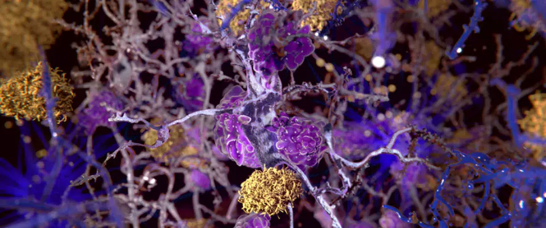
The presence of a gut dysbiosis in patients suffering from Alzheimer’s disease has already been proven. So too has the microbiota’s involvement in the cerebral accumulation of amyloid beta proteins associated with the disease. This new study aimed to investigate the signaling pathways through which patients’ gut microbiota contributes to this amyloid pathology.

In search of correlations

The study involved 89 individuals aged between 50 and 85 with cognitive performance ranging from normal to cognitive impairment with memory loss (whether or not associated with the disease). Amyloid deposits were measured by PET-scan and quantified in the various areas of the brain, while blood levels of molecules produced by the gut microbiota (lipopolysaccharides–LPS–and short-chain fatty acids–acetate, propionate, valerate, butyrate), pro- and anti-inflammatory biomarkers (including interleukins–ILs) and biomarkers of endothelial dysfunction (cell adhesion molecules–CAMs) were also measured.

Bacterial mediators implicated

Regardless of the brain area, amyloid deposition was positively correlated with blood levels of LPS, acetate, valerate, certain pro-inflammatory cytokines (e.g. IL1b, IL6) and many CAMs (e.g. P-selectin, PECAM-1), but negatively correlated with butyrate and IL10 (anti-inflammatory) levels. Lastly, some biomarkers of endothelial dysfunction were positively correlated with acetate, valerate, IL1b and IL4 levels, but again negatively correlated with levels of butyrate and IL10. The authors interpreted these correlations as evidence of a direct and indirect association between blood parameters linked to gut dysbiosis and amyloid pathology.

Inflammation, barrier function and Alzheimer’s

Therefore, the reduction in butyrate levels associated with an increase in the levels of acetate, valerate and LPS may compromise the integrity of the gut barrier, cause and maintain low-level systemic inflammation, and alter the blood-brain barrier, ultimately allowing pro-inflammatory compounds facilitating the pathological cascade of Alzheimer’s disease to enter the central nervous system. While highlighting that no causal link could be established from their data, the authors emphasize that the strength of the associations found supports this pathophysiological hypothesis. Lastly, it may be possible to develop prevention strategies for Alzheimer’s based on enriching the microbiota with beneficial bacteria or metabolites, once the microbial signature associated with the disease has been clarified.

Summary
Off
Sidebar
On
Migrated content
Activé
Updated content
Désactivé
Old content type
pro_article
Hide image
Off
News Neurology Gastroenterology

Microbiota transplant and type 1 diabetes mellitus: a trial in humans

Fecal microbiota transplantation is accompanied by changes in microbial metabolites and T cells involved in autoimmunity and may preserve residual function of pancreatic β-cells in type 1 diabetes mellitus.

The gut microbiota Is there a relationship between gut microbiota and circulating metabolites? Obesity: fecal virome transplant on trial Can statins combat intestinal dysbiosis?

Type 1 diabetes mellitus (T1DM) is an autoimmune disease that leads to the destruction of pancreatic β-cells. Studies in mice suggest that interactions between the gut microbiota and the innate immune system are involved in the development of the disease, the progression of which may be slowed by fecal microbiota transplantation (FMT).

Autologous versus allogenic transplantation

In a randomized controlled trial, patients recently diagnosed with T1DM received three FMTs by nasoduodenal tube at 0, 2 and 4 months, either from their own feces (autologous FMT, n=10) or from the feces of healthy donors (allogenic FMT, n=10). In the year following the first FMT, the researchers evaluated residual β-cell function (via C-peptide release in response to a test meal), as well as metabolic, immune and microbiota changes induced by the two types of FMT.

Pancreatic function preserved

Contrary to the researchers’ expectations, β-cell function was preserved in the autologous group one year after the first FMT. β-cell function deteriorated in the allogenic group, however, although less than in (sidenote: Overgaard AJ, Weir JM, Jayawardana K, et al. Plasma lipid species at type 1 diabetes onset predict residual beta-cell function after 6 months. Metabolomics 2018;14:158; Lachin JM, McGee PL, Greenbaum CJ, et al. Sample size requirements for studies of treatment effects on beta-cell function in newly diagnosed type 1 diabetes. PLoS One 2011;6:e26471 ) . According to the researchers, the benefits of FMT may be more pronounced and long-lasting where immunological compatibility between donor and host is high.

Desulfovibrio piger involved?

Changes in the microbiota were found to be associated with certain metabolic and immune changes. In the duodenum, the presence of Prevotella spp. was inversely correlated with residual β-cell function. In the colon, Desulfovibrio piger became significantly more abundant following autologous FMT only. Its abundance was associated with improved residual β-cell function and increased levels of plasma 1-arachidonoyl-GPC (A-GPC), a microbial metabolite associated with increased C-peptide production. In addition, the abundance of D. piger was negatively correlated with levels of certain T cells involved in autoimmunity. What was the significance according to the authors? D. piger may inhibit autoimmunity by suppressing these T cells via the production of A-GPC. From the multiple correlations found, the researchers have identified mechanistic leads that will need to be further investigated to clarify the effects of FMT on T1DM. They have also newly identified the therapeutic potential of certain bacterial species.

Summary
Off
Sidebar
On
Migrated content
Activé
Updated content
Désactivé
Old content type
pro_article
Hide image
Off
News Gastroenterology

Bees: gut microbiota is key to the unique scent shared by nestmates

Honey bees from the same hive can recognize each other based on a specific scent influenced by their gut microbiota. Intruders with the wrong microbiota and wrong scent beware!

Diet
Actu GP : Abeilles : le microbiote intestinal, clé du parfum identitaire de la ruche

As you probably know, Maya the Bee lives surrounded by her half-sisters, since the queen spends her life producing eggs to populate the hive. However, despite their genetic similarity, she and her sisters recognize each other by smell! What’s more, this study suggests that a bee’s scent–a signal of hive membership–is directly linked to the gut microbiota shared with its nestmates

Recognizing their own by smell

The honey bee’s body is covered with scent molecules. This allows the guards at the entrance to the hive to recognize hive members and stop intruders trying to sneak in and steal food. A research team has recently shown that the olfactory cues are based on shared characteristics of the gut microbiota (bacteria, fungi and viruses colonizing the digestive system), rather than genetic similarity. Bees from the same colony share several types of identical bacteria in the gut, giving them their common scent. Conversely, bees from a different colony, whose microbiota is home to distinct bacteria, emit a different scent.

Mechanisms involved

How to explain this influence of the microbiota? A number of theories have been put forward. According to one of them, the colony-specific scent is derived from the smell of the gut microbiota itself. However, this hypothesis seems unlikely as it goes against previous studies suggesting the involvement of molecules secreted by cells located under bees’ “skin”, to which the gut bacteria have no access. A second, more likely, theory suggests that the microbiota of honey bees quantitatively and qualitatively influences the production of scent molecules, for example, by providing their ingredients (or failing to do so). This scent recognition system is very useful to bees, but also has advantages for their gut bacteria: by rejecting bees with a distinct digestive flora, the hive also limits the entry of different bacteria, offering the organisms in the microbiota a quiet life, without competition.

Summary
Off
Sidebar
Off
Migrated content
Activé
Updated content
Désactivé
Old sources

Sources:

Vernier CL, Chin IM, Adu-Oppong BA et al. The gut microbiome defines social group membership in honey bee colonies. Science Advances. 2020. 6 (42), eabd3431. 

Old content type
article
Hide image
Off
News