
Modulating the skin microbiota with oral solutions

The existence of a gut-skin axis suggests the possibility of influencing the skin microbiota by modulating the gut microbiota. Pre- and probiotic oral solutions are therefore an option.

The skin microbiota Atopic dermatitis: nasal and skin microbiomes associated with disease severity Role of the microbiota in gut-brain communication Antibiotic exposure during first six years of life disrupts gut microbiota and impairs child growth
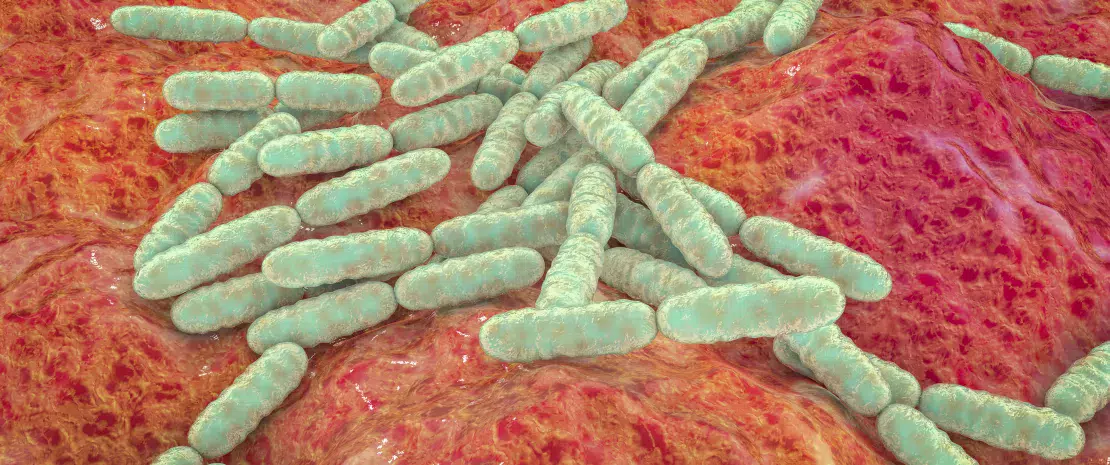
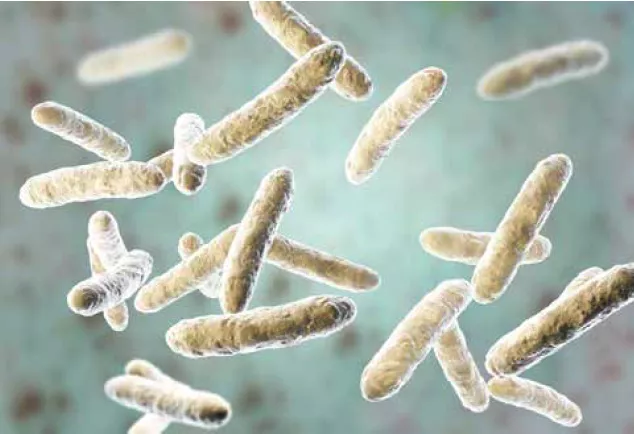
In numerous murine models, a Lactobacillus-enriched diet reduces skin sensitivity, rash, inflammation, dermatitis, etc., and improves skin phenotype (increased dermal thickness, enhanced folliculogenesis and increased sebocyte production).23 These beneficial probiotic effects have been confirmed by several interventional studies in humans involving lactobacilli and/or bifidobacteria.23 Managing skin diseases by modulating the gut microbiota will most likely involve probiotics (beneficial live bacteria), prebiotics (bacterial substrates) and symbiotics (combinations of pro- and prebiotics).23
Image
Legend

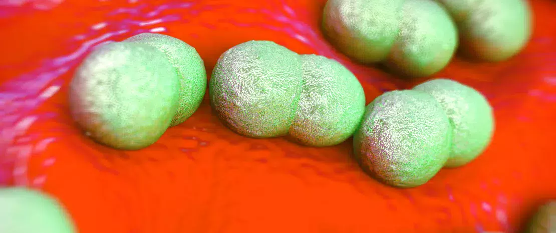
In mice, a Lactobacillus-enriched diet reduces skin sensitivity and improves skin phenotype.

A lack of adverse effects makes oral probiotics of even greater interest for the management of skin diseases.14

For example, in atopic dermatitis, daily consumption of probiotics (Bifidobacterium) and prebiotics (galacto-oligosaccharides) improves skin hydration in healthy adult women.14 To take another example, oral Lactobacillus supplementation reduces skin sensitivity and strengthens the skin’s barrier function in adults29 and children30. Several clinical trials have shown probiotics to have a positive effect when taken alone or in a cocktail (lactobacilli, bifidobacteria and/or S. thermophilus), with a reduction in lesions and severity in the case of acne.12,23 The positive effects of oral probiotics may be due to their ability to reduce systemic oxidative stress, regulate cytokines and reduce inflammatory markers.9 In the case of psoriasis, there are still few clinical data, but two studies in humans show beneficial effects: a reduction in inflammation markers with B. infantis; a reduction in the severity and appearance of lesions with B. longum, B. lactis and L. rhamnosus alongside a topical corticosteroid treatment.13 There were similar results for seborrheic dermatitis, with inflammation and symptoms relieved by oral L. paracasei.12 Some probiotics may even protect against skin cancer.16 However, clinical trials are still required to identify the most effective formulation of probiotic strains, the optimal duration of supplementation and the patients most likely to benefit.14

Summary
Off
Sidebar
Off
Migrated content
Activé
Updated content
Désactivé
Old sources


Old content type
pro_dossiers_article
Hide image
Off
Dossier detail

Modulating the skin microbiota via topical applications

Preventing and treating dysbiosis without eliminating pathogens: novel therapeutic strategies aim to rebalance the skin microbiota directly via topical applications, or indirectly via oral solutions that modulate the gut ecosystem.

The skin microbiota Atopic dermatitis: nasal and skin microbiomes associated with disease severity Role of the microbiota in gut-brain communication Antibiotic exposure during first six years of life disrupts gut microbiota and impairs child growth

Staphylococcus epidermidis

The first clinical trials seem to support the use of topical applications to rebalance the skin microbiota. However, further trials are needed to confirm these results.

In general, there have been few clinical trials evaluating the topical application of probiotics in skin diseases.12 For acne, creams containing S. epidermidis or bacteriophages of C. acnes that preferentially target pathogenic strains have shown positive results.12 The application of R. mucosa in patients with atopic dermatitis may reduce lesion severity, the need to use topical steroids and the presence of S. aureus.28,29 The limited availability of microbial candidates on the skin has forced researchers to also use other sources of microorganisms. Derived from thermal spring water, Vitreoscilla filiformis may be beneficial in seborrheic dermatitis: one study reported a reduction in erythema, desquamation and pruritus by soothing the inflammation.12 

Image

In acne, Nitrosomonas eutropha decreases lesion severity12, while the topical use of bacterial products (E. faecalis enterocins) reduces lesions by 60% compared to controls.12 An alternative strategy corrects the dysbiosis by using sucrose to promote the growth of S. epidermidis over C. acnes.9 Scientific data are scant for skin cancer and non-existent for rosacea. In murine models of UV-related cancers, a molecule produced by S. epidermidis was shown to inhibit tumor proliferation.12,16

Summary
Off
Sidebar
Off
Migrated content
Activé
Updated content
Désactivé
Old sources


Old content type
pro_dossiers_article
Hide image
Off
Dossier detail

A gut-brain-skin axis?

The skin microbiota Atopic dermatitis: nasal and skin microbiomes associated with disease severity Role of the microbiota in gut-brain communication Antibiotic exposure during first six years of life disrupts gut microbiota and impairs child growth

Should we go further than a gut-skin axis and include the brain also?

As early as 1930, dermatologists John Stokes and Donald Pillsbury25,26, suggested that emotional states such as anxiety or depression can alter the gut microbiota and induce local or systemic inflammation27. They recommended the use of fermented milk to reintroduce beneficial microorganisms.

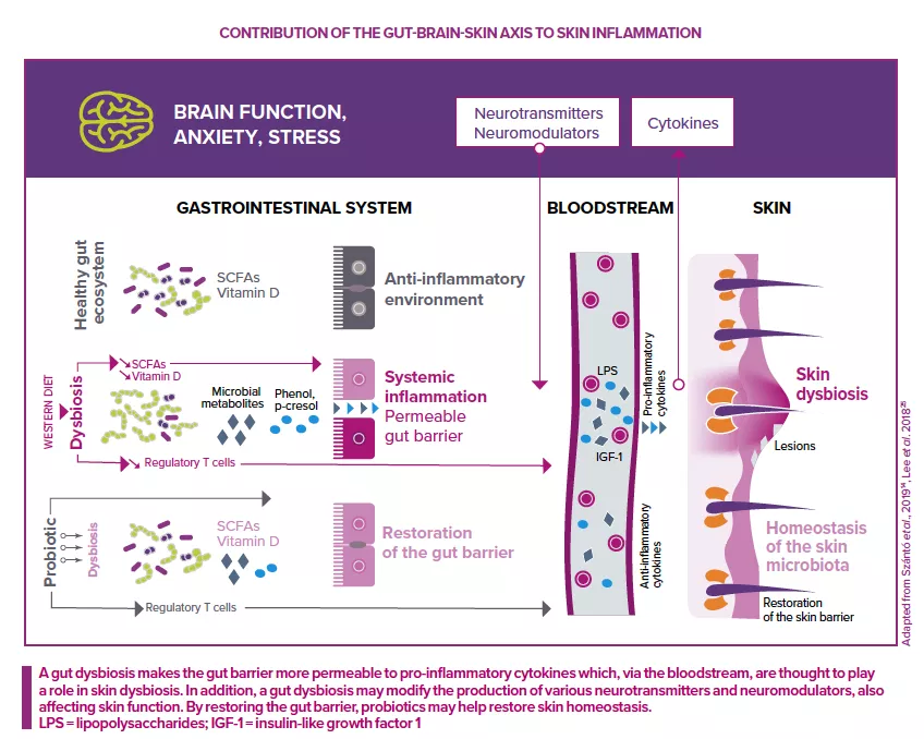
More precisely, stress leads to the secretion of neurotransmitters (serotonin, norepinephrine and acetylcholine). These neurotransmitters increase gut permeability, leading to local inflammation. At the same time, they also provoke systemic inflammation via the bloodstream.11,23

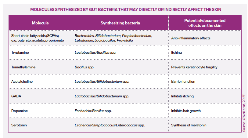
For example, stress hormone cortisol is thought to alter the composition of the gut microbiota and blood levels of neuroendocrine molecules (tryptamine, trimethylamine and serotonin), ultimately affecting the skin barrier and skin inflammation.25

Is the gut-brain-skin axis a two-way axis, i.e. can the skin in turn act on the gut via the nervous system?

Image
Gut-brain-skin axis

Acne and atopic dermatitis 

This gut-brain-skin axis is implicated in certain skin diseases. For example, upregulation and strong expression of substance P (a neurotransmitter and neuromodulator of the central and peripheral nervous systems) are observed in both acne and gut dysbiosis. Substance P is known to trigger the expression of many pro-inflammatory mediators implicated in the development of acne (IL-1, IL-6, TNF-α, PPAR-γ).22,23

The gut-brain-skin axis is also thought to be involved in atopic dermatitis.25 An altered gut microbiota may modify the production of various neurotransmitters and neuromodulators, affecting the functioning of the skin barrier and immune system, two key parameters of the pathophysiology of atopic dermatitis.25

Tryptophan produced by the gut microbiota is thought to cause skin itching, while lactobacilli and bifidobacteria may inhibit these sensations.25 Moreover, some researchers ask whether the gut-brain-skin axis is a two-way axis: can the skin in turn act on the gut via the nervous system?22

Summary
Off
Sidebar
Off
Migrated content
Activé
Updated content
Désactivé
Old sources


Old content type
pro_dossiers_article
Hide image
Off
Dossier detail

Psoriasis, atopic dermatitis, rosacea: gut-skin axis involved

Laced with blood vessels, packed with nerves, heavily involved in the immune system and massively colonized by microbial communities, the gut and the skin have a number of things in common.22 But that’s not all. Recent years have seen growing evidence for the existence of a link between the gut and the skin (the gut-skin axis) or even the gut-brain-skin axis.23


The skin microbiota Atopic dermatitis: nasal and skin microbiomes associated with disease severity Role of the microbiota in gut-brain communication Antibiotic exposure during first six years of life disrupts gut microbiota and impairs child growth

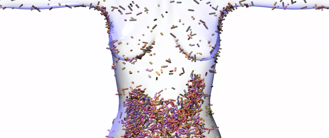
Human microbiome, conceptual image. Computer illustration showing the microbiome of the human body, including the colon (large intestine, lower centre). The microbiome is the collection of microbes (micro-organisms including bacteria, archaea, protists, fungi and viruses) found on and in the human body.

The gut microbiota appears to play an active role in the pathogenesis of various skin diseases, including psoriasis, rosacea and atopic dermatitis.

Three mechanisms are at play: the composition of the skin microbiota, the skin’s barrier effect and the skin’s immune response.

Molecules, gut bacteria and skin

Skin ulcers or psoriasis in patients with inflammatory bowel disease (IBD), dermatitis and psoriasis in celiac patients, a gut dysbiosis and H. pylori infection in people with rosacea... There are many examples of associations between digestive and skin conditions.22

Although the gut-skin axis is not fully understood, several explanations have been put forward.

Image
Axe intestins peau

COMPOSITION OF THE SKIN MICROBIOTA

The gut microbiota may influence the composition of the skin microbiota.23 Short-chain fatty acids (SCFAs, e.g. acetate, propionate) produced by the gut microbiota via fiber fermentation in the gut may modify the predominance of certain microorganisms or microbial profiles in the skin. For example, gut bacterium Propionibacterium (see table) mainly produces acetate and propionate. Propionic acid has an antimicrobial effect against certain skin pathogens, particularly methicillin-resistant Staphylococcus aureus.23 In contrast, commensal skin bacteria S. epidermidis and Cutibacterium acnes have been shown to tolerate wider shifts in SCFAs.23

INTEGRITY OF THE SKIN BARRIER

Children with atopic dermatitis also seem to suffer from a gut dysbiosis. A damaged gut barrier sees increased penetration by food antigens, bacterial toxins and pathogens.14 For example, gut bacteria, especially Clostridiales difficile, can produce free phenol and p-cresol, which can disturb the skin barrier and reduce keratin production.14,22,23

A low level of vitamin D has been associated with atopic dermatitis and psoriasis. Vitamin D may be regulated by the gut microbiota and may participate in a signaling mechanism between microbiota and host.14

In the case of acne, microbial metabolites may regulate various skin functions (cell proliferation, lipid metabolism, etc.) via other metabolic pathways.14

A high glycemic load, typical of adolescent meals in developed countries, influences insulin metabolism, ultimately triggering sebaceous gland hyperproliferation, lipogenesis and hyperplasia of keratinocytes, thereby contributing to the development of acne.14,23 This appears to be a two-way process, with the metabolic pathway in turn affecting the composition of the gut microbiota via the gut barrier. This may result in a vicious circle via a positive feedback cycle of inflammation.23

IMMUNE RESPONSE OF THE SKIN

The mechanisms by which the gut microbiota acts on the skin microbiota may also involve the modulating effect of gut microorganisms on systemic immunity.22 Some gut microbes and metabolites facilitate anti-inflammatory responses24. For example, SCFAs are thought to exert local and remote anti-inflammatory effects, particularly on the skin.22 Conversely, other metabolites may participate in the inflammatory loop and the appearance of skin diseases. For example, filamentous bacteria may promote the accumulation of pro-inflammatory Th17 and Th1 cells.23

In the case of rosacea, some authors suggest a link with Helicobacter pylori. This bacterium may exert pro-inflammatory effects via peptides.11,22

Other mechanisms have been mentioned in psoriasis, involving a decrease in beneficial species such as Faecalibacterium prausnitzii13 or Akkermansia muciniphila, with the latter thought to strengthen the integrity of the gut epithelium and protect against inflammatory diseases.1 Psoriasis patients whose blood contains bacterial DNA, have significantly higher levels of systemic inflammatory response markers, including IL-1β, IL-6, IL-12, tumor necrosis factor, and interferon γ.11

Summary
Off
Sidebar
Off
Migrated content
Activé
Updated content
Désactivé
Old sources


Old content type
pro_dossiers_article
Hide image
Off
Dossier detail

Non-pathological skin conditions associated with a dysbiosis

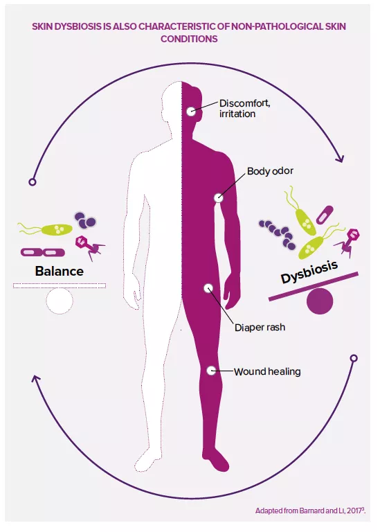
Changes in the skin microbiota can also be seen in non-pathological skin conditions. The skin is constantly exposed to various endogenous, exogenous and lifestyle factors that can affect the physical, mechanical or microbial properties of the skin barrier.19

The skin microbiota Atopic dermatitis: nasal and skin microbiomes associated with disease severity Role of the microbiota in gut-brain communication Antibiotic exposure during first six years of life disrupts gut microbiota and impairs child growth

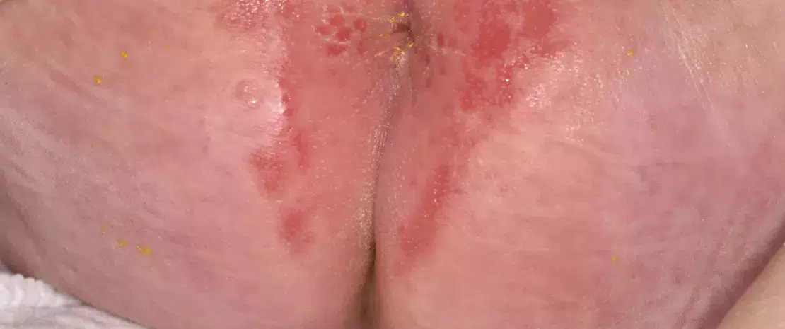
Nappy rash. Close-up of perianal dermatitis in a 1 month old female patient, showing red and inflamed skins. 

Discomfort, irritation, diaper rash

Sensitive skin “tightens”, tingles or burns in response to stimuli that would not normally cause such sensations. It is seen both in people with normal skin and in those with a disruption of the skin barrier.19 A hyperreactive cutaneous nervous system, the skin barrier and the skin microbiota are thought to be involved.19 An alteration of the stratum corneum in sensitive subjects may contribute to penetration by chemical, environmental and microbial agents associated with increased skin sensitivity.19

Diaper rash only affects skin exposed to diaper friction, excessive hydration and a variable pH, and in constant contact with urine and feces. Candida albicans and Staphylococcus aureus are potentially involved.20

Skin sensitivity may be linked to a hyperactive cutaneous nervous system, to the skin barrier and the skin microbiota.

Image
Cutaneous dysbiosis

Wound healing 

As a result of the physical tear of skin tissue, the wound healing process begins with inflammation that results from close cooperation between immune cells and bacteria involved in the process.21 Commensal bacteria such as Staphylococcus, Streptococcus, Pseudomonas and Corynebacterium have both positive and negative effects on wound healing. They stimulate the host immune system and reduce invasion by other pathogenic microorganisms, but this loss of microbial diversity is often accompanied by prolonged inflammation, which may slow wound healing.21

This close relationship between host and skin microbiota in wound healing processes could open the door to novel therapies, such as creams rich in antimicrobial peptides, biofilm-destroying probiotics or anti-inflammatory bacteria.12,21


Body odor

Human body odors result from the metabolization by bacteria of sweat components (amino acids, fatty acids and glycerols), leading to the production of malodorous molecules, e.g. the “sulfurous” or “sour” odor of acetic acid produced by Staphylococcus spp. in children and adolescents, or the “sour” odor of thiols produced by Corynebacterium and Staphylococcus spp. in adults.7 The repeated use of deodorants and antiperspirants alters bacterial diversity in the armpit, favoring staphylococci over Corynebacterium, which may have counterproductive effects in adolescents.7

Expert opinion
Could probiotics be a third option for dealing with body odors, in addition to the two classical strategies, alcohol-based deodorants and antiperspirants?
"I think it’s possible that the regular, long-term application of a body odor product containing live microorganisms could change the microbiota of the armpit so that it’s less prone to producing odors. However, I suspect this would have a very mild effect and would probably be less effective than the antimicrobial effect of alcohol. Also, probiotics would not be able to prevent underarm dampness (sweat production) with the same effectiveness as the aluminum chlorohydrate that blocks sweat pores in antiperspirants."
DR. MARKUS EGERT
Summary
Off
Sidebar
Off
Migrated content
Activé
Updated content
Désactivé
Old content type
pro_dossiers_article
Hide image
Off
Dossier detail

Skin diseases associated with a dysbiosis

The skin microbiota is a dynamic system in which microorganisms constantly compete to survive. Sometimes this balance breaks down, commensal bacteria become opportunistic pathogens1,4 and a dysbiosis results: it is a common feature in skin diseases (acne, psoriasis, dermatitis, etc.) and other non-pathological skin conditions (irritation, wounds, odors). However, it is not yet known whether dysbiosis is a cause or effect.2

The skin microbiota Atopic dermatitis: nasal and skin microbiomes associated with disease severity Role of the microbiota in gut-brain communication Antibiotic exposure during first six years of life disrupts gut microbiota and impairs child growth

Acne, psoriasis, rosacea... Many skin diseases are associated with a dysbiosis. This may have diagnostic or predictive value or even open up novel therapeutic approaches.

Skin cancer

Pathophysiology:

in many cutaneous neoplasms, dysbiosis appears to be involved in carcinogenesis.9,10,11,12 Conversely, a healthy microbiota may inhibit the development of tumors by regulating the immune system and controlling inflammation.

Role of the microbiota:

  • S. aureus, Streptococcus pyogenes, Pseudomonas aeruginosa, the β-human papillomavirus, the Epstein Barr virus and Malassezia or Candida fungi may induce a state of chronic inflammation, leading to cancer;16
  • link between S. aureus infection and severity of cutaneous T cell lymphoma.12

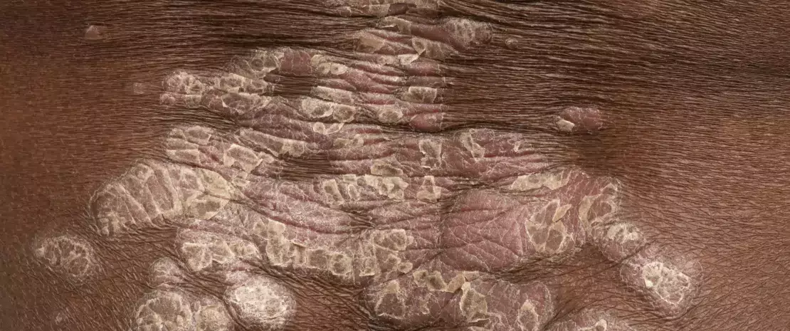
Psoriasis

Pathophysiology:

multifactorial immune-mediated disease, involving genetic factors, immune system disturbances and environmental triggers.13

Prevalence:

affects 2%-3% of the population, often appearing between 15 and 20 years of age11 with two common peaks of incidence (20-30 years of age and 50-60 years of age).13.

Role of the microbiota:

  • psoriasis patients see an alteration in the composition of their skin microbiota and a loss of microbial diversity11, which affects not only the lesions, but the skin microbiota as a whole.11
  • microorganisms associated with the disease still not clearly identified1,11, with numerous contradictory data. However, S. aureus thought to be more abundant and to participate in inflammation (by increasing the response of Th17 cells, which release pro-inflammatory cytokines);11
  • often associated with gut dysbiosis.14

Acne

Pathophysiology:

multifactorial chronic inflammatory disease involving hyperseborrhea, abnormal keratinization of follicular ducts and a dysbiosis of the skin microbiota associated with a predominance of virulent C. acnes phylotypes.9

Prevalence:

8th most common skin disease, affecting 9.38% of the world’s population (all ages), with higher prevalence in adolescents, reaching 35%-100% in some countries.10

Role of the microbiota:

  • loss of balance between the different C. acnes phylotypes (the more virulent phylotype IA1 becomes do minant and induces inflammation by activating the innate immune system);9
  • loss of reciprocal control between C. acnes (maintains acidic pH, inhibits the development of S. epidermidis) and S. epidermidis (anti-inflammatory activity, limits the proliferation of C. acnes);9
  • suspected secondary pro-inflammatory role (folliculitis) of opportunistic fungal species of the pilosebaceous apparatus (Malassezia and possibly Candida);11
  • additional effect of diet on acne severity (interaction with gut microbiota).9

Atopic dermatitis (eczema)

Pathophysiology:

chronic inflammatory skin disease with a strong genetic component involving a disruption of the skin barrier and immune system (inflammatory Th2 cells), resulting in increased susceptibility to infections and allergens.11,15

Prevalence:

up to 20% of infants and 3% of adults worldwide11, and up to 10% of adults in developed countries.14

Role of the microbiota:

  • patients see a loss of diversity in the skin microbiota11,12, both in lesions and healthy areas;
  • increase in content of staphylococci, with a proliferation of S. aureus linked to a lower production of antimicrobial peptides by keratinocytes via the influence of Th2 cells.15 Increased presence of S. epidermidis in less severe forms;12
  • a higher density of colonization with S. aureus correlated with more inflammation and increased disease severity.11

Seborrheic Dermatitis (SD) and Dandruff

Pathophysiology:

chronic skin disease involving a complex interaction between the Malassezia fungus, keratinocytes, and the inflammatory response induced by an altered lipid composition in the skin.12,18

Prevalence:

three peaks of incidence (early childhood, adolescence and from the age of 50 onwards). Half of adult population thought to be affected by DS and dandruff.11,18

Role of the microbiota:

  • hydrolysis by Malassezia of skin lipids into free fatty acids that trigger an inflammatory response;16
  • increased presence of Malassezia species, with M. restricta,M. globose and M. furfur the most commonly associated with seborrheic dermatitis. The first two species are the most virulent (they produce irritating oleic acids, leading to IL-8 and IL-17 activation);17
  • Actinetobacter, Staphylococcus and Streptococcus dominate microbiota in the lesions;11
  • correlation between disease severity and decreased bacterial diversity; no correlation with Malassezia abundance.12

Rosacea

Pathophysiology:

chronic inflammatory disease whose pathophysiology is not fully understood. Factors include neurovascular reactivity, genetic susceptibility, dysfunction of the innate immune responses, and comorbid gastrointestinal conditions.17

Prevalence:

between 0.9% and 10% of the population in the US and Europe.11

Role of the microbiota:

  • Demodex folliculorum (a sebaceous gland mite) stimulates the production of inflammatory peptides and cellular growth factors. This mite may also carry Bacillus oleronius, a pro-inflammatory bacterium;11
  • a variant of S. epidermidis, more virulent than the commensal bacterium, also thought to be involved;11
  • often associated with a gut dysbiosis.18
Sources

1 Ederveen THA, Smits JPH, Boekhorst J et al. Skin microbiota in health and disease: From sequencing to biology. J Dermatol. 2020 Oct;47(10):1110-1118.

2 Egert M, Simmering R, Riedel CU. The Association of the Skin Microbiota With Health, Immunity, and Disease. Clin Pharmacol Ther. 2017 Jul;102(1):62-69.

9 Dréno B, Dagnelie MA, Khammari A, et al. The Skin Microbiome: A New Actor in Inflammatory Acne. Am J Clin Dermatol. 2020 Sep;21(Suppl 1):18-24.

10 Heng, A.H.S., Chew, F.T. Systematic review of the epidemiology of acne vulgaris. Sci Rep 10, 5754 (2020). https://doi.org/10.1038/s41598-020-62715-3.

11 Ellis SR, Nguyen M, Vaughn AR, et al. The Skin and Gut Microbiome and Its Role in Common Dermatologic Conditions. Microorganisms. 2019;7(11):550.

12 Yu Y, Dunaway S, Champer J, et al. Changing our microbiome: probiotics in dermatology. Br J Dermatol. 2020;182(1):39-46.

13 Rigon RB, de Freitas ACP, Bicas JL, et al. Skin microbiota as a therapeutic target for psoriasis treatment: Trends and perspectives. J Cosmet Dermatol. 2021;20(4):1066-1072.

14 Szántó M, Dózsa A, Antal D et al. Targeting the gut-skin axis-Probiotics as new tools for skin disorder management? Exp Dermatol. 2019 Nov;28(11):1210-1218.

15 Langan SM, Irvine AD, Weidinger S. Atopic dermatitis. Lancet. 2020 Aug 1;396(10247):345-360.

16 Squarzanti DF, Zavattaro E, Pizzimenti S et al. Non-Melanoma Skin Cancer: news from microbiota research. Crit Rev Microbiol. 2020;46(4):433-449.

17 Tutka K, Żychowska M, Reich A. Diversity and Composition of the Skin, Blood and Gut Microbiome in Rosacea-A Systematic Review of the Literature. Microorganisms. 2020;8(11):1756.

18 Adalsteinsson JA, Kaushik S, Muzumdar S et al. An update on the microbiology, immunology and genetics of seborrheic dermatitis. Exp Dermatol. 2020;29(5):481-489.

Summary
Off
Sidebar
Off
Migrated content
Activé
Updated content
Désactivé
Old content type
pro_dossiers_article
Hide image
Off
Dossier detail

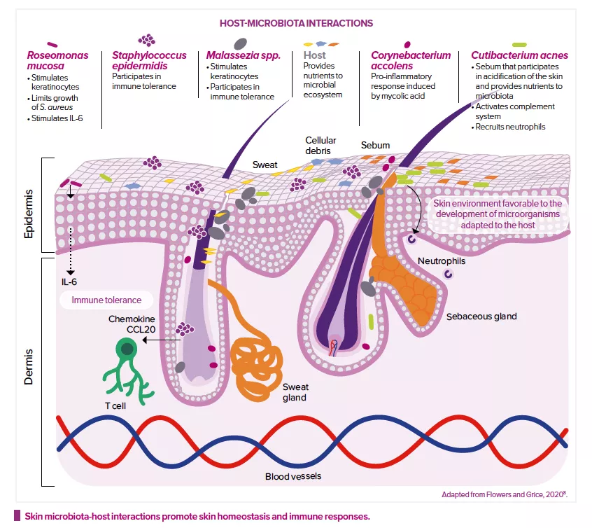
Functions of the microbiota and its interactions with the host

For a long time, the skin microbiota was considered a potential source of infection. Now we know it to be an important factor in host health2, even if its interactions with the body are only beginning to be understood.

The skin microbiota Atopic dermatitis: nasal and skin microbiomes associated with disease severity Role of the microbiota in gut-brain communication Antibiotic exposure during first six years of life disrupts gut microbiota and impairs child growth

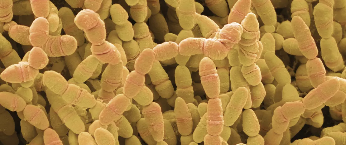
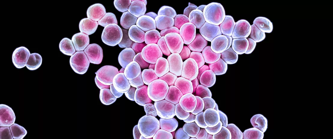
Staphylococcus epidermidis.

Reduced colonization by pathogens 

Although it remains difficult to define, a “healthy” skin microbiota is generally considered synonymous with a diversified flora and the presence of commensal bacteria.2 This balanced microbiota is thought to help protect against infection, limiting colonization by pathogens. For example, C. acnes, which lives in the sebaceous glands, releases fatty acids from sebum, contributing to the acidity of the skin, which in turn inhibits the proliferation of pathogens.8

Other bacteria secrete bacteriocins and other antimicrobial factors. For example, S. epidermidis releases a protease that destroys S. aureus biofilms, while nasal bacterium, Staphylococcus lugdunensis, produces an antibiotic peptide that acts against many pathogens, including S. aureus, Enterococcus faecalis, Listeria monocytogenes, Streptococcus pneumoniae, and Pseudomonas aeruginosa.2

Lastly, Corynebacterium striatum modifies the transcriptional program of S. aureus, repressing virulence-related genes and stimulating those associated with commensalism. 6,8 The skin microbiota thus maintains its balance not only by competitive exclusion but also via subtle interactions between microorganisms.6

Image

Modulation of the immune system 

The skin microbiota also plays a key role in the development and regulation of the innate and acquired immune systems.2 It modulates the expression of innate immune factors (interleukin IL-1α, antimicrobial peptides, etc.) produced by keratinocytes and sebocytes6, and even produces some of these factors itself.

For example, S. epidermidis can, in different situations, either stimulate or reduce inflammation: it inhibits the release of inflammatory cytokines by keratinocytes and the immune responses of injured skin cells; it reinforces the skin’s defense mechanisms against infection by increasing the expression of genes that encode for antimicrobial peptides; and it modulates the expression of skin T cells.2 S. epidermidis promotes tolerance towards the commensal microbiota, while adjusting immune responses to pathogens or those triggered during wound healing.8 Roseomonas mucosa, Malassezia spp. or Corynebacterium accolens can also modulate host and keratinocyte immune responses.8

Lastly, the genetic profile of bacteria also plays a role. Cutibacterium acnes strains from acne-prone skin have genes that encode for virulence factors, which could explain the higher pro-inflammatory activity observed. Conversely, strains from healthy skin, which do not have these factors, are thought to promote the production of anti-inflammatory cytokines.8

Summary
Off
Sidebar
Off
Migrated content
Activé
Updated content
Désactivé
Old sources


Old content type
pro_dossiers_article
Hide image
Off
Dossier detail

Factors affecting the skin microbiota

The skin microbiota of healthy individuals appears to be relatively stable over periods of a few months or years.3,4 However, its composition is still influenced by the host and its environment.

The skin microbiota Atopic dermatitis: nasal and skin microbiomes associated with disease severity Role of the microbiota in gut-brain communication Antibiotic exposure during first six years of life disrupts gut microbiota and impairs child growth

Staphylococcus epidermidis

HOST-RELATED EFFECTS

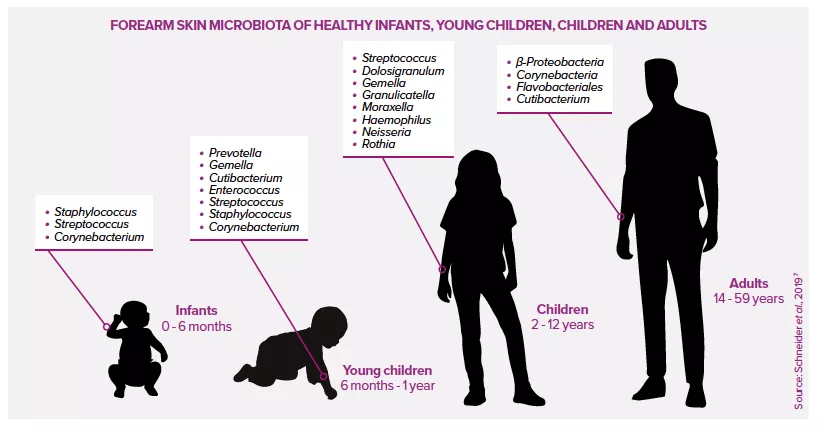
The composition of the skin microbiota is strongly influenced by the host, specifically by their age, sex, genes, immune status, concomitant health conditions (dermatological or otherwise), the skin area in question, interactions between microorganisms, diet and stress levels.2

The initial colonization of a newborn baby’s skin depends on the mode of delivery4,7: children born vaginally acquire vaginal bacteria (Lactobacillus, C. albicans), while those born by caesarean section acquire skin microbes (Staphylococcus, Streptococcus). Within a few hours of birth, sebum secretion increases sharply. This continues for several days before decreasing.2 The immature immune system facilitates colonization due to the lack of any inflammatory response.4

At puberty, the skin microbiota undergoes a profound restructuring due to hormonal changes that stimulate sebaceous secretions. It contains more lipophilic organisms (Cutibacterium, Malassezia), whereas previously it had been dominated by Firmicutes, Bacteroidetes and Proteobacteria, with a diverse fungal community.4

Image

ENVIRONMENTAL EFFECTS

Many external factors also influence the composition of the skin microbiota2, including lifestyle, domestic and personal hygiene, cohabitation, geographical location, sunlight, occupation (and work clothing), etc. For example, contact with other humans, but also with pets and objects (telephone, computer keyboard, classroom objects, etc.), modifies the skin microbiota and explains the similarities observed between the microbiota of members of the same household or group.3

Moreover, the conditions in a given environment affect the different areas of the skin to different degrees. For example, some skin areas (e.g. hand) have more physical contact, others are less exposed to ultraviolet light, etc.3,4 Despite this, the skin microbiota remains relatively stable in adulthood, suggesting reciprocal beneficial interactions between microorganisms and host.6

Summary
Off
Sidebar
Off
Migrated content
Activé
Updated content
Désactivé
Old sources


Old content type
pro_dossiers_article
Hide image
Off
Dossier detail

A unique set of skin microbiota for each individual

The fourth most populated microbial niche in the human body,2 the skin is home to a complex community of microorganisms.2 Bacteria, fungi, parasites and viruses live together on the skin in a unique balance specific to each individual, to the point where some investigators speak of an individual microbial fingerprint.5

The skin microbiota Atopic dermatitis: nasal and skin microbiomes associated with disease severity Role of the microbiota in gut-brain communication Antibiotic exposure during first six years of life disrupts gut microbiota and impairs child growth

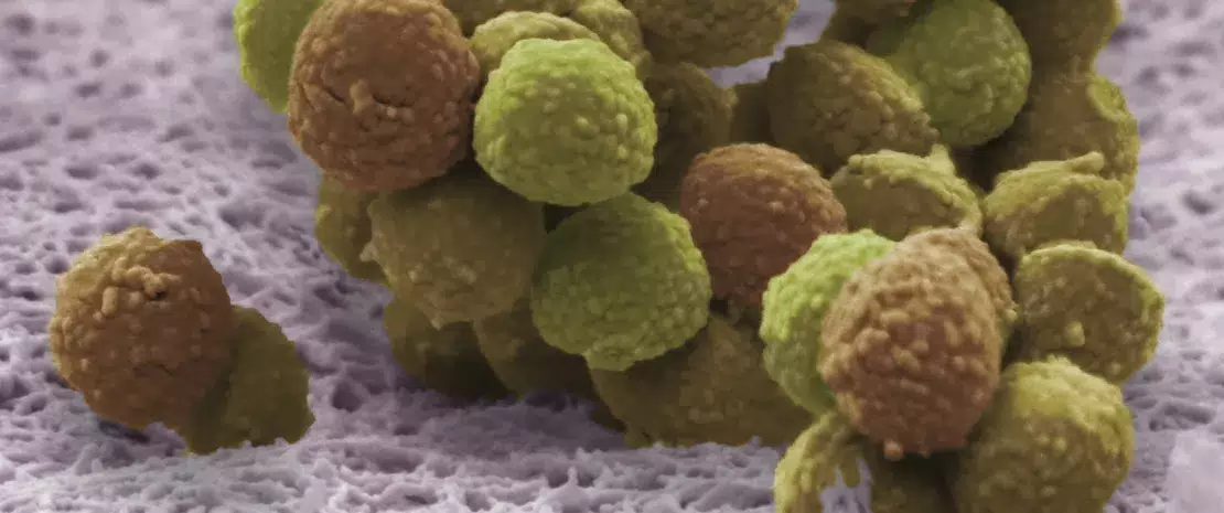
Staphylococcus epidermidis

Each individual is not characterized by one but by multiple skin microbiota. In fact, the skin microbiota varies “horizontally”, according to skin area (face, armpits, etc.), but also “vertically”, according to the layers that make up the skin epithelium.

BACTERIA, FUNGI, VIRUSES AND PARASITES

Although easily accessible, the skin microbiota remains poorly understood. Its density is believed to be low compared to that of the large intestine, instead resembling that of the small intestine, i.e. around 1011 bacteria.1 It is the fourth largest microbial niche in the body in terms of the number of microorganisms, just after the digestive tract, the oral cavity and the vagina.2

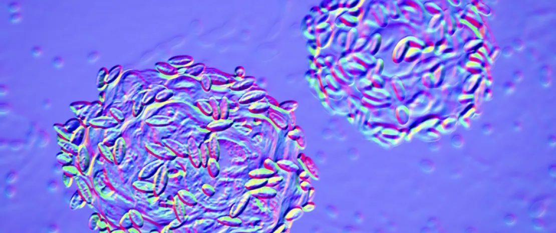
It hosts several bacterial phyla (Actinobacteria, Firmicutes, Proteobacteria and Bacteroidetes), archaea, and fungal species mainly from the genus Malassezia.2,3 Among the bacterial species identified are included Cutibacterium acnes and Staphylococcus epidermidis, although the strains present differ depending on the individual, the state of their skin (healthy or otherwise) and the sampling site.3,6

Lastly, although not well described,2 numerous viruses (papillomavirus, adenovirus, etc.) have been identified on the skin of healthy individuals, as well as phages that target C. acnes and S. epidermidis, suggesting the existence of a complex virome. Parasites (such as Demodex mites, etc.), few in number, are even more scantly described.3

“HORIZONTAL” VARIATION ACCORDING TO SKIN AREA

The skin is not a homogeneous habitat. The surface of the skin is acidic, salty and aerobic, whereas the invaginations of the hair follicles offer a lipid-rich and anaerobic environment6.

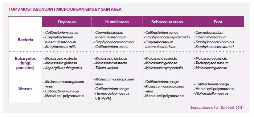
Three major niches are generally identified based on properties such as pH, temperature, humidity, perspiration levels and lipid content:1,3,4

  • sebaceous areas (face, chest, back) that secrete lipid-rich sebum;
  • dry areas (forearms, palm of the hand);
  • humid areas (armpits, elbow crease, nostril, back of the knee and groin), where numerous sweat glands participate in thermoregulation (sweat), acidify the skin and secrete antibacterial peptides.

Some authors distinguish a fourth area in the foot (nails, heel and space between toes)4 (see table).

These areas are separate ecological niches, each with a unique microbial community: the most exposed and dry areas, such as the hands, are the most diverse; the armpit, which is moist and rich in sweat, is dominated by Corynebacterium and Staphylococcus species; while lipid-rich areas, such as the face, display much less diversity (Cutibacterium bacteria, fungi of the genus Malassezia, and Demodex folliculorum mites).3

Microbiota also vary in density from one skin area to another: from 102 bacteria per cm2 on the fingertips or back, to 106 bacteria per cm2 on the forehead or in the armpits.2

Image

“VERTICAL” VARIATION ACCORDING TO SKIN LAYER

For a long time, it was thought that microbial life in the skin was limited to the epidermis, hair follicles, and sebaceous and sweat glands. However, microorganisms also seem to live in the deeper layers of the skin, i.e., the dermis and the underlying adipose tissue.2

On the skin’s surface, the deeper into the stratum corneum the fewer microorganisms are present.1

Then, from the surface to the subcutaneous regions, the microbiota changes and gradually loses its individual characteristics.4,5

In the dermis and subcutaneous adipose tissues, there seems to be more Proteobacteria while there are less Actinobacteria and Firmicutes than in the epidermis.2

Summary
Off
Sidebar
Off
Migrated content
Activé
Updated content
Désactivé
Old content type
pro_dossiers_article
Hide image
Off
Dossier detail

Using recombinant endolysins to treat bacterial vaginosis

A study has shown that by using recombinant endolysins encoded on a prophage it is possible to eliminate the bacterial biofilm responsible for bacterial vaginosis without damaging the beneficial bacteria of the vaginal microbiota. These are promising results.

The vaginal microbiota Vaginal microbiota: a marker for papillomavirus progression? The role of the vaginal, uterine and gut microbiota in endometriosis

35% Only 1 in 3 women know that bacterial vaginosis is associated with an imbalance in the vaginal microbiota

Bacterial vaginosis is a quite common disorder in women of reproductive age, with a prevalence estimated at 10%–30% worldwide. It is associated with an increased risk of infertility and complications during pregnancy. It is also a risk factor for contracting sexually transmitted diseases. The condition is characterized by an imbalance of the vaginal microbiota and a biofilm formed on the vaginal epithelium, which is initiated and dominated by Gardnerella bacteria. This biofilm is frequently refractory to antibiotic treatment. Antibiotics are effective in quickly reducing symptoms but are associated with a recurrence rate of up to 60% within six months of treatment. A new study has investigated (sidenote: Endolysins Bacteriophage enzymes that lyse the bacterial wall, allowing the release of phages. )  of the type 1,4-beta-N-acetylmuramidase encoded on Gardnerella (sidenote: Prophages Bacteriophage genomes integrated into the host genome. (Saussereau and Debarbieux 2012) )  as an alternative treatment.

Bactericidal effect 10 times higher than wild type

To this end, the authors generated several engineered endolysins via domain shuffling. They compared their bactericidal activity on Gardnerella strains to that of wild-type endolysins. The bactericidal activity of the recombinant endolysins was 10 times that of any wild-type enzyme. When tested against a panel of 20 Gardnerella strains (from (sidenote: G. vaginalis, G. leopoldii, G. piotii and G. swidsinskii ) ), the most active endolysin, called PM-477, showed superior efficacy compared to the antibiotics tested (metronidazole, tinidazole, clindamycin). Furthermore, PM-477 had no effect on beneficial lactobacilli or other species of vaginal bacteria. According to the authors, PM-477 is highly selective for Gardnerella and kills strains of each of the four main species without affecting beneficial lactobacilli or other species typical of the vaginal microbiota. The effect of PM-477 was confirmed by microscopy in mixed cultures of Gardnerella and lactobacilli. PM-477 (at 460 µg/mL for 5 h) lysed G. vaginalis and G. swidsinskii cells in monoculture, but also selectively lysed them in mixed cultures alongside lactobacilli without affecting the latter.

Efficacy in patient samples

To go further and analyze the efficacy of PM-477 in a physiological environment closely resembling the in vivo situation, the researchers treated vaginal swabs from 15 bacterial vaginosis patients and analyzed them by fluorescence in situ hybridization (FISH). They showed that in 13 of the 15 cases, PM-477 eradicated the Gardnerella bacterium and physically dissolved the biofilms without affecting the vaginal microbiota. For the authors, endolysins are a promising therapeutic alternative to antibiotics for the treatment of bacterial vaginosis. This is a significant finding since antibiotics are frequently a cause of recurrence and resistance in the treatment of the disease.

Summary
Off
Sidebar
On
Migrated content
Activé
Updated content
Désactivé
Old content type
pro_article
Hide image
Off
News