Metabolic syndrome represents a collection of metabolic problems associated with obesity, such as hypertriglyceridemia or high blood pressure, which increase the risk of diabetes and cardiovascular complications.
Metabolic syndrome is not, strictly speaking, a disease; its definition is the existence, in a single person, of abdominal obesity (waist measurement > 94 cm for men, 80 cm for women), associated with at least two of the following problems: abnormally elevated insulin level, hypertriglyceridemia, high blood pressure, hyperglycemia, or low HDL cholesterol (“good cholesterol”).
France spared
With 14 to 21% of people affected, compared with a quarter of Americans and almost 46% of Greeks, France has been somewhat spared the “epidemic” of metabolic syndrome. But this situation cannot last.
Unhealthy lifestyle, the primary risk factor
Other than likely genetic predispositions, it’s an unhealthy lifestyle that leads to metabolic syndrome. Junk food, paired with insufficient physical activity, causes metabolic dysfunction that leads to chronic inflammation, which itself causes metabolic disorders. It starts a vicious cycle, in which one contributor is imbalance in the microbiota or dysbiosis.
No visible signs
Other than obesity, metabolic syndrome has no visible signs since, once symptoms appear, it means that the syndrome has changed into a disease: type 2 diabetes, atherosclerosis, cardiovascular disease, etc.
Eat better, move more
For the time being, there is no treatment for metabolic syndrome. The only medical advice that works equally well as a prevention and a cure is a balanced diet with an emphasis on low glycemic index foods and regular and sustained physical activity. If the idea that probiotics and prebiotics as regulators of diet and weight is confirmed, it could also be factored into treatment for metabolic syndrome.
Our series returns with a 2nd episode devoted to intestinal microbiota, an indispensable ally for our health. Explore this fascinating world alongside Louise and Julie.
The MICROREVEAL series aims to raise awareness about the importance of microbiota in our daily lives. After a 1st episode focusing on imbalances in vaginal microbiota, journalist Louise Ekland is moving on to intestinal microbiota. Better known as “intestinal flora”, this microbiota is made up of more than 100 trillion microorganisms (bacteria, yeasts, viruses, and so on) and is one of the richest microbiota in our bodies.
Its role in diarrhea, among other things...
In this new episode, Julie suffers from diarrhea. To help shed light on this issue, Louise interviewed Dr. Alexis Mosca. Our intestinal microbiota is a genuine ecosystem that evolves over the course of our lives and varies according to several external factors – such as our diets or use of antibiotics – and internal factors – including our geographic origins. Its composition is thought to be associated with numerous illnesses (antibiotic-associated diarrhea, allergiesand so on.)
Do you know how to take good, regular care of yours?
Due to their antagonistic properties, bacteriophages have the potential to modulate the intestinal microbiota. One study has shown that virome transplantation can make the phenotype of obese recipient mice closely resemble that of lean donor mice.
In recent years, the link between gut dysbiosis and diseases such as metabolic syndrome, obesity and diabetes mellitus has become clear. Among potential solutions for these diseases, fecal microbiota transplant (FMT)1 is thought to have therapeutic potential. More recently, fecal virome transplant (FVT)–i.e. transplantation of the microbiota’s viral community–has been shown to be effective in eliminating the symptoms of recurrent Clostridioides difficile infection since the gut viral community,dominated by bacteriophages, is thought to play a key role in the structure of the microbiota.
FVT to normalize weight and metabolism
In order to assess the extent to which FVT can convert an initially obese phenotype into a lean one, certain physiological parameters (weight, blood sugar level) and the expression of key metabolic genes were measured in lean mice (LF)2 and mice fed on a fatty diet for 14 weeks (HF)2. The results of the analysis showed that FVT from an LF donor decreased weight gain and normalized blood sugar levels and energy homeostasis in HF mice. In particular, seven genes involved, among other things, in leptin signaling pathways, glucose metabolism and lipolysis, were impacted, suggesting a beneficial effect of FVT on weight and metabolism also.
Beneficial effect via intestinal modulation
Pre-treatment with ampicillin (Amp)2 in HF mice rendered FVT ineffective, suggesting that the beneficial effect of FVT involves a modulation of the intestinal microbiota. Indeed, the different treatments (FVT or FVT + Amp) on the HF mice had different scores on the Shannon index of viral and bacterial diversity. Unsurprisingly, Amp and/or the HF diet decreased bacterial diversity, which FVT restored to a level similar to that of the LF mice. As for viral diversity, it appears to be little impacted by the HF diet but increased strongly with the use of Amp, whether or not followed by FVT. The use of Amp apparently results in the induction of prophages3 that profoundly modify the bacterial and viral communities, which are only partially restored by FVT.
Microbiota and metabolome affected
FVT appears to have strongly influenced the bacterial and viral composition of the recipient mice’s intestinal microbiota. Indeed, the microbial profile of the HF + FVT group was significantly different from those of the HF and LF groups. This was due to the high viral diversity within the LF group and the difference between HF and LF diets. The same was true for the microbial profile of certain circulating metabolites associated with obesity, where significant differences between the HF + FVT group and the HF and LF groups were observed. Therefore, the transplanted viruses are thought to modulate the composition of the microbiota, leading to a phenotype that more closely resembles that of lean mice. This proof of concept encourages future studies on humans regarding the effectiveness of FVT as a treatment for diseases associated with a dysbiosis.
1) FMT is currently only used to treat recurrent Clostridioides difficile (formerly Clostridium difficile) infections.
2) Five treatment groups: a low fat (LF) control group of lean mice receiving a normal diet; a high fat (HF) group of mice receiving a high-fat diet for 14 weeks; HF + ampicillin (HF + Amp); HF + Amp + FVT; HF + FVT. Amp treatment: 24 hours prior to FVT treatment; FVT treatment: during weeks 6 and 7.
3) A prophage is a bacteriophage inserted into the genome of the infected bacterium
Lactobacilli are bacteria commonly found in dairy products, but they can also live in our nose. Certain, particularly hairy, species of lactobacilli may reduce the risk of sinusitis.
References to lactobacilli may make you think of yogurt (rich in these bacteria) or the intestinal microbiota, but did you know that these rod-shaped microorganisms which produce lactic acid (hence the name) are also present in our nasal passages?
Lactobacilli are more abundant in the nose in the absence of sinusitis
In particular, certain types of Lactobacillus casei (a species of lactobacillus) identified in healthy noses displayed multiple hairs allowing them to adhere strongly to the nasal walls. Like Spider-Man scaling a building, these specific lactobacilli can climb our nasal cavities even against wind (sneezing) and rain (runny noses). Once in place, L. casei inhibits infectious agents by preventing them from settling in the ground it has already occupied and hindering their growth via the lactic acid it produces.
A probiotic spray soon?
These initial results led the researchers to test a spray containing these “hairy lactobacilli” in 20 healthy volunteers. The results revealed that the bacteria seem able to settle, at least temporarily, in the nasal cavities. Therefore, the beneficial role of lactobacilli is far from being limited to the intestinal and vaginal microbiota. Their effect on the respiratory tract, hitherto largely unknown, gives hope for new approaches in the treatment of chronic respiratory diseases.
Researchers have identified a bacterial “signature” associated to cognitive decline and discovered how a Mediterranean and ketogenic diet (high-fat and low-carb) can slow down the progression of Alzheimer’s disease.
American researchers have thus tried to identify microbiome and brain markers of early stages of the disease, in order to assess the impact of diet on its development. To this end, they enrolled 17 subjects–11 patients with moderate cognitive disorder (early stage of the disease), and 6 healthy volunteers–who were alternately given two types of diet: one combining the principles of Mediterranean and ketogenic diet, and the other low-fat and high-carb. They also compared their gut microbiota, before and after these diets.
Bacterial “signatures” associated to cognitive decline
Even though microbial diversity was relatively similar between healthy volunteers and patients before and after adopting one of the two diets, the researchers identified, in the patient group, several markers (including the activity of various gut bacteria) which could be used to recognize a moderate cognitive decline. They also observed that both diets changed the gut microbiota of participants, but with very different effects according to the type of diet and the cognitive state of subjects. The scientists concluded that these results open the way to further studies to define new cognitive decline markers related to the gut microbiota and understand how these interactions with dietcould improve the status of high-risk patients.
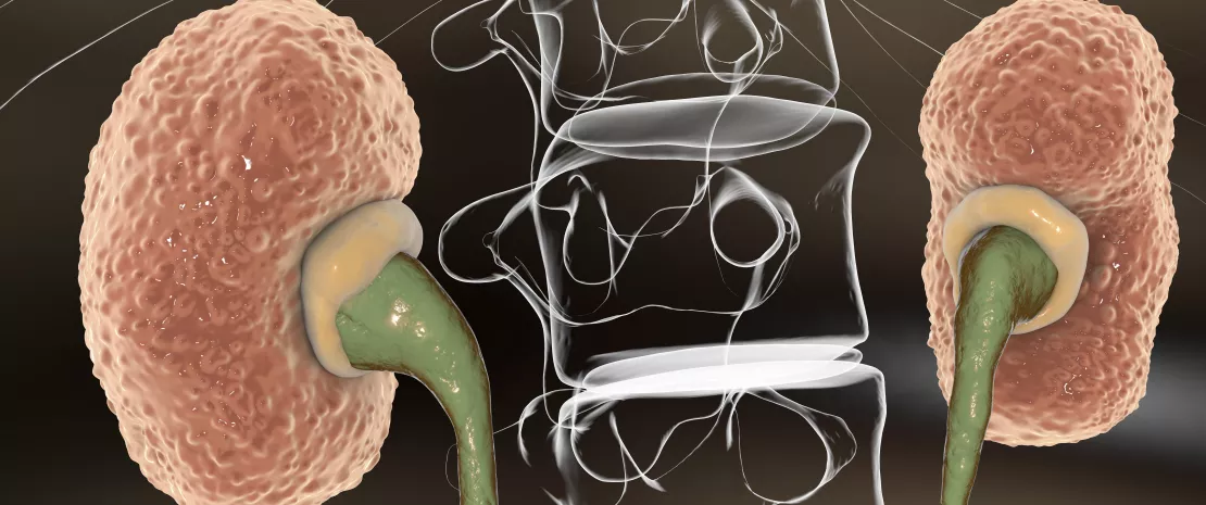
Intestinal bacteria and blood metabolites signal the progression of chronic kidney disease. In addition to the discovery of potential new biomarkers, investigation in this area is also revealing etiological leads.
Chronic kidney disease (CKD) is associated with specific changes in the gut microbiota and circulating metabolites. However, the functions of the microbiota and its complex relationship with the host’s metabolism during the progression of CKD are still only loosely understood. Hence this study involving 72 patients with CKD at different severity stages (26 mild, 26 moderate and 20 advanced cases) and 20 control subjects with normal renal function. Fecal samples were subjected to (sidenote:
Shotgun sequencing method is more accurate than 16S rARN.
), while blood metabolite profiling was performed, targeting bile acids (BA), short- and medium-chain fatty acids and uremic toxins.
A bacterial and metabolic signature
13 bacterial species and 6 circulating metabolites experienced significant changes (increases or decreases) from early to advanced stages of CKD, or at a specific stage or stages only. For example, Bacteroides eggerthii distinguished control subjects from patients in the early stages of CKD, while Prevotella sp. 885 correlated with urea excretion and reflected progression of the disease. Some gut bacteria can therefore act as useful biomarkers for the early diagnosis and monitoring of CKD. As regards metabolites, propionic acid decreased significantly in late stages of CKD, with its absence strongly signaling advanced patients.
Etiological leads
Bacterial genes associated with the biosynthesis of secondary BA were found to be more prevalent in the early stage of CKD, indicating that the conversion of primary BA into secondary BA by intestinal bacteria occurs at the onset of kidney function decline. The advanced stages of the disease were correlated with enrichment of pathways linked:
– on the one hand, to the metabolism of (sidenote:
Steroids, ether lipids, polyunsaturated fatty acids
) (probably involved in metabolic syndrome, which is often associated with dyslipidemia, known to be an etiological factor in CKD)
– and, on the other hand, to the biosynthesis of lipopolysaccharides (LPS, inflammatory endotoxins) Therefore, changes in the metabolism of the microbiota and inflammation in the host are thought to influence renal health.
Bacteria-metabolite links
The team identified gut bacteria linked to changes in circulating metabolites, suggesting the potential involvement of the intestinal microbiota in the development of CKD. For example, the notable decrease in B. eggerthii in CKD patients was correlated with the synthesis of secondary BA at an early stage of the disease. Similarly, the increased synthesis of LPS in late stages was partly attributed to an increase in Escherichia coli and other Enterobacteriaceae. These bacteria-metabolite links may indicate either that the bacterial species produces this metabolite or that the metabolite enhances/inhibits the growth of the bacterial species. In sum, this clearer understanding of the relationship between intestinal bacteria species and host metabolism at different stages of CKD provides potential etiological and diagnostic leads for the disease.
1A high throughput DNA sequencing technique, known as “random sequencing”, which allows large quantities of DNA to be sequenced in very short periods. This method can be used to sequence entire genomes, for example.
A recent study shed light on the beneficial mechanism of action of some gut bacteria on cholesterol levels in humans, thus exposing a new precious role of the gut microbiota on our health.
Hypercholesterolemia, i.e. elevated circulating cholesterol levels, is strongly associated to the development and progression of cardiovascular diseases, that are responsible for one out of every four deaths in developed countries1. Drugs such as statins are therapeutic strategies that lower blood cholesterol levels. Unfortunately, these molecules do not act on dietary cholesterol and can have many side effects. What if a new avenue for the treatment of hypercholesterolemia was found deep in our gut
Uncovering the mechanism at play in humans
Researchers recently identified within the gut microbiota the mechanism responsible for cholesterol metabolism by some bacteria, thus decreasing fecal and blood levels. The idea that some gut bacteria can break down cholesterol is not new since this bacterial activity was already well known for a hundred years. But the exact functioning could never be identified in humans because most of bacteria are hard to cultivate in petri dishes in laboratory settings, thus making their study extremely complicated.
A key player: IsmA gene
Using multiple analytical methods, the researchers identified within these bacteria the IsmA gene (Intestinal Steroid Metabolism A) which could be involved in intestinal cholesterol metabolism. People carrying this gene in their gut microbiota had reduced (55% to 75%) cholesterol contents in their stool compared to non-carriers. Blood cholesterol levels were also lower in carriers.
Towards new therapeutic strategies?
This new promising research could lead to new strategies targeting the gut microbiota: by introducing these cholesterol-metabolizing bacteria in the gut microbiota or by increasing their number using prebiotics, it could be possible to fight high blood cholesterol levels.
1 Goldstein, J.L., and Brown, M.S. (2015). A century of cholesterol and coronaries: from plaques to genes to statins. Cell 161, 161–172.
Stress-related intestinal dysbiosis may be involved in the development of depressive disorders. Such dysbioses may also limit the effectiveness of a family of antidepressants via alterations of the serotonergic pathway.
Current treatments for major depressive disorders, such as fluoxetine, a selective serotonin reuptake inhibitor, are only partly effective. Although the gut microbiota, which is sensitive to chronic stress, represents a therapeutic target for the treatment of depression, no study had evaluated whether it can affect the efficacy of antidepressants, until recently. This gap has now been filled thanks to the work of a French research team. Their goal was to evaluate whether an intestinal dysbiosis induced by chronic stress can induce metabolic changes that impact emotional behavior and responses to serotonergic drugs.
Transferring depression via fecal transplants
To find out whether depression is transmissible, the researchers transplanted the (imbalanced) gut microbiota of a mouse suffering from moderate chronic stress into healthy recipient mice previously treated with antibiotics. In doing so, they transferred most of the elements responsible for the mouse’s dysbiosis. Among the recipients, depressive-like behavior was observed, with a reduction in neurogenesis in the hippocampus and in serotonin levels (in the latter case, limitation of its synthesis and reuptake, and stimulation of its degradation). This process seems to involve tryptophan (a dietary amino acid that is a precursor to serotonin, the metabolism of which may be altered by dysbiosis), since lower levels of this compound were noted in the recipients’ serum. Lastly, the disturbances described were exacerbated by the transplant, with the recipient mice more affected than the donors, a discrepancy which may be explained by the decrease of a bacterial cluster in connection with lower tryptophan levels.
Resistance to antidepressants
Another notable effect of the fecal transplant was the altered antidepressant and neurogenic effects of fluoxetine in the recipient mice (but not in the donors). The antidepressant did not increase levels of serotonin in the hippocampus, failing to restore normal levels of neurotransmitter synthesis, reuptake, or degradation. However, treatment with an immediate serotonin precursor (5-HTP1, a hydroxylated tryptophan derivative) did restore serotonin levels in the hippocampus, improve neurogenesis and relieve depression.
A mechanism, a therapy, and a biomarker
Intestinal dysbioses may therefore explain the development of certain forms of depression and the lack of efficacy of fluoxetine (via alterations of the serotonergic pathway in the metabolism of tryptophan). According to the authors, modulating the microorganisms involved in the catabolism of tryptophan represents a potential therapeutic strategy. At the same time, the levels of tryptophan in the plasma may guide therapeutic choices by acting as a biomarker. Nevertheless, these results still require validation in humans.
One less pinch of salt could be enough to change the gut microbiota of hypertensive women. Their bacteria could increase the production of beneficial fatty acids, which, once in their bloodstream, could decrease their blood pressure and their arterial stiffness.
People with hypertension know that they have an increased risk of stroke, heart attack, and heart failure; and they must watch their diet and especially their salt intake. Because salt and hypertension are not a good mix. Based on studies carried out with mice, the mechanism connecting the two could be found in out gut. Moreover, since our entire diet has an impact on our microbiota, why not salt?
Salt-sensitive microbiota
British researchers thus wondered whether a salt-rich diet could modulate our gut microbiota. The experiment conducted on 145 untreated hypertensive subjects seems to prove them right: a decrease in salt intake, even modest, impacted the types of bacteria living in their intestines. Their altered gut microbiota produced more (sidenote:
Short chain fatty acids (SGFA)
Short chain fatty acids are a source of energy (fuel) for an individual’s cells. They interact with the immune system and are implicated in communication between the gut and the brain.
Silva YP, Bernardi A, Frozza RL. The Role of Short-Chain Fatty Acids From Gut Microbiota in Gut-Brain Communication. Front Endocrinol (Lausanne). 2020;11:25.), which are substances that enter the bloodstream and activate vascular receptors. This is greatly beneficial to hypertensive subjects: the increase in SCFA circulating in their bloodstream seemed to be directly correlated with a decrease in their blood pressure and pulse wave velocity, which is used to measure arterial stiffness. This beneficial effect could be related to anti-inflammatory properties that are attributed to these fatty acids of bacterial origin.
Only in hypertensive women
Another finding of the study is that the underlying mechanisms appear to be different between men and women. On closer examination, the change in blood SCFAs resulting from a low-salt diet was proven only in women, and it is still unclear why. In any case, every cook should make sure not to use too much salt in their recipes, especially if someone with high blood pressure (man or woman) is eating at their table: salt consumption remains too high all around the world, and it is recommended to reduce salt intake for everyone, and especially for those with hypertension.
Chen L, He FJ, Dong Y, Huang Y, Wand C et al. Modest Sodium Reduction Increases Circulating Short-Chain Fatty Acids in Untreated Hypertensives - A Randomized, Double-Blind, Placebo-Controlled Trial. Hypertension. 2020;76:73–79. WHO. Salt reduction. 29 April 2020. https://www.who.int/news-room/fact-sheets/detail/salt-reduction
Functional gastrointestinal disorders (FGDs), the most common intestinal afflictions, are a collection of chronic digestive symptoms that are not explained by any detectable anatomic anomaly.
Irritable bowel syndrome (IBS), the most common FGD
FGDs encompass a set of symptoms such as IBS, constipation, diarrhea, functional bloating, and non-specific FGDs.
IBS alone affects 10% of the population and is distinguished from other FGDs by abdominal pain associated with constipation, diarrhea, or alternations between them. It often presents with abdominal bloating and a higher level of stress than the general population.
52%
Just 1 in 2 people having suffered from a digestive condition involving the microbiota had made the connection
Irritable Bowel Syndrome (IBS) is a functional gastrointestinal disorder characterized by recurrent abdominal pain, that is associated with changes in stool frequency or stool form, in the absence of any organic disorder. Using ROME IV criteria, IBS is classified into four subtypes:
IBS with predominant constipation (IBS-C),
IBS with predominant diarrhea (IBS-D),
IBS with mixed bowel habits (IBS-M),
IBS, unsubtyped (IBS-U) which does not meet the criteria for IBS-C, D, or M
Psychiatric comorbidities, such as anxiety, depression and somatization are common in patients with IBS.
In very small children, FGDs represent the most common gastrointestinal reason for a doctor’s visit. This includes baby colic with regurgitation and constipation problems, IBS, and other less well characterized functional problems. Stomach pain, bloating, diarrhea, and constipation are commonly associated with FGDs and can have major consequences on a child’s daily life. Stress and anxiety can also favor or prolong certain symptoms, particularly pain.
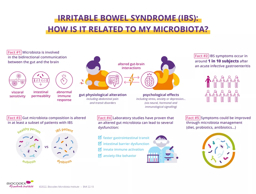
Disrupted communication between the intestines and the brain
The causes of IBS are still poorly understood. The risk of developing IBS increases five-fold after a bacterial infection causing acute diarrhea. It has been suggested that it could be related to dysfunction in the communication between the brain and the intestine, in conjunction with an imbalance in the intestinal flora. In the majority of cases, there is a (sidenote:
Dysbiosis
Generally defined as an alteration in the composition and function of the microbiota caused by a combination of environmental and individual-specific factors.
Levy M, Kolodziejczyk AA, Thaiss CA, et al. Dysbiosis and the immune system. Nat Rev Immunol. 2017;17(4):219-232.) among the bacterial species that make up the microbiota, with fewer positive bacteria and more harmful bacteria. This dysfunction causes intestinal motor problems: transit is slowed, the intestinal barrier is modified, and slight inflammation develops. It also causes hypersensitivity in the mucosa that makes normal phenomena, like the movement of intestinal gas, painful.
Have you heard of "dysbiosis"?
It refers to a breakdown in the delicate balance between the billions of microorganisms that make up our microbiota and in their relationship with our body.
For adults, in addition to a controlled diet, treatment options include antispasmodics, laxatives, and antidiarrheals. In children, relaxation and hypnosis techniques, which can relieve pain, are preferred. Sometimes antispasmodics are also prescribed. To modify the microbiota, there are promising data currently available about probiotics, particularly (sidenote:
Bifidobacterium
A genus of Y-shaped bacteria, most species of which are beneficial to humans. They are found in the gut of humans, and in some yogurts.