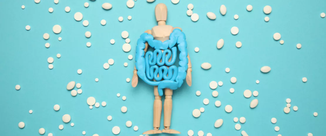
Introversion, social anxiety... alcoholics display alterations in social behavior that may facilitate relapse. Alcohol consumption can lead to a dysbiosis of the gut microbiota, which in turn is known to be a modulator of social behavior in rodents. Hence the theory that the gut microbiota may be involved in the sociability problems associated with alcoholism. To test this hypothesis, a team of researchers transplanted (FMT ) into mice the microbiota of alcoholic patients suffering from a dysbiosis (reduced bacterial count, reduced content of Faecalibacterium prausnitzii and increased content of Lachnospiraceae), increased gut permeability and psychological disorders (anxiety, alcoholic impulses, impaired sociability, etc.).
Microbiota was enough to modify behavior
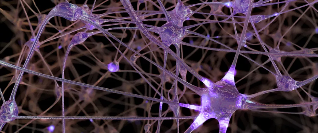
The results? The mice which received the transplant (FMT) showed a reduced interest in social interactions and more depressive-like behavior, as well as higher corticosterone levels, reflecting higher levels of stress. Disturbances of myelination and neurotransmission, as well as inflammation, were observed in the frontal cortex and striatum.
β-hydroxybutyrate, a metabolic mediator?
β-hydroxybutyrate (BHB), a ketone body produced by the liver that serves as an energy source for neurons may be involved in the behavioral and brain disorders observed. Reduced in the FMT mice, it is one of the metabolites distinguishing them from controls. Studies in other animal models and in humans supports the involvement of BHB. In mice, an increase in plasma BHB levels under the ketogenic diet improved social skills and myelination and reduced brain inflammation. In alcoholics, low plasma BHB levels were associated with higher levels of social anxiety, depression and alcohol craving, and lower white matter integrity (one of the determinants of which is myelination).
Is microbial ethanol involved?
But how would the microbiota influence plasma BHB levels? The microbiota of alcoholic patients produces ethanol, even with protracted alcohol withdrawal, an observation confirmed in FMT mice. The authors believe this alcohol may inhibit the Hmgcs2 enzyme and PPARα transcription factor, which are involved in the synthesis of BHB. Indeed, the expression of these two molecules is lower in FMT mice. Restoring the microbiota or ketone body metabolism is one clinical avenue that may result from this work: by favorably modulating the gut-brain axis, this may help limit relapse.