Gut microbiota and NASH course

A study recently demonstrated a link between gut microbiota and inflammatory state that promotes progression of non-alcoholic fatty liver disease to steatohepatitis through the production of short-chain fatty acids.

The gut microbiota Steatohepatitis: viral microbiota also involved Alcoholic hepatitis: towards new fungal targets? The Janus face of Antibiotics: Life Savers and Microbiota Disruptors
Photo : Gut microbiota and NASH course

Together with insulin resistance, non-alcoholic fatty liver disease (NAFLD) has become the most frequent hepatic disorder in Western countries, with a prevalence of around 25%. The first stage is fatty liver disease, which progresses to non-alcoholic steatohepatitis (NASH) in some patients under the influence of several factors, including a proinflammatory state. A German team recently published a study comparing three parameters from 32 patients with NAFLD (of which 18 with NASH) and 27 healthy volunteers: gut microbiota, fecal short-chain fatty acids (SCFA) and Th17/rTreg ratio in blood.

NASH-specific microbiota

Compared to patients with a less advanced stage of non-alcoholic fatty liver disease, those with NASH have a greater abundance of species from the Fusobacteriaceae family and Fusobacterium, Prevotella and Eubacterium genera, as well as a higher content of Fusobacteriaceae and Prevotellacea, two bacterial groups that probably produce SCFAs, compared to control subjects. The observed microbiotic profile thus characterizes two different subgroups corresponding to patients with or without NASH in the NAFLD group, and it is correlated to the results of hepatic biopsies. This could open the way to a new non-invasive method to monitor the disease, since hepatic biopsy is currently the only way to diagnose NASH. Moreover, fecal levels of acetate, propionate and butyrate–three SCFAs produced by the fermentation of dietary fiber in the GI tract–are higher in the NAFLD group than in the control group. A significantly higher level of butyrate is also observed in the NASH group compared to the control group.

The controversial role of SCFAs

Finally, the study focused on the comparison between levels of specific blood lymphocytes: anti-inflammatory resting Treg cells (rTreg) and pro-inflammatory Th17 cells. A study published by the same team had shown that the Th17/rTreg ratio was higher in the NASH subgroup than in patients with NAFLD. The current study brought to light a positive correlation between Th17/rTreg ratio and fecal levels of acetate and propionate. SCFAs are also known for their anti-inflammatory properties, but several studies, including this one, have raised the question of their proinflammatory role in some pathologies and under certain conditions.

Summary
Off
Sidebar
On
Migrated content
Activé
Updated content
Désactivé
Old content type
pro_article
Hide image
Off
News Gastroenterology

Fast sugars: bacterial disruptors in our intestines

Researchers discovered a new harmful effect of glucose and fructose on a bacterium deemed beneficial for our intestinal flora. It is a further argument for the reduction of added sugars in our diet.

The gut microbiota Obesity
Actu GP : Sucres rapides : des perturbateurs bactériens dans nos intestins

The WHO recommends that the amount of free sugars does not exceed 10% of the daily energy intake for adults and children. Free sugars are glucose, fructose, sucrose (white sugar) naturally present in honey, syrups, fruit juices and concentrates, table sugar, etc., and as additives in many processed foods. However, in the absence of fiber, contained in whole fruits and vegetables for instance, these fast sugars rapidly increase blood sugar levels as well as the risk of obesity, diabetes and cardiovascular diseases.

“Hindered” bacterium

Their impact seems to go beyond their absorption in the small intestine. A team of American researchers discovered that glucose and fructose, two common sugars in Western diet, could disrupt the growth of a bacterium abundantly found in the gut flora of healthy people with a balanced body weight: Bacteroides thetaiotaomicron, so called because some of its components look like the Greek letters “theta”, “iota” and “omicron”. Fructose and glucose are thus able to inactivate the production of a protein secreted by B. thetaiotaomicron that promotes the proper implantation of the bacterium in the gut microbiota.

Direct action on genes

This study was performed in vitro and in mice, and confirmed what other studies had previously postulated: glucose and fructose can reach the colon, main location of the intestinal flora. The new finding lies in the way they could destabilize the microbiota in the colon: not by serving as an energy source for some harmful bacteria, but by directly altering some bacterial genes, such as B. thetaiotaomicron. All this brings grist—or should we say sugar—to the mill of studies proving the harmful health effects of added sugar and confirms the importance of having a sufficient and regularly intake of fiber to mitigate its effects.

Summary
Off
Sidebar
Off
Migrated content
Activé
Updated content
Désactivé
Old sources

Sources:

GE Townsend, Dietary sugar silences a colonization factor in a mammalian gut symbiont, Proc Natl Acad Sci U S A. 2019 Jan 2;116(1):233-238

Old content type
article
Hide image
Off
News

Bacteria as restorers of works of art: future allies of heritage conservation?

An Italian team detailed the deterioration of a 17th-century Baroque painting caused by bacteria and fungi. They also identified three bacterial species that have the ability to counter such deterioration.

Diet
Actu GP : Bactéries restauratrices : futures alliées du patrimoine ?

When it comes to art, biodegradability is rarely compatible with durability. The deterioration of works of art and buildings by microorganisms is a threat dreaded by curators of historic pieces and sites worldwide. In the Santa Maria in Vado church in Ferrara, Italy, the 1620 painting (sidenote: The Coronation of the Virgin ) by the Italian artist Carlo Bononi, has suffered the ravages of time, natural disasters (2012 earthquake)… and microbes.

The perfect cocktail

Researchers from Ferrara made their contribution to the restoration of this work of art by analyzing the microbial life that had grown on the painting. By extracting samples from the painting–which was originally displayed on the ceiling and is now standing on the floor in a niche–the Italian team was able to observe two types of bacteria: Staphylococcus on the front, the most eroded section, and Bacillus on the back. This typical 17th-century painting is covered with natural pigments, which these bacteria love to feast on, especially red lac, red earth and yellow earth. Associated with ideal temperature, humidity and light conditions, it was the perfect cocktail to promote the growth of these “paint-eating” bacteria.

Each area has its own fungi

High heat and humidity also attract fungi: the scientists found in other darker areas Aspergillus and Penicillium molds. These two species are also found in museums and libraries housing old paintings and paper-based items. Lighter areas (yellowish light blue or pink), however, hosted Cladosporium spp., while the part resting on the floor was stained by Alternaria yeast.

Fighting fire with fire: rescuer bacteria

Identifying aggressors is one thing, finding saviors is another, although much more useful. The same Italian team was successful in their quest: they discovered that three species of Bacillus could prevent growth and harmful effects of microorganisms attracted to colors. These bacteria could become allies in the restoration of paintings and works of art subject to microbial attacks. We now have to make sure that they do not affect the paintings in any way. (Art)work in progress

Summary
Off
Sidebar
Off
Migrated content
Activé
Updated content
Désactivé
Old sources

Sources:

E caselli et al, Characterization of biodegradation in a 17th century easel painting and potential for a biological approach, PLoS One. 2018 Dec 5;13(12):e0207630

Old content type
article
Hide image
Off
News

Peripartum prophylactic antibiotic therapy decreases bifidobacterium levels in breast milk

Peripartum preventive antibiotic therapy changes the bacterial composition of breast milk. Levels of Bifidobacterium, bacteria that are beneficial to the development of newborns, are significantly reduced in the days following delivery.

The gut microbiota Microbiota, breastfeeding and early puberty Can fecal transplantation restore the microbiota of Caesarean-born infants? Infant microbiota: the breast milk feeding mode counts
Actu PRO : Une antibiothérapie prophylactique péri-partum appauvrit le lait maternel en Bifidobacterium

Prophylactic antibiotic therapy is necessary to reduce the risks of peripartum infections, which are the cause of 10% of maternal deaths and are associated to the death of nearly 1 million of newborns every year, according to the (sidenote: WHO recommendations for prevention and treatment of maternal peripartum infections- 2015 ) . However, these treatments are associated with adverse effects, including changes in the maternal microbiota that are likely to impact the child’s early colonization. This led a Brazilian team to study changes in breast milk bacterial populations, while focusing on bacteria from the Bifidobacterium genus. The main representatives of this genus (B. breve, B. adolescentis, B. bifidum, B. longum, and B. dentium in breast milk) are known for their beneficial effects on humans, especially through the production of short-chain fatty acids.

Significant decrease in Bifidobacterium on Day 7

Researchers compared samples of milk from 55 women who gave birth vaginally: 21 were preventively treated with broad-spectrum antibiotics (cefazolin, penicillin or clindamycin) and 34 treatment-naive women. Total bacterial concentration as well as detailed count of Bifidobacterium were determined by qPCR in samples taken on Days 7±3 and 30±4. The results did not show significant differences between the study groups in the total number of bacteria. An explanation for this finding might be the repopulation by bacteria that are resistant to the antibiotics used in the study. On the contrary, a significant decrease in levels of Bifidobacterium is observed in the milk of women treated prophylactically. This dysbiosis is at its peak on Day 7±3 but returns to normal over time and is not noticeable after one month.

Hailed as one of the greatest medical advances of the 20th century, antibiotics have saved millions of lives. But they also have an impact on our microbiota by inducing a dysbiosis. Let’s take a look at this ambivalence role:

The ambivalent role of antibiotics

By destroying the bacteria responsible for infection, antibiotics can also lead…

Dysbiosis gradual improvement: hypotheses

Researchers suggest a likely recolonization from the gut microbiota, trough the endogenous entero‑mammary pathway involving maternal dendritic cells able to capture commensal bacteria in the lumen. Other hypothesis: oligosaccharides present in breast milk might act as a substrate and promote Bifidobacterium growth. All we can say for sure is that if the mother is breastfeeding, peripartum prophylactic antibiotic therapy reduces the supply of beneficial bacteria to the child. This initial observation requires us to detail the impact of this temporary deficit on the development of the newborn’s gut microbiota and correlated functions, especially immune and inflammatory functions.

What is the World AMR Awareness Week?

Each year, since 2015, the WHO organizes the World AMR Awareness Week (WAAW), which aims to increase awareness of global antimicrobial resistance.
Held on 18-24 November, this campaign encourages the general public, healthcare professionals and decision-makers to use antimicrobials carefully, to prevent the further emergence of antimicrobial resistance.

Summary
Off
Sidebar
On
Migrated content
Activé
Updated content
Désactivé
Old content type
pro_article
Hide image
Off
News Gastroenterology

Gut microbiota, a reflection of our thyroid?

Our gut microbiota could reflect the state of our thyroid’s health, according to a Chinese study that brings to light the link between its composition and the risk of contracting thyroid nodules and cancer. A first step towards the development of potentially useful probiotics?

The gut microbiota Diet
Actu GP : Le microbiote intestinal, miroir de notre thyroïde ?


Thyroid diseases are steadily increasing, especially in women. Estrogens, body mass index, radioactivity, ethnicity or high consumption of iodine… there are many risk factors. Individuals with a thyroid disease have a thyroid hormone imbalance observed in the blood levels as well as a disrupted functioning. However, these hormones can impact the composition of the gut microbiota. On the contrary, the latter may be involved in the exchange of hormonal messages between gut and brain. Previous works have already established a link between gut microbiota on the one hand, and some autoimmune thyroid disorders ( (sidenote: Hashimoto's thyroiditis and Graves’ disease Two autoimmune diseases caused by an attack of the thyroid gland by the immune system, which considers it as foreign to the body. Hashimoto's thyroiditis is characterized by hypothyroidism; and Graves’ disease by hyperthyroidism. ) ) on the other hand. What about thyroid nodules and cancer?

Patients with thyroid disorders have a richer microbiota

To test this hypothesis, a Chinese team analyzed and compared the stool composition of 74 participants: 20 with thyroid cancer, 18 with nodules and 36 healthy volunteers. The results showed a greater abundance of intestinal bacteria in patients with a thyroid disorder compared to healthy individuals. Patients also had lower contents of Lactobacillus, involved in the selenium retention, a trace element that is key to a properly-functioning thyroid, and Butyricimonas, which produces a substance with beneficial effects on the gastrointestinal tract. Other bacteria were, however, more abundant: Neisseria and Streptococcus in case of nodules, as well as Clostridium in case of cancer. According to the authors, these species seem to play a role in the proper functioning of the thyroid and onset of these disorders.

Useful bacterial biomarkers

The researchers also observed a link between the abundance of some bacterial species and TSH (thyroid-stimulating hormone) and T3 (triiodothyronine) levels, two hormones indicating the potential presence of thyroid disorders. However, it is still impossible to explain if observed thyroid disorders lead to a specific microbiota, or if some bacterial species trigger these disorders. All these discoveries could make thyroid nodules and cancer diagnosis easier and lead to the development of probiotics useful to their treatment.


Summary
Off
Sidebar
On
Migrated content
Activé
Updated content
Désactivé
Old sources

Sources:

Zhang J et al. Dysbiosis of the gut microbiome is associated with thyroid cancer and thyroid nodules and correlated with clinical index of thyroid function. Endocrine 2019 Jun;64(3):564-574.


Old content type
article
Hide image
Off
News

Ulcerative colitis: an innovative fecal transplant protocol

Preservation of anaerobic microorganisms in fecal transplants could lead to a less intensive but just as efficient treatment as standard methods in patients with ulcerative colitis.

The gut microbiota Gut microbiota not yet considered “adult” at 5 years old? Role of the microbiota in gut-brain communication Role of antibiotics and microbiota in parkinson's disease
Photo : Ulcerative colitis: an innovative fecal transplant protocol


While the involvement of the gut microbiota in chronic inflammatory bowel diseases (Crohn’s and ulcerative colitis) has been proven, the fecal microbiota transplant (FMT) is still being investigated. There seems to be a significant donor effect: the more diversified the transplanted microbiota, the better are the results. In this context, Australian researchers developed an innovative technique.

Preserved anaerobic microorganisms, streamlined protocol

Scientists hypothesized that anaerobic microorganisms could be involved in the therapeutic effect of fecal transplants for the treatment of UC. They thus developed a preparation technique that preserves these species which are largely destroyed by standard methods performed in the presence of oxygen. Their administration protocol was less intensive than those previously used: one dose (from a mix of several donors) received by colonoscopy followed by two doses administered through enemas in a 7‑day period, compared to one colonoscopy and five enemas per week during 8 weeks in the (sidenote: Paramsothy et al., Multidonor intensive faecal microbiota transplantation for active ulcerative colitis: a randomised placebo-controlled trial. Lancet 2017 ) .

Effective protocol on corticosteroid dependency

Seventy-three patients with mild to moderate UC were included in this trial. In addition to their background treatment, participants received prednisolone (doses < 25mg/day). Primary endpoint was complete corticosteroid weaning on Week 8, when a new colonoscopy was performed as well as a clinical examination. In the group receiving FMT from donors (dFMT), remission rate was 32%, compared to 9% in the control group who received autologous FMT (aFMT). Average decrease in overall (sidenote: MAYO score https://www.igibdscores.it/en/info-mayo-full.html ) was 3.5 points in the dFMT group (4 patients attained a Mayo score of 0), vs. 1.2 points in the aFMT group. It should also be noted that the greater abundance of both anaerobic species (Anaerofilum pentosovorans and Bacteroides coprophilus) was strongly associated to an improvement of the disease in the dFMT group. This study confirms the benefits of FMT in the treatment of UC and concludes that an anaerobic preparation method could streamline therapeutic protocol and improve patients’ lives, while maintaining the same efficacy.


Summary
Off
Sidebar
On
Migrated content
Activé
Updated content
Désactivé
Old content type
pro_article
Hide image
Off
News Gastroenterology

Fatty liver disease: what are the benefits of whole-grain flour?

Could whole-grain wheat limit the risk of non-alcoholic fatty liver disease attributed to high fat diet?

The gut microbiota Obesity Liver diseases Diet
Actu GP : Maladie du foie gras : qu’apporte la farine complète ?

The main symptom of (sidenote: Metabolic syndrome Combination of several metabolic anomalies: high blood pressure, large waist circumference, increased blood triglyceride and glucose levels associated to a low blood level of “good” cholesterol. Alberti KG, Zimmet P, Shaw J. The metabolic syndrome--a new worldwide definition. Lancet. 2005 ; 366 (9491) : 1059-62. ) is the accumulation of fat (triglycerides) in the liver, which not only increases the risk of non-alcoholic fatty steatohepatitis (NASH), but may also alter the whole digestive system, thus leading to increased cardiometabolic risk factors (cardiovascular diseases and type 2 diabetes), including insulin resistance. Could replacing refined flour (white) with whole-grain flour (richer in fiber, minerals and vitamins) help limit these risks? Studies tend to confirm this hypothesis. However, underlying mechanisms remain poorly understood, thus illustrating the complexity of relations between the organs involved in the metabolism, especially gut, fatty tissue and liver.

Whole-grain vs. refined flour

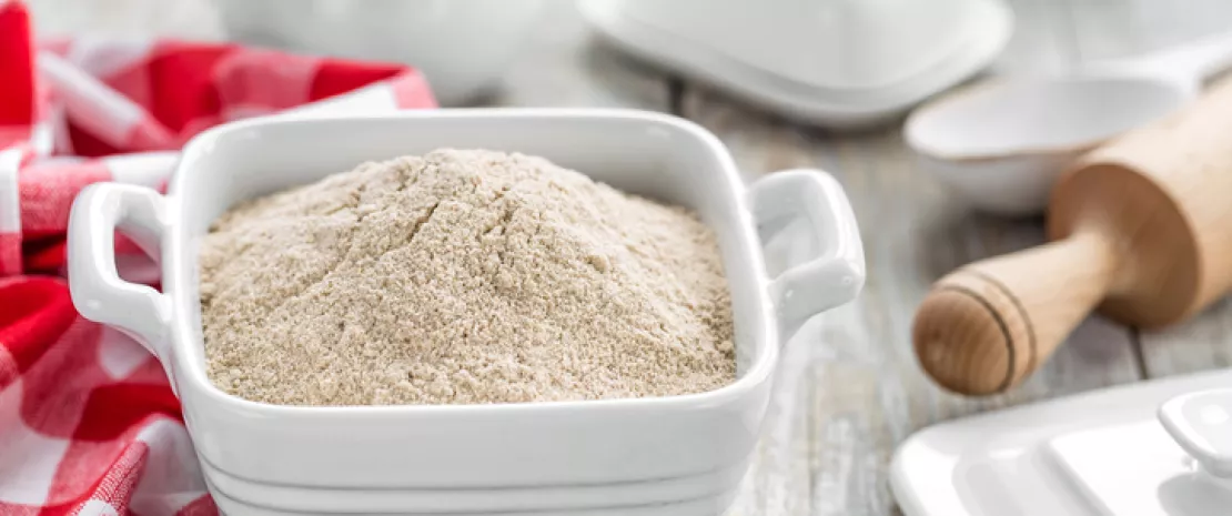
To assess the benefits of whole-grain flour in the digestive system (including liver, fatty tissue and microbiota), Dutch researchers compared its effects to those of refined flour in 50 overweight adults whose total cholesterol levels were slightly elevated. 12 weeks later, the results showed that the wheat refining process does not impact lipid blood levels (lipemia) nor sugar blood levels (glycemia), nor subcutaneous or deep abdominal fat. However, hepatic triglycerides levels were increased by 50%, driving up the proportion of participants with NASH in the refined-flour group from 33% to 44%. On the contrary, this rate decreased from 35% to 25% in the whole-grain-flour group. Researchers also observed a decrease in bacterial diversity and change in composition of the gut microbiota in the refined-flour group, although these alterations were not predictive of changes observed in the liver.

A treatment for NASH?

According to the scientists, these results tend to show the benefits of whole-grain wheat products as a preventive and therapeutic avenue for fatty liver disease. Its protective effects against metabolic disorders could be due to the hepatic benefits of some chemical components naturally present in whole-grain wheat, or to the fermentation of fiber by gut flora bacteria. The authors concluded that, were this true, it would confirm the hypothesis that there is a bidirectional communication between liver and gut.

Summary
Off
Sidebar
On
Migrated content
Activé
Updated content
Désactivé
Old sources

Sources:

Schutte S, et al. A 12-wk whole-grain wheat intervention protects against hepatic fat: the Graandioos study, a randomized trial in overweight subjects. Am J Clin Nutr 2018;108:1264–1274

Old content type
article
Hide image
Off
News

Spinal cord injuries and colorectal disorders: impact of the gut microbiota

According to a Chinese study, functional gastrointestinal disorders affecting patients with trauma-induced chronic spinal cord injuries could be related to gut microbiota disruptions and correlated to serum biomarkers.

The gut microbiota Kidney failure: impact of the gut microbiota Severity-specific signature of gut microbiota in chronic kidney disease Kidney transplant: is pre-operative dysbiosis a risk factor for diabetes?
Photo : Spinal cord injuries and colorectal disorders: impact of the gut microbiota

Colorectal disorders caused by chronic spinal cord injuries may have a significant impact on the quality of life of many paraplegic and quadriplegic patients. Disruptions of the autonomous nervous system can lead to gastrointestinal disorders (discomfort, bloating, flatulence), to a combination of chronic constipation and fecal incontinence, and require laxatives or techniques that encourage bowel movements with or without assistance. In some patients, disruptions are so severe that the wish to improve this neurological gastrointestinal disorder exceeds that of treating urinary incontinence or sexual dysfunctions, or even the loss of walking ability.

Disability-associated bacterial profiles

A Chinese team analyzed the stools of 43 men with chronic traumatic spinal cord injuries (23 paraplegic and 20 tetraplegic) and 23 healthy men. The gut microbiota of disabled subjects was different from that of control subjects. It was less diversified and contained higher levels of Bacteroides, Blautia, Lachnoclostridium and Escherichia-Shigella, among others. Moreover, changes were observed in the bacterial profiles of paraplegic (overabundance of Acidaminococcaceae, Blautia, Porphyromonadaceae, and Lachnoclostridium) and tetraplegic patients (overabundance of Bacteroidaceae and Bacteroide) compared to control subjects. Reduced levels of Alistipes also seem to be associated to longer defecation times in tetraplegic patients.

Blood lipid and glucose levels are impacted

To complete their observations, researchers studied correlations between these changes in bacterial populations and some environmental factors such as age, BMI, and various serum markers (CRP, glucose, liver enzymes, blood lipids, uremia and uric acid, creatinine, etc.). Bacteria from the Bacteroides genus, which are more abundant in tetraplegic patients, were associated with low levels of HDL, probably because of the lack of physical activity. On the contrary, bacteria from the Dialister genus, which are more abundant in healthy subjects, were negatively correlated to blood lipids (LDL, triglycerides and total cholesterol). High blood concentrations of these factors could thus be a sign of worsening colorectal disorders. Megamonas were associated with lower glycemia but also with longer defecation time and increased bloating, probably due to the fermentation of undigested carbohydrates by these same bacteria in the colon. A low level of Prevotella was also related to lower glycemia (thus to a beneficial role), even if other studies have shown pro-inflammatory effects. These results need to be completed by other analytical tools (more detailed analysis of bacterial communities, serotonin assay), by including women in cohorts, and by studying the impact of immobility itself, which may also generate dysbioses.

Summary
Off
Sidebar
On
Migrated content
Activé
Updated content
Désactivé
Old content type
pro_article
Hide image
Off
News Oncology Gastroenterology