
Reconstitution of the gut microbiota of antibiotic-treated patients by autologous faecal microbiota transplant

Commented articles - Adult's section

By Pr. Harry Sokol
Gastroenterology and Nutrition Department, Saint-Antoine Hospital, Paris, France
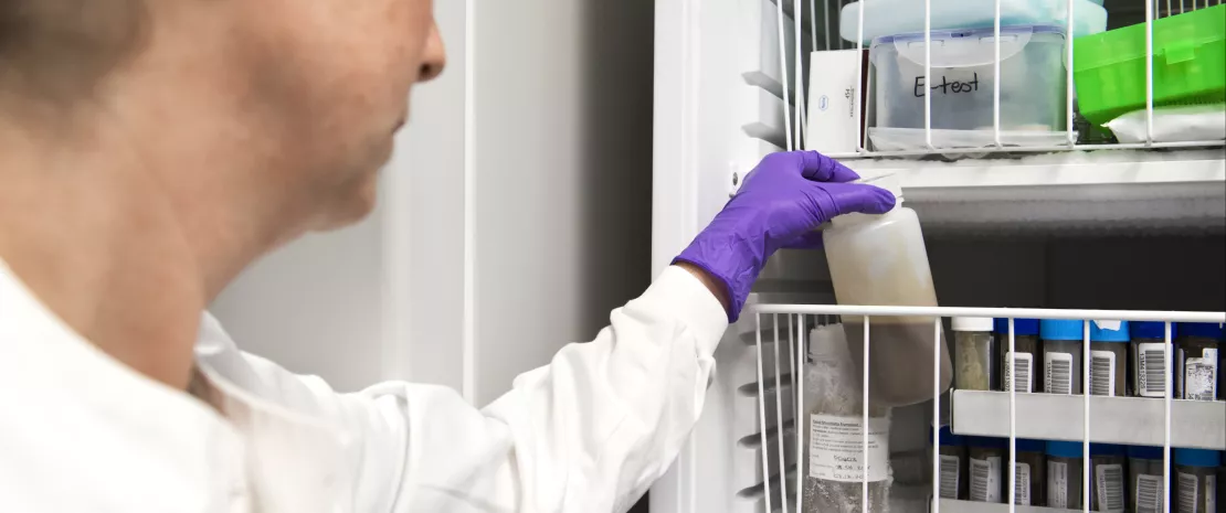
Faecal transplant preparation. A technician storing a prepared sample of human faecal matter in cold storage.

Commentary on the original publication by Taur et al. (Science Translational Medicine 2018)

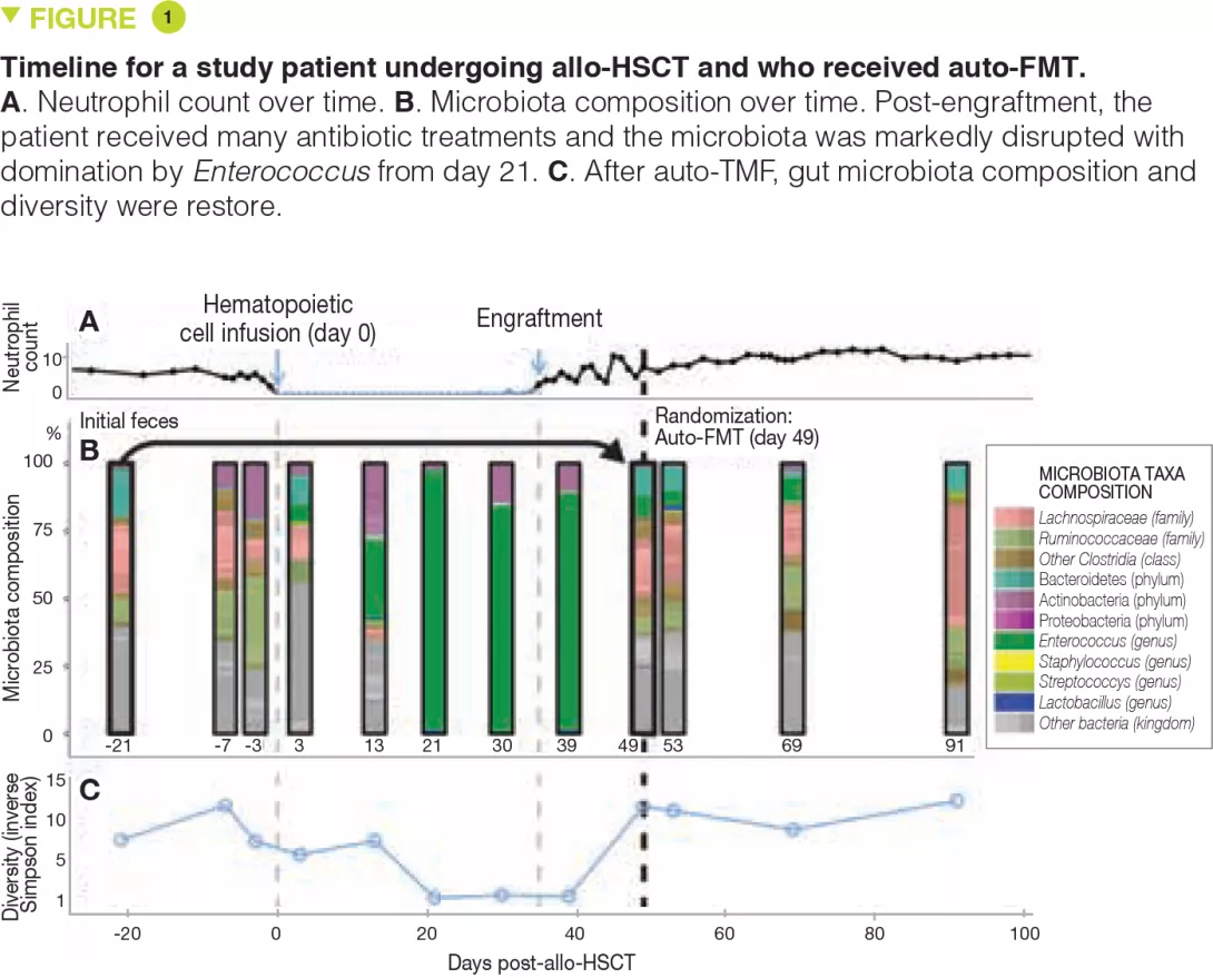
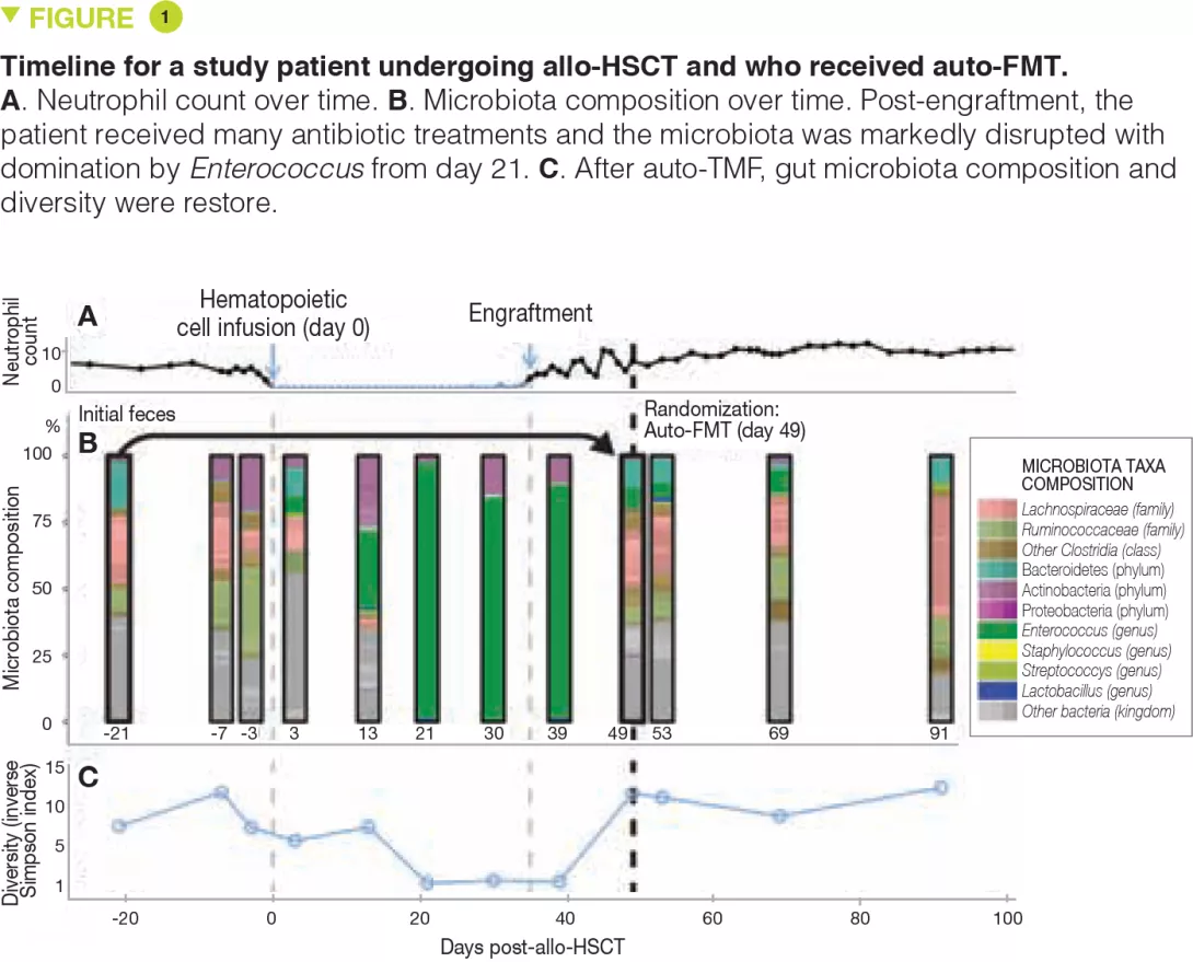
Antibiotic treatment can deplete the commensal bacteria of a patient’s gut microbiota and, paradoxically, increase their risk of subsequent infections. In allogeneic hematopoietic stem cell transplantation (allo-HSCT), antibiotic administration is essential for optimal clinical outcomes but significantly disrupts intestinal microbiota diversity, leading to loss of many beneficial microbes. Although gut microbiota diversity loss during allo-HSCT is associated with increased mortality, approaches to re-establish depleted commensal bacteria have yet to be developed. A randomized, controlled clinical trial has been initiated to compare autologous faecal microbiota transplantation (auto-FMT) versus no intervention; the intestinal microbiota profiles of 25 allo-HSCT patients (14 who received auto-FMT treatment and 11 control patients who did not) were analysed. Changes in gut microbiota diversity and composition revealed that the auto-FMT intervention boosted microbial diversity and re-established the intestinal microbiota composition that the patient had before antibiotic treatment and allo-HSCT. These results demonstrate the potential utility of faecal sample banking for auto-FMT for posttreatment remediation of a patient’s gut microbiota after microbiota-depleting antibiotic treatment during allo-HSCT.[1]

What do we know about this subject? 

Antibiotic treatment damages the intestinal microbiota and increases the risk of gastrointestinal infection. Although this effect has been recognized for more than 60 years, remediation of the antibiotic-depleted gut microbiota has yet to become standard clinical practice. In patients undergoing allogeneic hematopoietic stem cell transplantation (allo-HSCT), antibiotics are routinely given to treat or reduce the risk of serious infection. Prospective studies of allo-HSCT patients demonstrated that the intestinal microbiota is markedly altered during treatment, with profound loss of obligate anaerobic bacteria including immunomodulatory species such as those belonging to the Clostridia class and Bacteroidetes phylum [2]. The clinical consequences of these alterations are also apparent in allo-HSCT: disruption of beneficial obligate anaerobes correlates with complications that include systemic infection with vancomycin-resistant Enterococcus (VRE), Clostridium difficile infection, and graft-versushost disease (GVHD).[2, 3] Overall, patients who lose gut microbiota diversity at the time of hematopoietic stem cell engraftment have higher rates of transplant-related death.[4]

What are the main insights from this study? 

Allo-HSCT patients remain immunocompromised for many months after engraftment, and although immunocompromised patients, including allo-HSCT recipients, have undergone heterologous FMT without side effects,[5] the authors reasoned that autologous FMT would be safer by minimizing the risk of exposure to potentially pathogenic microorganisms not previously encountered by the patient. The authors initiated a randomized, controlled clinical trial to determine the feasibility of auto-FMT for restoring the gut microbiota and for decreasing complications related to allo-HSCT. Here, they present an analysis of the gut microbiota compositional changes in 25 patients enrolled and randomized from whom faecal samples were longitudinally collected.

The authors first confirmed in their cohort of 753 patients (3,237 longitudinally collected faecal samples) that allo-HSCT and the different associated antibiotic treatments induced a marked decrease in gut microbiota diversity, reaching a nadir 5 days after allo-HSCT, which persisted for at least 6 weeks and which in most patients had not recovered at day 100 post-allo-HSCT.
Key points

  • The intestinal microbiota is markedly disrupted during allo-HSCT and this disruption can play a role in the associated complications.

  • Auto-FMT is a feasible and effective strategy for reconstitution of the microbiota after the disruption caused by allo-HSCT.

  • The consequences of microbiota reconstitution in terms of haematological outcomes remain to be evaluated.

Image

As part of the randomized study, faecal samples from patients collected before allo-HSCT were frozen at -80°C and stored. One to 5 weeks (mean, 13 days) after allo- HSCT, upon engraftment (defined by recovery of the neutrophil count to > 500/mm3), patients were re-evaluated and another faecal sample was taken. If there was a paucity of bacteria from the Bacteroidetes phylum, patients were randomized. The results of microbiota analyses from the first 25 evaluable patients (14 from the auto-FMT group and 11 from the control group) are presented. Auto-FMT was administered as a single retention enema after colonic preparation with polyethylene glycol, similar to the preparation for a colonoscopy. The authors show that auto-FMT not only restores the diversity of the intestinal microbiota but also its composition pre-allo-HSCT.

What are the consequences in practice?

microbiota and disruptions thereof play a role in the common infectious and non-infectious complications encountered during allo-HSCT. This first study shows that the collection and storage of a patient’s faecal samples prior to allo-HSCT to be re-administered after engraftment is a feasible and effective strategy to reconstitute the microbiota. It remains to be seen whether patients who underwent auto-FMT have better outcomes with regard to these complications and if they have a better overall survival. If the efficacy of this strategy is confirmed, it could also be considered in other situations where significant microbiota disruption is expected, such as broad spectrum or prolonged antibiotic treatment or anticancer chemotherapy.

Conclusion

Although the benefits in terms of haematological outcomes and overall survival still need to be evaluated, auto-FMT is a promising strategy to re-establish the intestinal microbiota after the disorders induced by the antibiotic treatment associated to the allo-HSCT.

Summary
Off
Sidebar
Off
Migrated content
Désactivé
Updated content
Désactivé
Hide image
Off
Commented article

Digestive cancers and intestinal microbiota: from oncogenesis to response to treatment

Overview

By Pr. Iradj Sobhani
Gastroenterology, Université Paris Est Créteil (UPEC) and Henri Mondor University Hospital, Créteil, France

With an increasing prevalence that has made it the leading cause of death in many western countries, cancer is now regarded more as an emergent disease arising from environmental factors than a disease caused by constitutional genetic aberrations, which are now known to be much less common than previously thought. A paradigm shift favoured by the development of molecular biology techniques, a better understanding of the underlying mechanisms and the identification of associated biomarkers. For several decades, epidemiologists have observed a connection between cancer and diet, which has made the intestinal microbiota – and thus dysbioses – a focal point in the study of cancer. This connection has now found a mechanistic explanation involving energy metabolism, inflammation and immunity: influenced by diet, some bacteria can affect tumour progression, the response to cancer treatments and the side effects of these treatments.

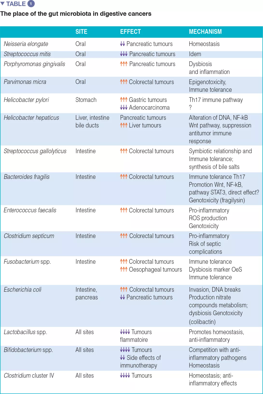
The associations between certain cancers and dysbiosis, the mechanisms by which the intestinal microbiota can promote human cancers, and an inventory of diagnostic and/or therapeutic biomarkers, particularly in anticancer immunotherapy, are summarized in Table 1.

As in the case of obesity and diabetes, it is important to identify bacterial markers for diagnostic purposes but also to study bacterial functions in order to better understand the impact of the environment on these cancers.

In obese individuals, for example, an imbalanced diet in terms of quantity and quality can rapidly alter the intestinal microbiota and the functions of its constituent bacteria.[1] By characterizing the intestinal microbiota in these individuals, it can be possible to identify a specific dysbiosis and thus assess the probability of success or failure of a corrective diet. Many emergent diseases such as cancer have undergone similar developments and are benefiting from new avenues of pathophysiological research.

Image
Image

Oesophagus - Stomach

In physiological conditions, the oesophageal microbiota is similar to that of the oral cavity, with an abundance of Firmicutes, Bacteroidetes, Actinobacteria, Proteobacteria and Fusobacteria and predominance of the genus Streptococcus in the oesophagus. In gastroesophageal reflux disease (GERD) and Barrett’s oesophagus (BE), conditions which promote preneoplastic changes, the microbiota composition is closer to that of the stomach, with an abudance of Bacteroidetes, Proteobacteria and Fusobacterium. Paradoxically, Helicobacter pylori, a gastric bacterium known to be a cofactor in the development of gastric neoplasias (cancer and MALT lymphoma), appears to exert a protective role against oesophageal adenocarcinomas which are on the rise in western countries. In reality, different bacterial species in addition to H. pylori, such as Pasteurella stomatis, Dialister pneumosintes, Slakia exigua, Parvimonas micra and Streptococcus anginosus, are implicated in the development of gastric tumours. Recent work suggests that Enterobacteriaceae, especially Ruminococcus, might play an important role in the immune escape of gastric and oesophageal adenocarcinomas.[2]

Colorectal cancer (CRC) and model of tumor immune escape

Since the first descriptions of an association between colonic dysbiosis and CRC,[3, 4] the hypothesis that the oral flora participated in the dysbiosis implicated in CRC has been updated in light of original work on the link between oral-gut transmission and colon dysbiosis.[5] Disruption of the bacterial equilibrium often occurs at the expense of beneficial species like Bifidobacteria and Lactobacilli, which help maintain the immune response.[2] Since these bacteria can no longer provide a counterweight to pro-inflammatory bacteria, an asymptomatic chronic inflammation, long been known to promote oncogenic processes, develops in the colonic mucosa. Now, the current western diet (high in animal protein and sugars) is known to favour pro-inflammatory bacteria at the expense of anti-inflammatory bacteria. In contrast, a Mediterranean diet (rich in plant fibre) limits these damaging effects.[6] When fibre intake is insufficient, the bacteria recruited as a result of excessive consumption of animal protein and fat erode the mucosa, taken as a source of fibre, and expose the intestinal epithelium to potentially virulent bacteria (Figure 1). At the cellular level, the main biological signalling pathways such as the Wnt and the canonical NF-kB (nuclear factor-kappa B) pathway, respectively responsible for cell renewal and higher production of pro-inflammatory cytokines, are stimulated by this diet.[6] This phenomenon can be likened to a shift of the immune response toward tolerance, due to an overabundance of other bacterial populations such as Parvimonas micra and Streptococcus fragilis.[7] In animals, Bacteroides fragilis and Escherichia coli, which are present in overabundance in advanced CRC (TNM stage III or IV), maintain an inflammatory state in the colonic mucosa and promote tumour development.[8]

Hepatocellular carcinoma (HCC)

Primary liver tumours develop through a chronic process including cirrhosis, itself the result of hepatitis B or C virus infection. Epigenetic mechanisms resulting from the action of microorganisms lead to extinction of certain key genes such as p16 (INK4A), glutathione S-transferase P 1 (GSTP1), CDH1 (E-cadherin), Ras association domain containing protein 1 (RASSF1A), p21 (WAF1/CIP1), all of which are hypermethylated by HBV, as well as the Suppressor of Cytokine Signalling 1 (SOCS-1) and the STAT1 gene, hypermethylated by HCV. These genes delay the occurrence of cancer but hypermethylation inhibits their expression. Bacteria can intervene to promote these processes: Helicobacter hepaticus increases cancer risk, either directly by activating the Wnt and NK-kB pathways, or by facilitating the HVC-induced process. Just like environmental factors (viruses, chemical pollutants, etc.), certain enterobacteria such as E. coli, have been identified as cofactors for activation of the carcinogenic process. In metabolism, a disruption of Firmicutes/Bacteroidetes populations, a known risk factor for obesity, enhances the risk for HCC by crowding out protective species such as Lactobacillus, Bifidobacterium, Parabacteroides and Oscillibacter.[9, 10]

Pancreatic cancer 

Patients with pancreatic cancer have a high density of Enterobacteriaceae, Pseudomonadaceae, Moraxellaceae and Enterococcaceae in tumour tissue, while Acinetobacter, Aquabacterium, Oceanobacillus, Rahnella, Massilia, Delftia, Deinococcus, and Sphingobium are abundant in the duodenal lumen. As in CRC, the dysbiosis linked to this cancer also includes changes in the oral flora, characterized by an overabundance of Porphyromonas gingivalis and an underabundance of Neisseria elongate or Streptococcus mitis. One more example of the link between intestinal dysbiosis and gastrointestinal cancers. As far as treatment is concerned, it is important to note that Gammaproteobacteria can increase resistance to gemcitabine, the standard of care for pancreatic cancer.

Antitumor immune response and dysbiosis

Axenic (germ-free) animals develop fewer tumours, probably due to immune tolerance and less reactive inflammatory activity, which could be explained by the absence of a physiological microbiota. The microbiota can contribute to cancer development through different mechanisms: first of all, activation of inflammation by dysbiosis and reorientation of the immune system; production of genotoxins (colibactin, fragilysin) and virulence factors by bacteria able to directly alter host DNA; induction of oxidative stress by production of reactive oxygen species (ROS); and finally, by bacterial production of secondary metabolites (secondary bile acids, etc.). In the colon model, for example, there are four distinct subtypes corresponding to different metabolic, immune or inflammatory pathways.[11] In the CRC subtype with T lymphocyte infiltration, the T cells have a reduced ability to express cytokines or attack target cells due to persistent stimulation by tumour antigens. This phenomenon is known as T cell exhaustion. It is the most common mechanism of immune escape. Regardless of the initial recruitment of lymphocytes – cytotoxic or facilitator – the tumour continues to grow.[9] Regulatory T cells (Tregs) will facilitate the immunosuppressive effect by producing factors such as TGF-β. Tregs are preferentially recruited in the exhaustion phase. Furthermore, the intratumoral density of Tregs is a negative prognostic marker. By producing immunosuppressive cytokines (IL-10 and TGF-β), Tregs interfere with the specific action of cytotoxic T cells which normally target the tumour. In particular, Tregs increase the immune down-regulating protein CTLA-4 or CD152 (cytotoxic T-lymphocyte-associated protein 4) of these T cells. This protein has become a target of modern immunotherapies. Tregs act with the help of Th17 cells and STAT3 (Signal Transducer and Activator of Transcription 3), involved in the process of carcinogenesis in various organs. Th17 cells produce pro-inflammatory cytokines (IL-17 and IL-23) which promote tumour growth by increasing the production of Th1 cytokines and that of a chemokine (C-X-C motif) ligand 9 and 10 (CXCL9 and CXCL10). Th17 cells have similar characteristics to stem cells and can self-renew. The cytokine environment at the site of the tumour affects the different models of Th17 cell expression: in colorectal, hepatocellular and pancreatic cancer, tumour infiltration by Th17 cells is an unfavorable prognostic marker because it promotes immune tolerance to the tumour. The dysbiosis in the mucosal lining modulates the expression of IL17, IL-23, STAT3.

Microbiota and cancer treatments 

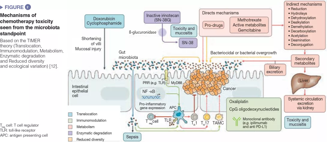
The intestinal microbiota has been shown to modulate the response to anticancer chemotherapy and immunotherapy in mouse models and in humans. Lung and kidney cancers and melanoma have been studied in clinical trials. This effect is never attributable to a single species: it is always a reflection of the impact of the intestinal microbial community as a whole on immunity or a function shared by different bacterial species. These bacterial communities influence the response to therapy in terms of side effects/toxicity and resistance to treatment (Figure 2). For example, proteobacteria, particularly Mycoplasma hyorhinis, possess cytidine-deaminase activity which metabolizes gemcitabine and thereby reduces its efficacy. Likewise, cyclophosphamide has variable antitumor effects according to dose; its efficacy is modulated by gram-positive (including Enterococcus hirae) and gram-negative species (including Barnesiella intestinihominis).[12]

Anticancer immunotherapies have been successfully used in malignant melanoma. These are the most promising therapies involving immune checkpoint inhibitors targeting PD-1 and CTLA-4. It was first noted that in metastatic renal or lung cancer patients, the use of antibiotics could modulate the activity of anti-PD-1 or anti-PD-L1 immunotherapies.[13] Subsequently, a large American study in metastatic melanoma treated with immunotherapy found that a good response to treatment (longer progression- free survival and overall survival) depended on the colonic microbial composition: faecal microbiota transplantation from the patients to recipient mice showed that intestinal dysbiosis was indeed the root cause of the variability in response to anti-PD-1 immunotherapy.[14, 15]

These data are to be compared with data on TLR4 polymorphisms that are related to the variable response to immunotherapy. TLR (toll-like receptors) are transmembrane or cytosolic receptors which belong to the large family of receptors of the innate immune system (PRR, pattern recognition receptors), expressed by epithelial cells and immune cells in the intestine. Binding of a TLR with a microbial ligand triggers an intracellular signalling cascade which usually leads to an inflammatory response through activation of NF-kB. Host immune status has proved to be the main factor in the response to all antineoplastic treatments, directly and by alterations of the intestinal microbiota. It should also be noted that other therapeutic techniques such as radiotherapy and surgery are also affected by the microbiota: ionizing radiation is less toxic to axenic mice compared to conventional mice; postoperative healing of patients after colon cancer surgery depends on the type of dysbiosis.

Image

Conclusion

The colonic microbial composition is influenced by environmental factors and can affect the development and progression of malignant tumours through metabolic, inflammatory and immune pathways. Studies are under way to better understand resistance to and toxicity of anticancer treatments. It is likely that modulation of the intestinal microbiota will become a way to optimize anticancer therapy in the future.

Sources

1 David LA, Maurice CF, Carmody RN, et al. Diet rapidly and reproducibly alters the human gut microbiome. Nature 2014 ; 505 : 559–63.

2 Cianci R, Franza L, Schinzari G, et al. The interplay between immunity and microbiota at intestinal immunological niche: The case of cancer. Int J Mol Sci 2019 ; 20 : 501.

3 Sobhani I, Tap J, Roudot-Thoraval F, et al. Microbial dysbiosis in colorectal cancer patients. Plos One 2011 ; 6 : e16393.

4 Zeller G, Tap J, Voigt AY, et al. Potential of fecal microbiota for early-stage detection of colorectal cancer. Mol Syst Biol 2014 ; 10 : 766.

5 Prodan A, Levin E, Nieuwdorp M. Does disease start in the mouth, the gut or both? eLife 2019 ; 8: e45931.

6 O’Keefe SJ, Li JV, Lahti L., et al. Fat, fibre and cancer risk in African Americans and rural Africans. Nat Commun 2015 ; 6 : 6342.

7 Purcell RV, Visnovska M, Biggs PJ, et al. Distinct gut microbiome patterns associate with consensus molecular subtypes of colorectal cancer. Sci Rep 2017 ; 7: 11590.

8 Tjalsma H, Boleij A, Marchesi JR, Dutilh BE. A bacterial driver-passenger model for colorectal cancer: beyond the usual suspects. Nat Rev Microbiol 2012 ;10 : 575–582.

9 Tilg G, Schmiderer A, Djanani A. Gut microbiome-immune crosstalk affects progression of cancer. Transl Gastroenterol Hepatol 2018 ; 3 : 34.

10 Guinney J, Dienstmann R, Wang X, et al. The consensus molecular subtypes of colorectal cancer. Nat Med 2015 ; 21 : 1350-6.

11 Sivan A, Corrales L, Hubert N, et al. Commensal Bifidobacterium promotes antitumor immunity and facilitates anti–PD-L1 efficacy. Science 2015 ; 350 : 1084–9.

12 Alexander J, Wilson ID, Teare J, et al. Gut microbiota modulation of chemotherapy efficacy and toxicity. Nat Rev Gastroenterol Hepatol 2017 ; 14 : 356–65.

13 Routy B, Le Chatelier E, Derosa L, et al. Gut microbiome influences efficacy of PD-1-based immunotherapy against epithelial tumors. Science 2018 ; 359 : 91-7.

14 Matson V, Fessler J, Bao R, et al. The commensal microbiome is associated with anti-PD-1 efficacy in metastatic melanoma patients. Science 2018 ; 359 : 104-8.

15 Gopalakrishnan V, Spencer C, Nezi L, et al. Gut microbiome modulates response to anti-PD-1 immunotherapy in melanoma patients. Science 2018 ; 359 : 97-103.

Summary
Off
Sidebar
Off
Migrated content
Désactivé
Updated content
Désactivé
Hide image
Off
Article

The gut microbiome and chronic inflammatory diseases

Press review

By Pr. Markku Voutilainen
Turku University Faculty of Medicine; Turku University Hospital, Department of Gastroenterology, Turku, Finland

IBD

3D illustration of Pasteurella multocida bacteria. This is a Gram-negative, non-motile, penicillin-sensitive coccobacillus belonging to the Pasteurellaceae family.

The authors have reviewed gut microbiota alterations in chronic inflammatory diseases.[1] Chronic inflammatory bowel diseases (IBD), rheumatoid arthritis (RA), ankylosing spondylitis (AS), psoriasis/psoriatic arthritis (Ps/PsA) and systemic lupus erythematosus (SLE) are the major chronic immune-mediated diseases (IMID) affecting globally 5–8% of the population. Environmental stimuli initiate pathological immunological response in genetically susceptible individuals. Gut microbiome may start aberrant immune responses.

Ulcerative colitis and Crohn’s disease are the two most common types of IBD in the western world, but their prevalence has increased globally. IBD is a chronic and incurable disease with low mortality most often diagnosed at a young age, which leads to compounding prevalence of IBD. IBD patients have an increased risk for other immune mediated diseases such as Ps, RA, AS and primary sclerosing cholangitis.

IBD patients have increased number of Proteobacteria (e.g., adherent-invasive Escherichia coli), Pasteurellaceae, Veillonellaceae, Fusobacterium and Rumincoccus gnavus. IBD patients typically have lowered number of Clostridium groups IV and XIVa, Bacteroides, Suterella, Roseburia,  Bifidobacterium and Faecalibacterium prausnitzii. Of the fungi, Saccharomyces cerevisiae is lowered. Of viruses, Caudovirales are higher in IBD patients.

Similarly to IBD, patients which multiple sclerosis have lower abundance of Faecalibacterium suggesting that this could be a sign of systemic inflammation. RA probably starts at the oral or gut mucosa and autoimmunity to citrullinated proteins is a typical phenomenon. Also in RA patients, a reduction of Faecalibacterium and increase in Actinobacteria was reported. We do not know whether gut dysbiosis is a cause or effect of RA. Of viral infections, parvovirus B 19 and hepatitis C are associated with increased RA risk. Also patients with AS, Ps/PsA, and SLE reportedly have altered gut microbiome profile.

In the gut, protective bacteria increase beneficial metabolites like butyrate and polysaccharide A stimulating regulatory T-cell production. Decreased abundance of these bacteria is typical for IMIDs leading diminished immune tolerance. Dysbiosis and increased gut permeability may stimulate dendritic cells at the gut mucosa resulting the production of inflammatory cytokines. Increase in xenobiotic metabolites (e.g. methane) stimulates TH 17 cells, which play important role in IMID pathogenesis. Stimulation of proteases may generate production of autoantigens typical for IMIDs. A decrease in butyrate- producing bacteria is typical for IBD and other IMIDs.

Long-term diet influences to gut microbiome profile, but also acute changes are detected. A change from animal-based diet to a plant-based diet alters gut microbiome within one day. The single diet components studied include animal, whey and pea protein, high/low fat, high saturated/ unsaturated fat, lactose, artificial sweetener, fiber, resistant starch, probiotics and polyphenols. Mediterranean and vegetarian diet increase gut bacterial diversity, whereas western and gluten-free diet may decrease microbial diversity. Decreased bacterial diversity and loss of short-chain fatty acid producing bacteria are associated with IBD.

Dysbiosis profiles are common for IMIDs, but some dysbiosis subtypes are specific for single disease. Possibly a set of microbial metabolites produced by a variety of microbial compositions could be involved in the pathogenesis of IMIDs. Microbiome’s functional profile may be the decisive factor in the IMID pathogenesis.

IBD, other IMIDs and metabolic diseases are associated with westernized lifestyle (diet, increased sanitation). Hygiene practices and the use of antibiotics may lead to unfavorable alterations of the microbiome, which could cause disorders in the maturation and function of the immune system. Probiotics and antibiotics are not effective treatments for IBD. Also only one third of ulcerative colitis patients reach remission after fecal microbial transplantation. The authors conclude that dysbiosis may not be specific for IBD but generally modulate the immune system. People may be genetically programmed to respond to immune changes in different organ systems leading to different IMIDs.

Summary
Off
Sidebar
Off
Migrated content
Désactivé
Updated content
Désactivé
Hide image
Off
Press review

The mediterranean diet, gut microbiota and noncommunicable diseases

Press review

By Pr. Markku Voutilainen
Turku University Faculty of Medicine; Turku University Hospital, Department of Gastroenterology, Turku, Finland

Computer illustration of Bacteroides sp. bacteria. These are rod shaped, obligate anaerobic, Gram-negative, saccharolytic bacteria. 

Mediterranean diet is characterized by high intake of vegetables, fruits, legumes, nuts, seeds, wholegrain cereals, moderate consumption of fish, and low intake of saturated fat, meat and dairy products with not more than moderate consumption of alcohol – in the first instance red wine. The diet of some people living in the Nordic countries resembles Mediterranean diet. People consuming Mediterranean diet have lower morbidity and mortality for cardiovascular diseases, and the diet has preventive and therapeutic effect on metabolic syndrome, obesity, type 2 diabetes, inflammatory diseases and some cancers.

Human gut is colonized by over thousand microbial species (bacteria, viruses, archaea, unicellular eukaryotic species) that contain over three million different genes (the human genome contains 23 000 genes). Microbiota ferments non-digestible dietary fibers and endogenous intestinal mucus which promotes the growth of microbes producing short-chain fatty acids (butyrate, propionate, acetate).

Dysbiosis is linked to localized inflammation of the gut mucosa, deterioration of gut physiology and metabolic disorders. Dysbiosis associates with many gastrointestinal and extraintestinal diseases. There is, however, significant interpersonal variation of the microbiota between persons with same disease and the microbial population is highly variable between different diseases.

Animal studies have shown that diet has a strong effect on gut microbiome. Mediterranean diet contains complex carbohydrates which are fermented by healthy microbiota producing short chain fatty acids. Mediterranean diet has beneficial effects on microbiota and its metabolomic profile. Increased gut microbiota diversity has also been reported even after moderate intake of red wine. Mediterranean diet increases the abundance of Bacteroides and decreases Firmicutes. In persons with increased adherence to Mediterranean diet, the concentration of fecal butyrate and propionate is higher. A high Bifidobacteria/ Escherichia coli ratio associates with good gut equilibrium and health is detected in persons adhering to the Mediterranean diet. This diet also increases levels of Faecalibacterium prausnitzii and certain clostridial species, and capacity of gut microbiota to metabolize food polyphenols.

Mediterranean diet has been suggested for the treatment of patients with metabolic diseases (type 2 diabetes, obesity, non-alcoholic fatty liver disease), because it may reverse dysbiosis and metabolomic profile disturbances often detected in these patients[2]. However, more data are needed on the fluctuation and temporal patterns of gut microbiota in relation to the Mediterranean diet. We need also better understanding of the mechanisms by which diet modifies microbiota and how dysbiosis is involved in the pathogenesis of non-communicable diseases.

Summary
Off
Sidebar
Off
Migrated content
Désactivé
Updated content
Désactivé
Hide image
Off
Press review

Gut microbiota and age-related frailty

Press review

By Pr. Markku Voutilainen
Turku University Faculty of Medicine; Turku University Hospital, Department of Gastroenterology, Turku, Finland

The authors have reviewed the role of gut microbiota and dysbiosis in the development of age-related frailty.[1] The physical manifestations of frailty are weight loss, muscle weakness, fatigue, sedendary lifestyle and slow gai. Muscle weakness is due to sarcopenia characterized by loss of both muscle mass and function, e.g. strength and power. The prevalence of sarcopenia is 5–13% and 11–50% in persons aged 60–70 and over 80 years, respectively.

Aging is characterized by increased inflammatory responses, endothelial dysfunction, changes of the immune system an increased nitrosative stress. With increasing age, the gut microbiome shows decreased biodiversity and increased number of pathogens. The changes are typical for persons aged over 65 years, and are attributable to altered diet composition caused by reduction in appetite, loss of dentition and chewing efficiency, swallowing disorders and malabsorption. Changes in microbiota are not uniform, but may be associated with geographical location, habitat, lifestyle (smoking, alcohol consumption), physical activity, the use of antibiotics and other medication as well as genetic factors. The most typical age-related microbiome changes are the diminution of butyrate-producing bacteria (Bifidobacteria, Firmicutes) and the increase of Bacteroides. There is a tendency to increasing number of opportunistic pathogens that may increase gut permeability. However, interindividual heterogeneity is wide.

Dysbiosis is associated with reduced muscle protein synthesis (anabolic resistance) leading to sarcopenia. The reduction of the short-chain fatty acids may have a central role in the disordered muscle energy and protein metabolism. Dysbiosis may also reduce the bioavailability of dietary amino acids and disturb vitamin metabolism of skeletal muscle cells. The main mechanisms of dysbiosis-induced sarcopenia are anabolic resistance, inflammation, disturbed mitochondrial metabolism, oxidative stress, and insulin resistance.

Deficient nutrition and physical inactivity have central role in the pathogenesis of sarcopenia and they also have major impact on gut microbiota. Conversely, gut dysbiosis may modulate systemic inflammation, muscle protein synthesis, insulin sensitivity and energy metabolism. At present, there is no evidence of specific microbiota composition of sarcopenic patients. The present review, however, supports the concept that gut microbiota mediates the effects of nutrition on muscle cells (“gut-muscle axis”).

Summary
Off
Sidebar
Off
Migrated content
Désactivé
Updated content
Désactivé
Hide image
Off
Press review

ESPGHAN: main contributions on intestinal microbiota in children

Congress review

By Pr. Patrick Bontems
Université Libre de Bruxelles, Brussels, Belgium

ESPGHAN was created over 50 years ago and organises an annual congress attended by more than 4,000 participants coming from over 100 different countries.

Development of microbiota at birth 

Exchanges between the mother and child determine the development of microbiota after birth. Any disruption to these exchanges increases the risk of developing certain disorders.[1] The main causes of dysbiosis induced during the neonatal period are caesarean birth, the use of antibiotics and the absence or premature discontinuation of maternal breastfeeding (before the age of 4-6 months). Given that some indications for a caesarean and a reasonable use of antibiotics cannot be called into question, the promotion of maternal breastfeeding remains a priority for paediatricians.

Several studies presented have strengthened this notion. For example, 267 children were monitored by Sakwinski et al. up to the age of two years. This longitudinal study showed that the risk of suffering a respiratory infection in non-breastfed infants was 3.84 times higher. The protective effect of breastfeeding is due to its modulation of the microbiota, since breast milk encourages the constitution of microbiota with a predominance of Bifidobacteria, as restated by Berger et al. The Berger study analysed stools collected from exclusively breastfed children in the United States, Belgium, Italy, Philippines and Bangladesh. The study showed that the predominance of Bifidobacteria was only present in 17% of infants in the United States, compared to an average of over 70% in other countries. This difference could be due to the composition of breast milk, to maternal microbiota, or to other environmental factors.

The composition of the microbiota of premature babies is disrupted following the separation of the mother and child. Administering breast milk can reduce such disruptions.[2] Thus, the oropharyngeal administration of colostrum stimulates the presence of Bifidobacteria, as shown by Feferbaum et al. In addition, the pasteurisation of colostrum results in an increase in Proteobacteria compared to raw colostrum. In a study conducted by Yamashiro et al. in Japan, the concomitant administration of colostrum and Bifidobacterium breve appeared to increase the colonisation of Bifidobacteria in the digestive tract and improve growth in premature babies.

Human milk oligosaccharides (HMO)

HMOs are the third component of breast milk.[3] HMOs are mainly galacto-oligosaccharides that have an effect on the microbiota.[4] In recent years, infant formulas have been supplemented with certain HMOs.[5] Many studies have been presented on this subject during the congress. For example, Binia et al. reported that the absence - due to a genetic variation - of 2’-fucosylated HMO in breast milk resulted in a higher frequency of respiratory infections. Sprenger et al. reported the findings of a controlled randomised study showing that this protective effect was due to the microbiota being richer in Bifidobacteria. Tomasi et al. studied the cognitive abilities of mice based on the presence or absence of 6’-sialyllactose. The memory and spatial orientation of mice improved when this HMO was present in the feed of young mice.

Faecal transplantation

Faecal transplantation is a therapy used to modulate and restore/rebalance the microbiota of a recipient in cases of dysbiosis. The primary indication recognised at present is refractory or recurrent Clostridium colitis. A Chinese study conducted by Zhang et al. on 11 children reported a 64% efficacy after a single administration, with the other cases improving after 2-3 administrations. In another presentation, these same authors warned against the risk of such transplantations, especially in immunosuppressed patients. Adverse reactions were reported in 25% of patients - reactions that were particularly severe in two cases including one fatality.

Symbioses

The development of symbioses probably offers a more reproducible solution for the future (no donor variation) and is potentially less dangerous than faecal transplantation. During the congress, the benefit of symbiosis was illustrated by a study conducted in Russia by Larkova et al. (food allergies) and by Lin et al. (non-alcoholic hepatic cirrhosis). In the latter study in mice, the authors demonstrated the protective effect of symbiosis in preventing fibrosis and steatosis in high-fat diets. In addition, probiotics alone retain measurable clinical effects. During the congress, a controlled randomised trial versus a placebo arm conducted by Bastruk et al. highlighted the efficacy of a strain of Lactobacillus in improving the symptoms associated with cow’s milk allergy. Nardi et al. restated the efficacy of certain probiotics in reducing the duration of acute gastroenteritis; Moretti et al. showed their effect in reducing the adverse digestive reactions of antibiotics, while Nocerino et al. reported their impact on functional digestive disorders in infants.

Microbiota and digestive tract disorders 

Dyspepsia symptoms are very common and proton pump inhibitors often prescribed. Acharyva et al. showed that 60% of children with digestive symptoms suggestive of oesophageal or gastric conditions presented with intestinal fermentation (SIBO). The authors suggest that a glucose test should be performed in the event of a negative gastroscopy in such patients.

Several authors have highlighted the role of the microbiota in Crohn’s disease, cystic fibrosis and lactose intolerance. However, a systematic review carried out by Bezawada et al. failed to demonstrate the role of the microbiota in autism. Equally, Lukasik was unable to demonstrate the link between the neonatal administration of antibiotics and autism.

Summary
Off
Sidebar
Off
Migrated content
Désactivé
Updated content
Désactivé
Hide image
Off
Congress review

Focus on the 4th biennial meeting of the ESNM

Congress review

By Pr. Fernando Man
Gastro Health, Buenos Aires, Argentine

Bandeau-article4-NL08

The 4th Biennial Meeting of the European Society of Neurogastroenterology and Moltility (Neurogastro 2019) was held from September 5th-7th in Lisbon, Portugal. More than 400 physicians and researchers from all over the world contributed to an oustanding meeting. Many late breaking investigational research and exciting conferences were presented.

Irritable bowel syndrome and microbiota 

Irritable bowel syndrome (IBS) is a chronic disorder associated with pain and changes in fecal consistency and frequency of bowel habits. The influence of gut microbiota composition has been proposed as a target for study.[1] A decreased alpha-diversity with increased ratio Firmicutes/ Bacteroidetes and increased Streptococcus and Ruminococcus has been described. Even though the treatment with probiotics in IBS is not ready for prime time, many studies have shown promising results. Small intestinal bacterial overgrowth (SIBO) has been associated with IBS in a subset of patients and lactulose/ glucose breath test are used to diagnose SIBO.

During a Biocodex workshop entitled “Microbiome Based Strategies in IBS” Professor Magnus Simren emphasized that gut microbiota is altered in a subset of patients with IBS. An integrated framework of the pathophysiology of IBS has been proposed implying that the gut microbiota may interact with gut immune system, the epithelial barrier and the gut-brain axis. A specific gut microbial signature might be linked to IBS symptom severity. Also probiotics may produce changes in visceral hypersensitivity, neuromotor dysfunction, dysbiosis, disruption of the intestinal barrier and low grade inflammation. In fact, most meta-analyses favour the use of probiotics in IBS.[2] The problem remains in determining which probiotic is useful in each patient.

In a placebo controlled trial, a strain of Bifidobacterium has shown to be superior to placebo in the global assessment of symptom relief in IBS of all subtypes.[3] The proposed mechanism is a normalization of the balance of anti and pro-inflammatory cytokines Il-10 and Il-12.

The low FODMAP diet has been recently proposed. The long-term effect in microbiota composition and nutritional consequences deserve further studies. Responsiveness to a low FODMAP diet intervention may be predicted by fecal bacterial profiles.

Many exciting studies addressing IBS and microbiota were presented during the meeting.

Fecal microbial transplantation (FMT) has become a promising candidate in IBS treatment. However, a recent meta-analysis [4] showed no difference between placebo and FMT. The availability of a super- donor (healthy young athlete, 3 times-in-life antibiotic use) as shown in a study presented by M. El Salhy might be the key for better results.

In a poster presented by V. Passananti, Bifidobacterium infantis showed improvement in symptoms in non-responders to low FODMAP diet. The results were similar for severity and frequency of pain and abnormal distention. Also the number of severe cases of IBS was reduced by half. There was also a significant reduction of anxiety (p < 0.005) and depression (p < 0.006) scores.

The effect of a probiotic Saccharomyces boulardii (Sb) alone or multispecies (Lactobacillus casei, L. rhamnosus, L. acidophilus, L. bulgaricus, Bifidobacterium longum and B. brevis) was studied in 53 patients with bloating and abdominal pain and was presented by D. Vera in a study. Both probiotics showed a decrease in bloating and pain with a greater effect of Sb in abdominal pain relief (p < 0.001).

The impact of Saccharomyces boulardii (Sb) in IBS D with SIBO positive patients was evaluated by L. Bustos Fernandez. A trend to a greater decrease in breath test in Hydrogen excretion AUC from baseline in the Sb group was found in patients with an improvement of the IBS-SSS score and normalization of the Bristol Stool Scale as compared to control group. Faecalibacterium prausnitzii was more abundant coincidently with marked clinical improvement with Sb; stool consistency normalization (+ 120%), negative SIBO with improved IBS-related symptoms (+ 400%) and reduction of abdominal pain (- 76.5%). Mycobiota analyses showed significant modifications in Sb and phylogenic related lineage (Saccharomyces [+ 27%], Debaryomyces [– 88%]) and Filobasidium [> 1,000%]). In addition, the genus Penicilium and upper related lineage were 100 times more abundant in SIBO negative samples after Sb treatment.

Microbiota and obesity 

The role of microbiota and obesity has gained great interest suggesting that certain microbiota signatures might increase the capacity of energy harvest.

P. Enck emphasized that a poor diversity in gut microbiota might also be used as a biomarker for obesity and that a specific microbiota signature drives individuals to prefer high caloric intake. The altered Firmicutes/ Bacteroidetes ratio has been proposed as a condition but is this not specific to obesity. This has not been confirmed in recent meta-analysis.[5]

In order to be relevant as a putative biomarker for obesity, the microbiota composition must be responsive to weight change, which is not always observed in bariatric surgery and conversely, changing the microbiota should induce weight change. Pre/probiotic nor FMT use have not accomplished with this aim.

Even though the gut microbiota is known to be involved in obesity, it not possible today to find a reliable signature as biomarker. Clinical trials in humans are interfered by daily nutrition and other factors such as probiotics, exercise and FMT.

Microbiota and brain gut axis

The gut microbiota plays a role in determining mental health and this has been targeted with the so called psychobiotics. The use of probiotics, prebiotics, diet, FMT and altering microbial consortia and metabolites represent an exciting field of investigation in stress-related disorders.

G. Clarke presented studies showing that the gut microbiota can modulate the amygdala volume and that a dendritic hypertrophy in basolateral amygdala neurons is observed in germ free animals. Serotonin and tryptophan, a serotonin precursor, play a role in the brain-gut microbiota axis. The microbiota can regulate the hippocampal serotoninergic system and tryptophan depletion normalizes depression-like behaviors. Also microbiota alteration is associated with stress induced despair behavior in rats and restoring the intestinal Lactobacillus levels normalized the stress induced behavior and ameliorated the serotonin production. A reduced microbial diversity is also present in depression with reduced Prevotella. Anhedonia-like behaviour, anxiety and tryptophan metabolism profile can be transferred via gut microbiota. The work presented by G. Clarke shows that B. longum could play an antidepressant role in rats and reduce stress response in healthy, healthy volunteers.

Summary
Off
Sidebar
Off
Migrated content
Désactivé
Updated content
Désactivé
Hide image
Off
Congress review

What to take away?

Commonly hailed as one of the most important advances of the 20th century, antibiotics have saved millions of lives.

A new generation of antibacterial agents? A plasmid capable of killing pathogenic bacteria Can statins combat intestinal dysbiosis? Antibiotic exposure during first six years of life disrupts gut microbiota and impairs child growth

They also had, however, a deleterious impact on microbiota:

  • antibiotic-induced dysbiosis, which is associated with both short- and long-term health consequences;
  • host-specific pool of antimicrobial resistance genes and organisms developing as a result of the misuse or overuse of antibiotics.


This points to the need for antibiotics to be handled with care, and that a more rational use of antibiotics be adopted.

Antibiotic-induced dysbiosis can affect every human microbiota:

  • Gut microbiota:
    Diarrhea, its main short-term side effect, occurs in up to 35% of patients receiving antibiotics;1,2,3

  • Urogenital microbiota:
    Following antibiotic treatment, between 10 and 30% of women go on to develop vulvovaginal candidiasis;4

  • Cutaneous microbiota:
    60% of patients treated for acne have macrolid-resistant Cutibacterium acnes strains;

  • Ears-nos-throat microbiota:
    Antibiotics administered for upper respiratory tract infections increase by a factor of 2.6 the incidence of acute otitis media;

  • Lung microbiota:
    Broad-spectrum antibiotics used for treating lung infections are regarded as one of the principal contributors to the overall burden of antibiotic resistance.

What to do?

To prevent dysbiosis:

  • adopting a more diverse diet, high in fiber: diet has a considerable influence on the composition of the intestinal microbiota;5
  • using probiotics6: when administered in adequate amounts these live microorganisms (yeasts or bacteria) confer a definite health benefit on the host;7
  • using prebiotics: substrates that are selectively utilized by host microorganisms and which thereby confer health benefits.8

To promote the reconstruction and the functionality of a dysbiotic microbiota:

  • using probiotics (yeasts or bacteria) may be helpful;6
  • considering fecal microbiota transplantation to treat recurrent Clostridioides difficile infection only.9

To combat antimicrobial resistance:

  • explore phage therapy:10 phages, the natural predators of bacteria, were used to treat bacterial infections before the advent of antibiotics;
  • investigate CRISPR-Cas9:11: these “molecular scissors” could be used to implement corrections to genes;
  • consider nanomaterial-based therapies:12: the physical properties of certain nanomaterials endow them with the capability to target biofilms.
Sources

1 McFarland LV, Ozen M, Dinleyici EC et al. Comparison of pediatric and adult antibiotic-associated diarrhea and Clostridium difficile infections. World J Gastroenterol. 2016;22(11):3078-3104.

2 Bartlett JG. Clinical practice. Antibiotic-associated diarrhea. N Engl J Med 2002;346:334-9.

3 Theriot CM, Young VB. Interactions Between the Gastrointestinal Microbiome and Clostridium difficile. Annu Rev Microbiol. 2015;69:445-461.

4 Shukla A, Sobel JD. Vulvovaginitis Caused by Candida Species Following Antibiotic Exposure. Curr Infect Dis Rep. 2019 Nov 9;21(11):44.

5 Levy M, Kolodziejczyk AA, Thaiss CA et al. Dysbiosis and the immune system. Nat Rev Immunol. 2017;17(4):219-232.

6 Guarner F, Sanders ME, Eliakim R, et al. World Gastroenterology Organisation Global Guidelines. Probiotics and Prebiotics. 2017

7 FAO. Probiotics in Food: Health and Nutritional Properties and Guidelines for Evaluation (Food and Agriculture Organization of the United Nations, Rome, 2006)

8 https://isappscience.org/for-scientists/resources/prebiotics/

9 Cammarota G, Ianiro G, Tilg H, et al. European consensus conference on faecal microbiota transplantation in clinical practice. Gut 2017 ; 66 : 569-80.

10 Langdon A, Crook N, Dantas G. The effects of antibiotics on the microbiome throughout development and alternative approaches for therapeutic modulation. Genome Med. 2016;8(1):39.

11 Gholizadeh P, Köse Ş, Dao S, et al., How CRISPR-Cas System Could Be Used to Combat Antimicrobial Resistance. Infect Drug Resist. 2020 Apr 20;13:1111-1121.

12 Makabenta JMV, Nabawy A, Li CH et al. Nanomaterial-based therapeutics for antibiotic-resistant bacterial infections. Nat Rev Microbiol. 2020 Aug 19.

Summary
Off
Sidebar
On
Migrated content
Activé
Updated content
Désactivé
Old sources


Old content type
pro_dossiers_article
Hide image
Off
Dossier detail Pulmonology Pediatrics Otorhinolaryngology Gastroenterology

Clinical and biological predictors of response to standardized pediatric colitis therapy: a prospective multicenter study

Commented articles - Children's section

By Pr. Emmanuel Mas
Gastroenterology and Nutrition Department, Children’s Hospital, Toulouse, France

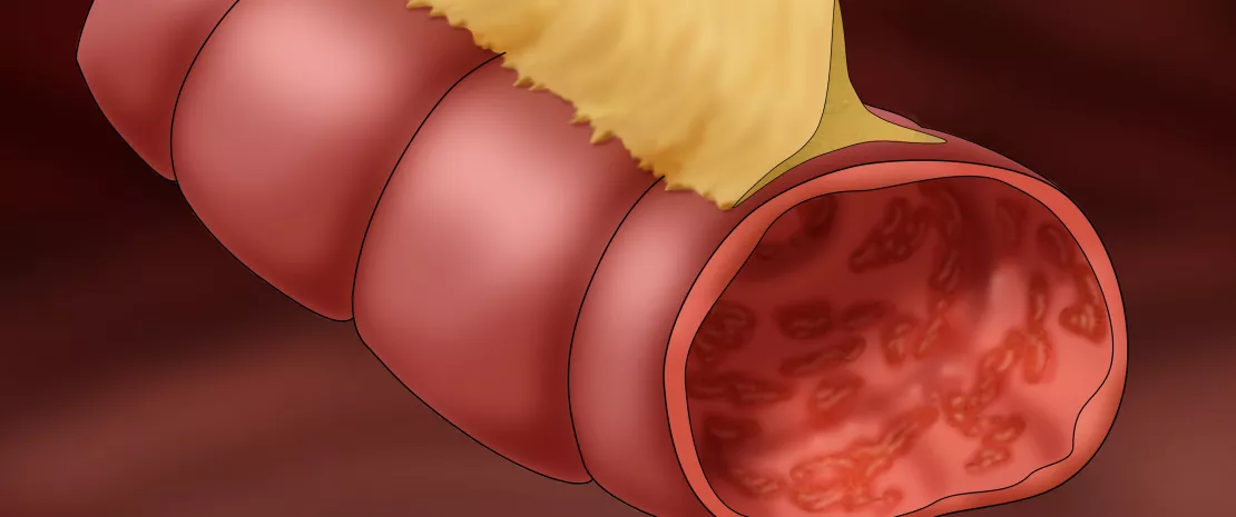
Section of the large intestine with Ulcerative Colitis. Illustration.

Commentary on the original publication by H yams et al. (Lancet 2019) [1]

Due to a lack of evidence-based data on therapeutic efficacy, there is uncertainty in the choice of treatment regimens in children who are newly diagnosed with ulcerative colitis (UC). This is why the authors hypothesized that clinical, transcriptomic, and microbial factors prior to treatment could predict the course of the disease. This inception cohort study recruited patients aged 4 to 17 years with newly diagnosed ulcerative colitis in 29 sites in the United States and Canada. Patients initially received mesalazine or corticosteroids with a pre-established protocol for escalation to immunomodulators (thiopurines) or biologic therapy with anti-TNF. RNA sequencing was used to define rectal gene expression before treatment, and 16S sequencing to characterize rectal and fecal microbiota. The primary outcome was corticosteroid-free remission at week 52 with no therapy other than mesalazine.

What do we already know about this subject?

Clinical, biological and/or endoscopic scores such as the PUCAI (pediatric ulcerative colitis activity index), are used to classify the severity of pediatric UC. Disease severity can be scored as mild (PUCAI 10-30), moderate to severe (35-60) or severe/fulminant (≥ 65). 5-aminosalycilates may be effective in mild forms, while corticosteroids are used in moderate disease, although some children will become steroid-dependent or refractory and require escalation of therapy to immunomodulators or anti-TNFα.

However, there is no prospective study evaluating the response to standardized therapy in newly diagnosed UC.

Image

What are the main insights from this study? 

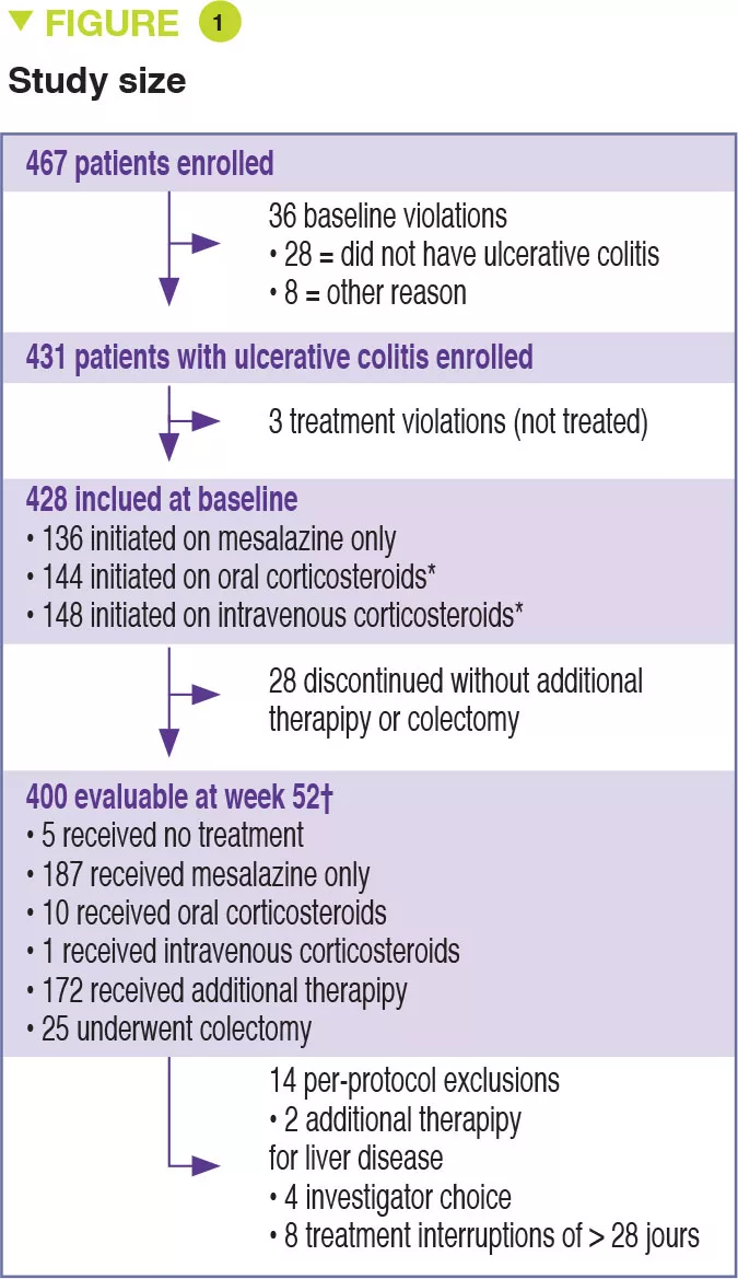
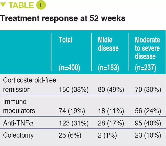
Between 2012 and 2015, 467 children aged 4 to 17 years were recruited into this prospective multicenter study conducted in 29 sites in the United States and Canada (Figure 1). The primary outcome was week 52 remission, defined by a PUCAI score < 10, with no therapy other than mesalazine (no corticosteroids and no colectomy). Baseline severity was defined as mild (therapy with mesalazine or oral corticosteroids with PUCAI < 45), or moderate to severe (oral corticosteroids with PUCAI ≥ 45 or intravenous corticosteroids). In addition to the usual clinical and biological parameters, this study also assessed gene expression from rectal biopsies as well as the rectal and fecal microbiota before treatment. 428 children started therapy: mean age was 12.7 years, 50% female, 42% with mild disease (mean PUCAI 31.9 ± 12.1 SD) and 58% with moderate to severe disease (mean PUCAI 62.9 ± 13.2 SD). At week 52, 150 (38%) of 400 evaluable participants achieved corticosteroid-free remission, of whom 80 (49%) had mild disease and 70 (30%) had moderate to severe disease (Table 1).

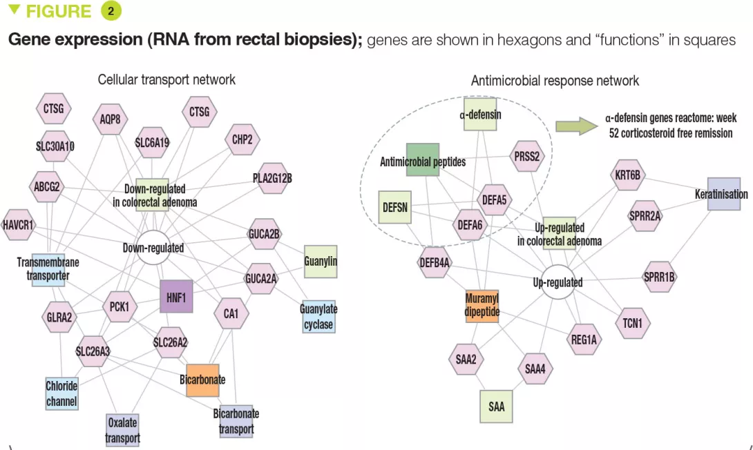
The clinical, biological, and endoscopic parameters that were associated with corticosteroid- free remission were validated in an independent prospective cohort of 307 children. It should be noted that corticosteroid- free remission was achieved by week 16 for moderate to severe disease (after which it can no longer be achieved) whereas it could be obtained up to week 52 in mild forms. Moreover, even patients with very severe disease were able to achieve week 52 corticosteroid-free remission (41/133 [31%] with PUCAI ≥ 65) and, conversely, some patients with mild disease were receiving anti-TNFα at week 52 (13/90 [14%] with PUCAI < 35). The authors identified 33 genes that were differently expressed between patients with moderate to severe disease who achieved corticosteroid-free remission (n = 51) or not (n = 101). Among these genes, 18 genes associated with cellular transport and ion channels were up-regulated and 15 genes involved in the antimicrobial response were down-regulated (Figure 2). The antimicrobial α-defensin signaling pathway showed the strongest negative correlation with week 52 corticosteroid-free remission. However, all 33 genes were negatively associated with the need for therapeutic escalation to anti-TNFα.

Image

Key points

  • The therapeutic decision depends on UC severity (PUCAI, Mayo scores) but also on treatment response by 4 weeks.

  • Other criteria (Hb, rectal eosinophil count, 25OH vitamin D) should also be taken into consideration.

  • Incorporating new parameters (gene expression and microbiota) into treatment decisions could facilitate the development of personalized medicine in the future.

Image

What are the consequences in practice? 

It is important to keep in mind the predictors of corticosteroid-free remission and therapeutic escalation as determined from multivariate analyses.


Predictors of week 52 corticosteroid-free remission were:

  • PUCAI < 45, Hb ≥ 10 g/dL;
  • remission by 4 weeks;
  • low expression of antimicrobial genes;
  • increased relative abundance for Ruminococcaceae and decreased for Sutterella.

Predictors of escalation to anti-TNFα therapy were:

  • total Mayo score ≥ 11;
  • eosinophil count in rectal biopsies < 32 per field at high magnification;
  • 25OH vitamin D < 20 ng/mL; - Hb < 10 g/dL;
  • no remission by 4 weeks;
  • decrease in genes involved in transport and antimicrobial genes;
  • decreased relative abundance of Oscillospira.

Conclusion

This study highlights the parameters that should be taken into consideration to guide the choice of therapy in children who are newly diagnosed with ulcerative colitis. Analysis of rectal gene expression and microbiota could both help predict the response to treatment and identify new therapeutic targets.

Summary
Off
Sidebar
Off
Migrated content
Désactivé
Updated content
Désactivé
Hide image
Off
Commented article

Antibiotic resistance: the lung microbiota pays a heavy price

Broad-spectrum antibiotics used for treating lung infections are regarded as one of the principal contributors to the overall burden of antibiotic resistance.

The pulmonary microbiota

Historically, the lungs of healthy individuals were considered sterile; the description of the LRT microbiota (Lower Respiratory Tract, from larynx to alveoli of the lungs)1 is a recent achievement.2,3 Along with viral and fungal communities, six bacterial phyla dominate a healthy lung microbiota: Firmicutes, Bacteroidetes, Fusobacteria, Proteobacteria, Acidobacteria, and Actinobacteria.1,2,4

“In Western populations, the treatment of lung infections is a primary driver of antibiotic resistance.4”

Antibiotics saved millions of lives but their misuse or oversuse now raises serious concerns for health, notably with the further emergence of antimicrobial resistance. Each year, the the World Health Organization (WHO) organizes the World AMR Awareness Week (WAAW) to increase awareness of this public health issue. Let’s take a look at this global threat that required urgent action:

Microbiota at the forefront of antibiotic resistance

The largescale and sometimes inappropriate use of antibiotics is making them in…

A loss of diversity in the lung microbiota

Microbial dysbiosis is observed in a range of respiratory disorders, including lung infection, asthma, chronic obstructive pulmonary disease (COPD) and cystic fibrosis (CF).5,6 But only few studies have explored the direct effects of antibiotics on lung microbiota. Recent investigation has shown that azithromycin treatment decreased bacterial diversity in patients with persistent uncontrolled asthma;1 however clinical benefits are still controversial.1,7,8 In COPD patients, azithromycin treatment lowered alpha diversity;1 in those suffering from CF, antibiotics appear to be the primary drivers of decreased airway microbiota diversity.5

The gut-lung axis

Respiratory diseases, chronic lung disorders and microbial infections are often accompanied by intestinal symptoms.12 Indeed, the intestinal ecosystem undergoes change during the course of several lung diseases.12 While the underling mechanism remains unclear, reciprocal influence between the gut and the lungs could, in part, explain why antibiotic-induced dysbiosis of the gut microbiota in early-life may be a risk factor for subsequent allergic rhinitis and asthma.1,12

The plague of broad-spectrum antibiotics

While the misuse of antibiotics is known to lead to the emergence and selection of resistant bacteria, antibiotic prophylaxis, without a microbial diagnosis, is still widely used to treat lung infections.4 Of the 12 antibiotic-resistant ‘priority pathogens’ listed by the WHO, 4 affect lungs: Pseudomonas aeruginosa, Streptococcus pneumoniae, Haemophilus influenzae and Streptococcus aureus.4,9 There is agreement among the scientific community as a key route to minimize antimicrobial resistance that the disease management of lung infections needs to be improved.4,10,11

Promoting research, raising awareness

  • The Global Alliance Against Respiratory Diseases (GARD), launched by the WHO in 2006 to help combat chronic respiratory diseases, asserts: “Physicians worldwide now face situations in which infected patients cannot be treated adequately because the responsible bacterium is totally resistant to available antibiotics.11”

  • At the European level, the ERS (European Respiratory Society) is involved in promoting scientific research, providing access to resources and raising awareness among the public and political decision makers. “Science, education and advocacy are at the core of everything we do.” Its latest monograph, ‘The lung microbiome’,13 reviews the different components of the respiratory microbiome, examines how diseases (asthma, COPD, cancer…) emerge and discusses new developments and therapies.

What is the World AMR Awareness Week?

Each year, since 2015, the WHO organizes the World AMR Awareness Week (WAAW), which aims to increase awareness of global antimicrobial resistance. Held on 18-24 November, this campaign encourages the general public, healthcare professionals and decision-makers to use antimicrobials carefully, to prevent the further emergence of antimicrobial resistance.

Sources

1 Hufnagl K, Pali-Schöll I, Roth-Walter F, et al. Dysbiosis of the gut and lung microbiome has a role in asthma. Semin Immunopathol. 2020;42(1):75-93.

2 Barcik W, Boutin RCT, Sokolowska M, et al. The Role of Lung and Gut Microbiota in the Pathology of Asthma. Immunity. 2020;52(2):241-255.

3 Mathieu E, Escribano-Vazquez U, Descamps D, et al. Paradigms of Lung Microbiota Functions in Health and Disease, Particularly, in Asthma. Front Physiol. 2018;9:1168. Published 2018 Aug 21.

4 Cookson WOCM, Cox MJ, Moffatt MF. New opportunities for managing acute and chronic lung infections. Nat Rev Microbiol. 2018;16(2):111-120.

5 Zhao J, Schloss PD, Kalikin LM, et al. Decade-long bacterial community dynamics in cystic fibrosis airways. Proc Natl Acad Sci U S A. 2012;109(15):5809-5814.

6 Chung KF, Huffnagle GB, Huang YJ. The lung microbiome in obstructive airways disease: potential pathogenetic roles. In: Cox MJ, Ege MJ, von Mutius E, eds. The Lung Microbiome 2019 (ERS Monograph). Sheffield, European.

7 Gibson PG, Yang IA, Upham JW, et al. Efficacy of azithromycin in severe asthma from the AMAZES randomised trial. ERJ Open Res. 2019;5(4):00056-2019. Published 2019 Dec 23.

8 Chung KF. Airway microbial dysbiosis in asthmatic patients: A target for prevention and treatment?. J Allergy Clin Immunol. 2017;139(4):1071- 1081.

9 WHO. Global priority list of antibiotic-resistant bacteria to guide research, discovery, and development of new antibiotics. 25 february 2017

10 Lim WS, Baudouin SV, George RC, et al. BTS guidelines for the management of community acquired pneumonia in adults: update 2009 Thorax 2009;64:iii1-iii55.

11 Forum of International Respiratory Societies. The Global Impact of Respiratory Disease – Second Edition. Sheffield, European Respiratory Society, 2017.

12 Marsland BJ, Trompette A, Gollwitzer ES. The Gut-Lung Axis in Respiratory Disease. Ann Am Thorac Soc. 2015;12 Suppl 2:S150-S156.

13 The Lung Microbiome. Edited by Cox MJ, Ege MJ, and von Mutius E. 2019. Monograph of European Respiratory Society.

Summary
Off
Sidebar
On
Migrated content
Activé
Updated content
Désactivé
Old content type
pro_dossiers_article
Hide image
Off
Dossier detail Pulmonology Gastroenterology Pediatrics