
The gut microbiome and neurodegeneration

Overview

By Pr. John F. Cryan
Department of Anatomy and Neuroscience, University College Cork, Cork, Ireland

Photo : The gut microbiome and neurodegeneration

The past decade has seen an explosion of research in the role of the gut microbiota in modulating brain health and disease. Although most research has focused on stress-related disorders, such as anxiety, depression and irritable bowel syndrome, a growing body of research, albeit largely preclinical, also implicates the microbiota as a disease moderator in neurodegenerative diseases such as Alzheimer’s disease and Parkinson’s disease. In tandem, research has shown that the microbiome plays a critical role in key brain processes involved in neurodevelopment, neuroinflammation, and aging. Currently, research is heavily focused on a better understanding of the precise mechanisms of how the gut talks to the brain and how it may lead to increased susceptibly to brain disorders.

In medicine, the disciplines of neurology and microbiology have largely matured along distinct parallel trajectories, only interfacing in pathological situations when direct infections of the central nervous system occur. However, over the past decade, there has been a revolution in biomedicine with the realisation that the gut microbiota (the trillions of bacteria that reside within the gut) plays a key role in maintaining homeostasis and in programming the major body systems, including even the brain.

A growing body of research is focused on illuminating the bidirectional communication pathways between gut bacteria and the central nervous system, the microbiota– gut–brain axis, however, this is a field in its relative infancy [1]. Changes in the microbiome, its metabolites, and its interaction with the gut-brain axis are associated with a wide array of illnesses including brain disorders. Studying the microbiome requires close collaborative efforts of clinicians with basic scientists and bio-informaticians and works best when traditional discipline barriers between neurology, gastroenterology, and microbiology are broken down.

In preclinical research, a number of experimental models have proven essential to evaluate the microbiome within the context of brain and behaviour, including prebiotic and probiotic intervention, antibiotic administration, faecal transplantation, and the use of germ-free and gnotobiotic animals [1]. In clinical research, most data, especially in the area of neurology, have relied on cross-sectional studies of the microbiome in patients with disease versus healthy age-matched individuals.

The microbiota gut-brain axis mechanisms of communication

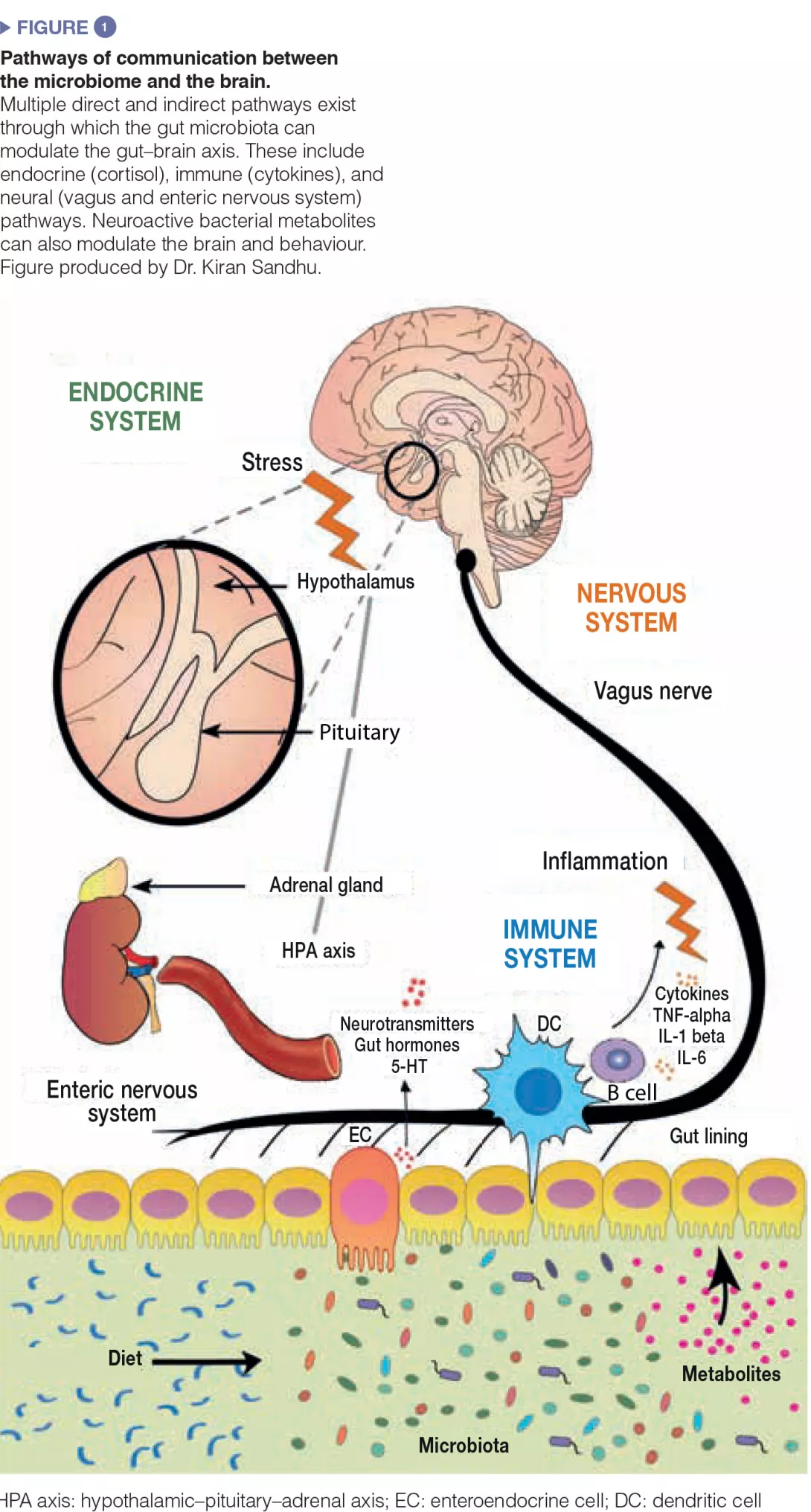
Much experimental effort is being placed at trying to dissect the pathways of communication between the gut and the brain. The gut bacteria influences central processes through a variety of mechanisms (Figure 1). Firstly, the microbiota’s ability to synthesize neurotransmitters (i.e. γ-amino butyric acid (GABA), noradrenaline, and dopamine) is an important avenue of communication. Secondly, microbes play a key role in activation of the immune system which can play a fundamental role in ageing, neurological disorders, and neurodegeneration. Finally, microbes produce metabolites including short-chain fatty acids (SCFAs) which are essential for the gut, the immune system, and potentially brain health. Moreover, the gut microbiota and the brain are linked through the vagus nerve, and through the modulation of key dietary amino acids, such as tryptophan.

Given the close association between the gut microbiota and the brain, it is not at all surprising that gut bacteria play key roles in neurological and psychiatric diseases. The strongest evidence for a role of the microbiome in brain function derives from germ-free mice. Studies from a number of research groups in Canada, Sweden, and Ireland have shown that, in germ-free animals, the brain fails to develop normally in the absence of the gut microbiome [2]. Moreover, fundamental brain processes, such as myelination, adult neurogenesis, and microglia activation, have also been shown to be critically dependent on microbiota composition.
Image

Microbiota & Ageing

The relationship between the microbiome and the ageing brain is also receiving much attention, which is of particular interest in the field of neurology as many neurological and neurodegenerative disorders occur in old age. Once again, the concept of linking the microbiome to healthy ageing is not new, and this was championed over 100 years ago by Nobel prize-winning immunologist, Elie Metchnikoff, who observed that the villagers in a certain region of Bulgaria lived unusually long lives, a fact he attributed to the presence of lactic acid bacteria in their diet. We have recently revisited Metchnikoff’s original study [3], and shown that the behavioural deficits in aged animals coincide with changes in the microbiome. Moreover, the ELDERMET study has shown that the composition of the gut bacteria in the elderly correlated with their overall health – frailty and the immune system [4]. The greater the microbiome diversity, the better the health outcomes. These investigators went one step further to investigate what is driving the diversity of the microbiome, which they determined to be a diverse diet. As people eat processed bland food (often in nursing homes), the diversity of their microbiomes is reduced, whereas those with a diet rich in fruit and vegetables have better outcomes [4].

A decline in microbial diversity is associated with a concomitant increase in microglial activation which is correlated with brain mass differences in the mouse. This contributes to an age-associated inflammatory response known as “inflammaging”, which in turn has been associated with neurodegenerative diseases such as Alzheimer’s (AD) and Parkinson’s disease (PD). Furthermore, the microbiome has been shown to regulate microglia activation; germ-free mouse brains were shown to express defective microglia, which was partially rescued upon restoration of the microbial community to control levels [5].

Parkinson's disease

There is a growing realisation that the aetiology of PD may actually originate in the gut [6]. Indeed, α-synuclein, the protein aggregate hallmark of PD pathology in the brain, has also been identified in the mucosal and submucosal nerve fibres and ganglia of Parkinsonian patients, with some preclinical evidence even suggesting that α-synuclein in the gut can be transported to the brain via the vagus nerve. Moreover, functional gut symptoms, such as constipation, often occur as prodromal symptoms years before any motor symptoms emerge. Since Scheperjans and colleagues first showed that there are specific alterations in microbiome composition in PD [7], many more studies have emerged [8]. However, to date, there is no consensus as to whether a specific microbial signature exists. When mice were colonized with the microbiota of PD patients via faecal microbiota transplantation, they developed motor deficits and neuroinflammation; two hallmark symptoms of PD [9]. Additionally, symptoms improved when the mice were treated with antibiotics. These studies implicated short-chain fatty acids as drivers of the neuroinflammatory processes in PD [9]. The vagus nerve is particularly well placed as the conduit for signals from the gut to the brain, involving either microbial or prionlike translocation of α-synuclein. Indeed, epidemiological studies based on Danish and Swedish patient registries have shown that truncal vagotomy is protective against PD. Although there has been much excitement in the field, caution is needed when examining the available data as it is largely derived from small cohorts and lacks a longitudinal perspective. Many more mechanistic studies are needed to understand how changes in the microbiota can moderate both the motor and non-motor symptoms of PD and its co-morbidities [10].

Alzheimer's disease

The concept that microbes may play a role in the pathophysiology of AD is not new and the notion that amyloid, the aggregation of which is one of the key hallmarks of AD, may act as an antimicrobial peptide in the brain is an intriguing concept [11]. However, it is ethically difficult from the perspective of Koch’s postulate to prove whether there is any infective cause of neuroinflammation and neurodegeneration. As in PD, the relationship between gut proteins and brain health is receiving increased attention with the realization that amyloid-like proteins can be produced by bacteria which has been shown to increase α-synuclein pathology in aged rats and worms [12]. Much more work is needed to validate such strategies in humans.

Recently, cross-sectional studies have identified that Escherichia/Shigella bacterial taxa, which are associated with mediating inflammation, were increased in faecal samples from AD patients relative to control subjects. Moreover, the microbiota changes correlated with pro-inflammatory cytokine levels in whole blood [13]. Such results suggest a causal link between dysregulation of the microbiota and systemic inflammation, which may initiate or exacerbate neurodegeneration in the brain in AD. However, these are still relatively small studies and much more research is needed in larger cohorts to assess the causal relationship between the gut microbiome and AD.

In parallel, a number of transgenic mouse models of AD have been shown to have an altered microbiome [14]. Seminal studies in germ-free mice showed that there is a marked absence of amyloid plaque buildup and neuroinflammation when microbes are not present [14]. Similarly, chronic treatment of APP/PS1 transgenic mice with an antibiotic cocktail reduced microglial and astrocyte accumulation surrounding amyloid plaques in the hippocampus and led to a decrease in insoluble A plaques [15]. Together, these studies unequivicolly place the microbiome as a regulator of key molecular components of AD.

Future perspectives

It is clear that the microbiome is critically important for the appropriate development and maintenance of brain function. Moreover, as outlined above, there is accumulating evidence from both animal and clinical studies implicating the microbiome in a variety of neurological and neurodegenerative diseases. Given the marked effects of the microbiota in regulating brain function, it is plausible that its composition affects the progression, susceptibility, and treatment of almost all neurological disorders. Nonetheless, there are marked gaps in our knowledge regarding the role of the microbiome in other neurodegenerative diseases, such as amyotrophic lateral sclerosis or Huntington’s disease, and caution is needed to not over-interpret such studies. The field needs to move away from corelative studies towards mechanistic causal approaches. Moreover, more interventional studies are needed using probiotic strains and prebiotics, and even faecal microbiota transplants may potentially be important in the field. It is possible that similar approaches could target different disorders, for example, the modulation of T lymphocyte signalling to the brain may be useful in dampening down the neuroinflammatory status in patients following stroke, as well as in patients with AD and during ageing.

With regards to clinical neurology, many patients are on multiple medications and there is a growing understanding of the relationship between the microbiome and drug action. Thus, all studies should aim to differentiate between the impact of drugs and that of disease on the microbiome. Moreover, temporal studies with presymptomatic individuals will be important to determine the potential role of the microbiome as a biomarker of disease.


Conclusion

Diet is perhaps one of the greatest factors that influences the microbiome. As many neurological disorders affect appetite, swallowing, and diet in general, it is essential to have good nutritional data for all human studies in the future. In addition, this will further enable a better understanding of the relationship between diet, the microbiome, and the brain, which is critical both in early life and as we age.

Sources

1 Cryan JF, Dinan TG. Mind-altering microorganisms: the impact of the gut microbiota on brain and behaviour. Nat Rev Neurosci 2012 ; 13 : 701-12.

2 Luczynski P, McVey Neufeld KA, Oriach CS, et al. Growing up in a Bubble: Using Germ-Free Animals to Assess the Influence of the Gut Microbiota on Brain and Behavior. Int J Neuropsychopharmacol 2016 ; 19.

3 Scott KA, Ida M, Peterson VL, et al. Revisiting Metchnikoff: Age-related alterations in microbiota-gut-brain axis in the mouse. Brain Behav Immun 2017 ; 65 : 20-32.

4 Claesson MJ, Jeffery IB, Conde S, et al. Gut microbiota composition correlates with diet and health in the elderly. Nature 2012 ; 488 : 178-84.

5 Erny D, Hrabe de Angelis AL, Jaitin D, et al. Host microbiota constantly control maturation and function of microglia in the CNS. Nature Neurosci 2015 ; 18 : 965-77.

6 Braak H, de Vos RA, Bohl J, et al. Gastric alpha-synuclein immunoreactive inclusions in Meissner’s and Auerbach’s plexuses in cases staged for Parkinson’s disease-related brain pathology. Neurosci Lett 2006 ; 396 : 67-72.

7 Scheperjans F, Aho V, Pereira PA, et al. Gut microbiota are related to Parkinson’s disease and clinical phenotype. Mov Disord 2015 ; 30 : 350-8.

8 Keshavarzian A, Green SJ, Engen PA, et al. Colonic bacterial composition in Parkinson’s disease. Mov Disord 2015 ; 30 : 1351-60.

9 Sampson TR, Debelius JW, Thron T, et al. Gut Microbiota Regulate Motor Deficits and Neuroinflammation in a Model of Parkinson’s Disease. Cell 2016 ; 167 : 1469-80 e12.

10 Lionnet A, Leclair-Visonneau L, Neunlist M, et al. Does Parkinson’s disease start in the gut? Acta Neuropathol 2018 ; 135 : 1-12.

11 Itzhaki RF, Lathe R, Balin BJ, et al. Microbes and Alzheimer’s Disease. J Alzheimers Dis 2016 ; 51 : 979-84.

12 Friedland RP. Mechanisms of molecular mimicry involving the microbiota in neurodegeneration. J Alzheimers Dis 2015 ; 45 : 349-62.

13 Cattaneo A, Cattane N, Galluzzi S, et al. Association of brain amyloidosis with pro-inflammatory gut bacterial taxa and peripheral inflammation markers in cognitively impaired elderly. Neurobiol Aging 2017 ; 49 : 60-8.

14 Harach T, Marungruang N, Duthilleul N, et al. Reduction of Abeta amyloid pathology in APPPS1 transgenic mice in the absence of gut microbiota. Sci Rep 2017 ; 7 : 41802.

15 Minter MR, Zhang C, Leone V, et al. Antibiotic-induced perturbations in gut microbial diversity influences neuro-inflammation and amyloidosis in a murine model of Alzheimer’s disease. Sci Rep 2016 ; 6 : 30028.

Summary
Off
Sidebar
Off
Migrated content
Désactivé
Updated content
Désactivé
Hide image
Off
Article

Fecal transplantation - ready for prime time?

Congress review

By Pr. Danny De Looze
Department of Gastroenterology University Hospital Gent, Belgium

Photo : Fecal transplantation - ready for prime time?

Despite the fact that we still do not know all the secrets and mysteries about the gut microbiota, a lot of hope is put on treating GI diseases with intestinal microbiota. Fecal microbiota transplantation seems to be the Holy Grale. But is it? At 2018’s UEGW in Vienna numerous lectures were dedicated to it. An overview.

The ideal donor 

Despite the fact that up until now nobody really knew how to precisely define the normal gut microbiota (“eubiosis”), we do know that high microbial diversity and gene richness are of key importance in the host-microbiota equilibrium. Therefore, the ideal fecal donor should be screened for bacterial richness. A surrogate marker for this property was proposed by Marie Joossen (Leuven, Belgium) who pointed out that the presence of Blastocystis hominis correlates with a higher microbial richness.[1] This finding – if confirmed by others – may change our current practice to avoid carriers of this commensal as fecal donors. Enriching the donor’s microbiota by prebiotics or using multiple donors may also ensure (theoretically) a higher baseline diversity of the donated material. This was also observed by Karakan et al. (Ankara, Turkey) who performed an open trial in ulcerative colitis with an overall complete response rate of 32% which was particularly influenced by a high bacterial diversity in the donated fecal material.

The new brown gold 

Strict adherence to current guidelines for selection of donors for fecal material necessitates the rejection of most donors. Terveer et al. (Leiden, the Netherlands) report that only 3,5% of possible donors are suitable at the end of the line [2]. The main reasons for not being accepted as a donor are: age above 50, high BMI and carrier status of non-pathogenic germs (Blastocystis hominis, Dientamoeba fragilis) and MDROs (multidrug resistant organisms).[2]

Clostridium Difficile 

The main indication for fecal microbiota transplantation remains refractory infection with Clostridium difficile (C. difficile). Ianiro et al. (Rome, Italy) showed, in a retrospective series of 282 C. difficile patients comparing antibiotic treatment and fecal transplant, that this latter treatment resulted in significant shorter hospital stay, significantly lower mortality and specifically less sepsis-related mortality.

Antonio Gasbarrini (Rome, Italy) therefore suggests that the time has come to promote fecal microbiota transplantation as the first line therapy in C. difficile infection.

Expanding the scope of FMT 

Antonio Gasbarrini gave a nice overview of promising indications for fecal micro- biota transplantation. It has been shown that fecal transplants restore the human microbiota better than probiotics after antibiotic-induced dysbiosis in humans. This was also the case following combined chemotherapy and antibiotic induced dysbiosis in the setting of hematological stem cell therapy and in patients with liver cirrhosis receiving antibiotics. Mice models even show evidence for restoration of immune function and intestinal integrity after chemotherapy induced intestinal damage. Therefore, Antonio Gasbarrini makes the case for pre-emptive stool conservation for subsequent autologous fecal microbiota transplantation, e.g. following antibiotic treatment or bone marrow transplantation. In vivo evidence from clinical trials must be awaited before this futuristic but not unrealistic strategy can be implemented. Anyhow, it seems quite logical that collecting one’s own stool for autologous transplantation later in life, is the way to go.

Ulcerative colitis 

In 3 out of 4 published randomized controlled trials fecal microbiota transplantation was superior to placebo in refractory ulcerative colitis (UC) patients [3]. Mean remission rate in these studies, however, was only 25-30 % and Rainer et al. (Graz, Austria) presented a study with similar complete remission rates, showing no added value of administering fresh stool in these patients. Nevertheless, up until now there was no standardized stool transplantation protocol in UC. Remission rates of 30% seem low but to put things in perspective: this is also the remission rate that is achieved with the expensive and widely used biologicals [...]. The importance of colonic microbiota in UC was demonstrated by Herrera-de Guise et al. (Barcelona, Spain) who showed that patients in long-term stable remission (more than 5 years) present with an abundance of Akkermansia muciniphila and Faecalibacterium prausnitzii, similar to healthy controls. These authors even suggest that we should think of a paradigm shift in treating UC: our therapeutic endpoint should perhaps no longer be immune suppression, but we should aim to reach eubiotic microbiota characteristics.

Irritable bowel syndrome 

Irritable bowel syndrome (IBS) is certainly the condition for which the expectations of a cure by fecal microbiota transplantation are very high in both patients and health care practitioners. Still, conflicting results from randomized controlled trials [4, 5] do not currently support a widespread use of this treatment in IBS. Intestinal dysbiosis is present in IBS but a clear causality between these microbial changes and symptoms are lacking. Halkjaer et al. (Copenhagen, Denmark) performed a randomized controlled trial in 52 adult IBS patients; an increase in biodiversity (comparable to the donors) was observed in the patients being actively transplanted.[5] Unfortunately, the placebo group had a significantly better clinical outcome at 3 and 6 months than the patients receiving fecal capsules.[5] In a small cohort of 16 IBS patients, Holster et al. (Orebro, Sweden) were also unable to demonstrate efficacy for fecal microbiota transplant vs. placebo. They also studied rectal sensitivity by means of a barostat and showed no difference between the active and control groups, concluding that changing the microbiota does not contribute to the visceral hypersensitivity in IBS.

Bacterial pills 

Ianiro et al. performed an open label trial in C. difficile infection with a synthetic microbiota suspension (10 patients only) demonstrating efficacy. Khanna et al. (Rochester, USA) have also demonstrated efficacy in prevention of C. difficile infection’s relapse with a non-frozen, lyophilized, orally administrated microbiota restoring drug (RBX7455) in an open-label phase 1 trial. These are promising results that need to be confirmed in large scale randomized trials.

Conclusion

The UEGW 2018 added more insight into the desired eubiotic characteristics of fecal material and the promising new indications for fecal microbiota transplantation that may arise the upcoming years. Up until now C. difficile infection remains the only clear-cut indication for this treatment which is currently not ready for prime time in other conditions.

Explain fecal transplantation to your patients with this dedicated content: 

Summary
Off
Sidebar
Off
Migrated content
Désactivé
Updated content
Désactivé
Hide image
Off
Congress review

Feeding practices from birth to 12 months: impact on the gut microbiota and the risk of being overweight

Commented articles - Children's section

By Pr. Emmanuel Mas
Gastroenterology and Nutrition Department, Children’s Hospital, Toulouse, France

Photo : Feeding practices from birth to 12 months: impact on the gut micro- biota and the risk of being overweight

Comments of the original article by Forbes et al. (JAMA Pediatr 2018)

The aim was to characterize the association between breastfeeding, microbiota, and risk of overweight during infancy.
The study included 1,087 infants; fecal microbiota at M3 to M4 and/or M12 were characterized by 16S ribosomal RNA sequencing. At M3, infants who were exclusively formula fed had an increased risk of overweight. At M12, microbiota profiles differed significantly according to feeding practices at M6; among partially breastfed infants, formula supplementation was associated with a profile similar to that of nonbreastfed infants, contrary to the introduction of complementary foods without formula. Breastfeeding may be protective against overweight by modulating the gut microbiota. Subtle microbiota differences emerge after brief exposure to formula in the hospital. Formula feeding appears to stimulate changes in microbiota that are associated with overweight, whereas other complementary foods do not.[1]

What is already known about this topic? 

From birth, certain factors influence the development of obesity. Breastfeeding has a protective effect, partly because breast milk is low in protein. The gut microbiota (GM) must also be taken into consideration because it is involved in food absorption and energy metabolism. The GM is formed during the first 2 to 3 years of life, and the method of feeding (breast milk vs. infant formula milk) is one of the main factors that modulates GM composition. The GM of obese adults is less diverse and has a higher ratio of Firmicutes/Bacteroidetes.

What are the main results of this study? 

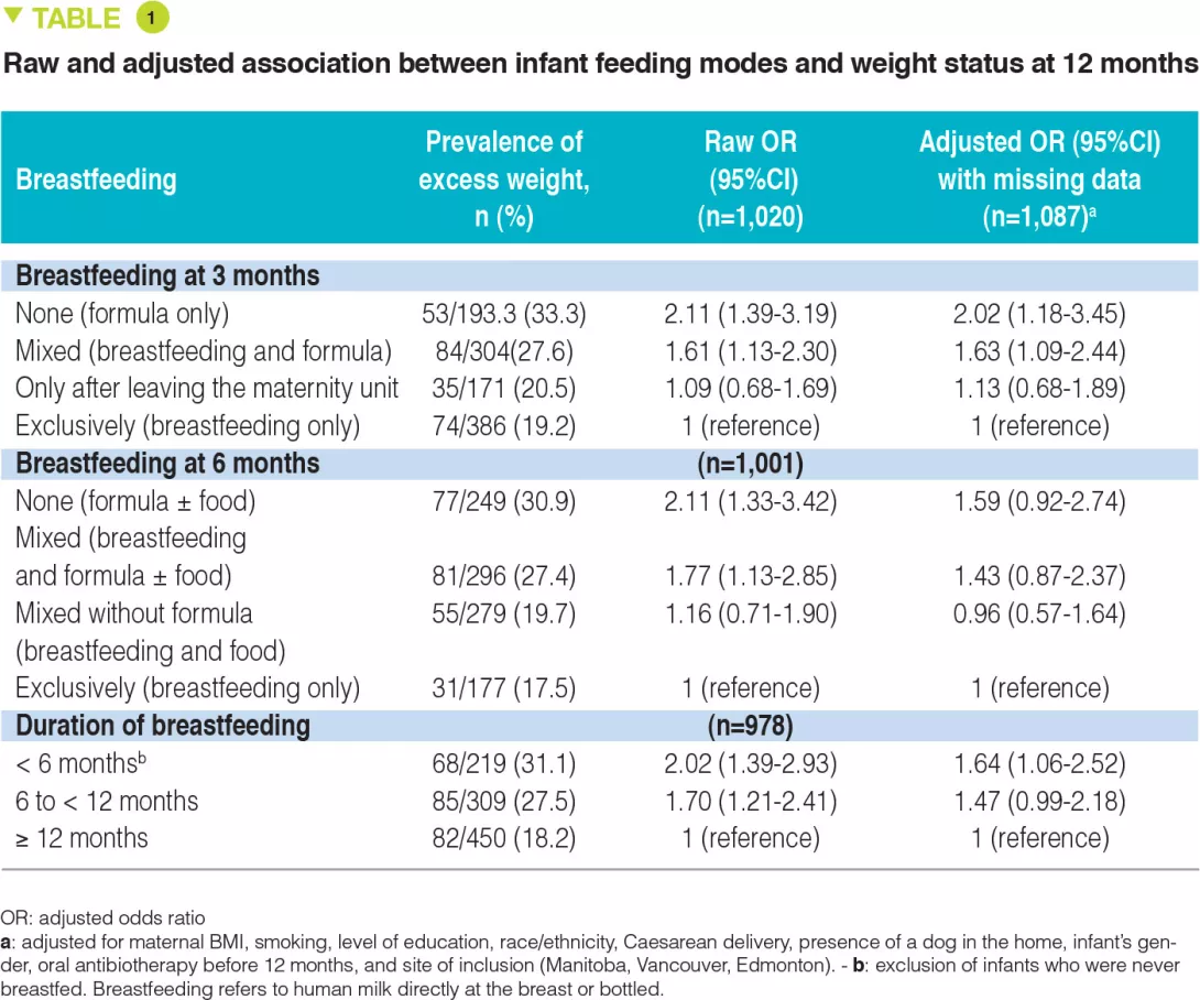
This study is based on data from 1,087 infants included in the CHILD birth cohort (Canadian Healthy Infant Longitudinal Development). Infant faecal samples were collected for microbial analysis at 3 to 4 months (n=996), 12 months (n=821),  and at both time points (n=730). Mothers completed questionnaires on the method of feeding at 3 and 6 months, which made it possible to define different groups according to breastfeeding practices (Table 1). Among these infants, 74.2% were delivered vaginally, and 39.8% of mothers were overweight or obese. Rates of exclusive breastfeeding were 53.8% at 3 months and 17.6% at 6 months.

At 3 months, exclusive breastfeeding protected against the risk of being overweight at 12 months (defined by measured weight: expected weight ratio > 85th percentile) compared to exclusively formula-fed infants: 19.2% vs. 33.3%, respectively, with no significant effect of adjustment (Table 1). At 6 months, formula milk in addition to breastfeeding increased the risk of being overweight at 12 months, but solid foods did not have the same effect. Prolonged breastfeeding was found to confer a protective benefit.

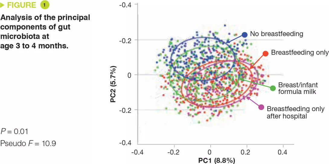
As expected, GM richness and diversity at age 3 to 4 months differed according to the method of feeding, and the composition was significantly different between exclusively breastfed and non-breastfed infants (Figure 1). An increase in breastfeeding was accompanied by an increase in relative abundance of Bifidobacteriaceae and Enterobacteriaceae and a decrease in Lachnospiraceae, Veillonellaceae, and Ruminococcaceae.

By 12 months, the GM had become more homogeneous but there were still differences relative to the method of feeding at 6 months, i.e. increased richness for infants at least partially fed with formula milk, and a relative abundance of Actinobacteria and Proteobacteria that was higher in exclusively breastfed infants and lower in non-breastfed infants.

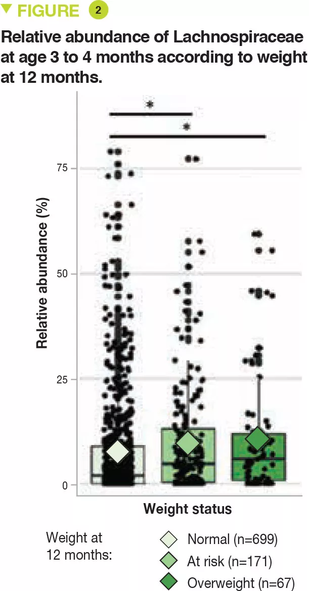
Greater GM richness at 3 to 4 months correlated with an increase in excess weight or a risk of becoming overweight at 12 months, particularly with regards to Lachnospiraceae, with a median relative abundance of 5.9% (overweight), 4.7% (risk of becoming overweight), and 1.9% (normal weight) (p=0.01) (Figure 2).

Image

Key points

  • Breastfeeding protects against becoming overweight at 1 year.

  • This effect is mediated by the composition of the gut microbiota.

  • Studies are needed to determine whether this effect persists over a longer term.

What are the practical consequences?

This study firstly demonstrates the benefit of breastfeeding on gaining excess weight at 1 year, and secondly that this benefit is related to modulation of the GM.

Additionally, it is important to promote exclusive breastfeeding from birth, by limiting supplementation with infant formula milk in the maternity unit. This benefit is enhanced by prolonged breastfeeding. Whereas the use of formula milk has a negative impact in infants, this is not the case for solid foods.

Image
Image

Conclusion

Breastfeeding, especially when prolonged, has a protective effect against becoming overweight at 1 year. Infant formula milk, even if used to supplement breastfeeding, increases GM richness and diversity at age 3 to 4 months, particularly Lachnospiraceae, and increases the risk of becoming overweight at 1 year.

Summary
Off
Sidebar
Off
Migrated content
Désactivé
Updated content
Désactivé
Hide image
Off
Commented article

Resilience of healthy adult gut microbiota following antibiotic exposure

Commented articles - Adults' section

By Pr. Harry Sokol
Gastroenterology and Nutrition, Saint-Antoine Hospital, Paris, France

Comments of the original article of Palleja et al. (Nature Microbiology 2018)

To minimize the impact of antibiotics, gut microorganisms harbour and exchange antibiotics resistance genes, collectively called their resistome. Using shotgun sequencing-based metagenomics, we analysed the partial eradication and subsequent regrowth of the gut microbiota in 12 healthy men over a 6-month period following a 4-day intervention with a cocktail of 3 last-resort antibiotics: meropenem, gentamicin and vancomycin. Initial changes included blooms of enterobacteria and other pathobionts, such as Enterococcus faecalis and Fusobacterium nucleatum, and the depletion of Bifidobacterium species and butyrate producers. The gut microbiota of the subjects recovered to near-baseline composition within 1.5 months, although 9 common species, which were present in all subjects before the treatment, remained undetectable in most of the subjects after 180 days. Species that harbour β-lactam resistance genes were positively selected for during and after the intervention. Harbouring glycopeptide or aminoglycoside resistance genes increased the odds of de novo colonization, however, the former also decreased the odds of survival. Compositional changes under antibiotic intervention in vivo matched results from in vitro susceptibility tests. Despite a mild yet long-lasting imprint following antibiotics exposure, the gut microbiota of healthy young adults are resilient to a short-term broad-spectrum antibiotics intervention and their antibiotics resistance gene carriage modulates their recovery processes.[1]

What is already known about this topic? 

The human gut microbiota forms a complex and balanced ecosystem. Perturbations of this ecosystem can play a role in the development of infections, obesity, diabetes as well as neurological and inflammatory disorders. It is estimated that antibiotics have added 2 to 10 years to our life expectancy, but early exposure to these drugs has also been associated with noxious metabolic, inflammatory and neurological effects, both in animal models and in humans. When microbial communities are exposed to antibiotics, not only do they react by shifting their composition, but also by evolving, optimizing and disseminating antibiotic resistant genes (ABR genes) which collectively form the resistome.[2] The human gut microbiota is a reservoir of ABR genes which are exchanged between the resident strains, thereby propagating resistance.[3] The development and spread of antibiotic-resistant bacteria constitute a serious threat to public health. Only a few studies have investigated the effects of specific antibiotics on intestinal ecosystems and their associated resistomes. In previous work it was shown that antibiotic administration induces a decrease in microbiota diversity and an in- crease in the carriage of ABR genes.[4, 5] However, the effects of a combination of antibiotics on the microbiota and the role of ABR genes in microbial persistence have not yet been studied. In this study, 12 healthy men aged 18 to 40 years received a cocktail of three last-resort antibiotics (vancomycin, gentamicin and meropenem). The authors analysed the impact of this treatment on the gut microbiota by shotgun sequencing of faecal samples taken before and at four time points over a 6-month period following antibiotic administration.

Image

Cornerstone of the modern therapeutic arsenal, antibiotics saved millions of lives. On the other hand, their excessive and sometimes inappropriate use can lead to the emergence of multiple forms of resistance in microorganisms. Each year, the World Health Organization (WHO) organizes the World AMR Awareness Week (WAAW) to increase awareness of this public health issue. Read the dedicated page:

Microbiota at the forefront of antibiotic resistance

The largescale and sometimes inappropriate use of antibiotics is making them in…

What is the World AMR Awareness Week?

Each year, since 2015, the WHO organizes the World AMR Awareness Week (WAAW), which aims to increase awareness of global antimicrobial resistance. Held on 18-24 November, this campaign encourages the general public, healthcare professionals and decision-makers to use antimicrobials carefully, to prevent the further emergence of antimicrobial resistance.

What are the main results of this study? 

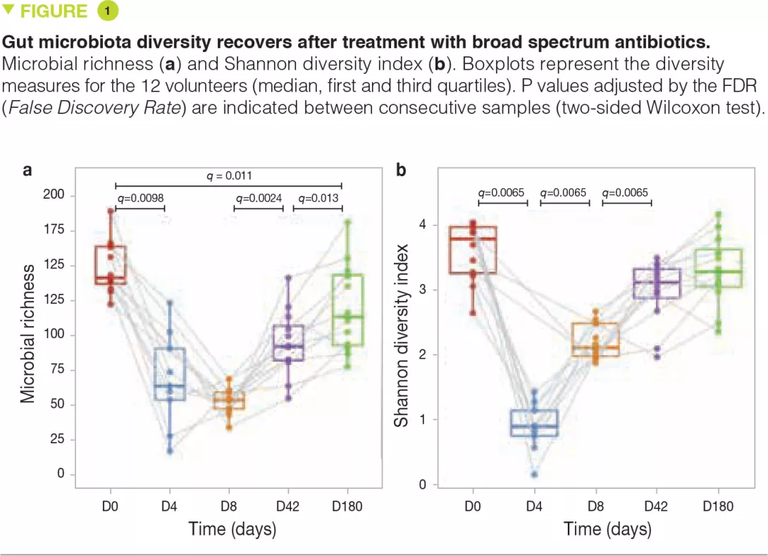
At D4, immediately after the intervention, microbiota diversity and richness were notably reduced compared to baseline. However, despite the use of very broad spectrum antibiotics, many species were still detectable at D4 (Figure 1a). By D8, microbiota diversity (measured by the Shan- non index) had considerably increased, suggesting that surviving microorganisms had begun to regrow (Figure 1b). At 6 months, microbiota diversity was almost completely restored to baseline levels but this was not the case for richness, suggesting that some strains had been permanently (or at least extendedly) eradicated.

Some of the early observable changes included blooms of normally subdominant commensals like Escherichia coli, Veillo- nella spp., Klebsiella spp., Enterococcus faecalis and Fusobacterium nucleatum and a sharp depletion of butyrate-producers like Faecalibacterium prausnitzii, Roseburia hominis, Anaerostipes hadrus, Coprococcus spp. and Eubacterium spp. These shifts in microbiota composition were no longer significant at D42.

The authors then investigated the role of ABR genes in microbiota recovery. In particular, they found that β-lactamase-harbouring metagenomic species had significantly higher odds of survival (OR = 1.64 [1.24-2.17]) at D8. In addition, metagenomic species not detected at baseline had better odds of subsequent de novo colonization if they harboured ABR genes against one of the three antibiotic classes used.

Key points

  • The intestinal microbiota of healthy young adults is resilient to 4 days of broad-spectrum antibiotic treatment with an approximate 6-month recovery of most bacterial communities.

  • The recovery of individual species is modulated by ABR gene carriage.

  • The impact of prolonged or repetitive antibiotic treatment requires further study, especially in paediatric populations.

What are the practical consequences? 

These findings indicate that the gut micro- biota of healthy young adults is resilient to a 4-day intervention with broad spectrum antibiotics with recovery of the majority of bacterial communities after about 6 months. The recovery of individual species is modulated by carriage of ABR genes. Further studies are needed to assess the impact of repetitive perturbations and/or over longer periods and to determine whether these findings also hold true in children whose immune system and microbiota are immature. It is possible that repetitive use of antibiotics over long periods selects for bacteria carrying ABR genes at the expense of other commensals, with prolonged or permanent effects on the microbiota. In such cases, corrective intervention with exogenous supply of microorganisms could be considered. The effects of antibiotics on the intestinal microbiota are therefore important and their use must be rationalized.

Conclusion

Broad spectrum antibiotics negatively impact the gut microbiota in an immediate, significant and durable manner for some species. In healthy young adults, the gut microbiota is resilient but near-complete recovery takes about six months. Modulated by ABR gene carriage, the capacity of species to regenerate is more favourable to diversity than to richness.

Summary
Off
Sidebar
Off
Migrated content
Désactivé
Updated content
Désactivé
Hide image
Off
Commented article Gastroenterology Off

Microbiota and celiac disease

Overview

By Pr. Elena F. Verdu
Farncombe Family Digestive Health Research Institute, McMaster University Hamilton, Canada

Environmental factors are suggested to contribute to celiac disease pathogenesis, an autoimmune disease triggered by the ingestion of gluten. Clinical studies show alterations in the composition of the microbiota in celiac disease patients. Although some consistent findings across studies have been established, no celiac disease microbial signature has been defined. Using reductionist and gnotobiotic* animal models, recent research suggested that bacterial strains from celiac disease patients may have more pathogenic or inflammatory potential. Targeting the microbiota with the use of specific probiotics, shown to modify pathogenic mechanisms critical for celiac disease, could be an attractive therapeutic approach to complement the gluten-free diet.

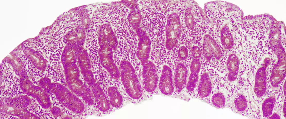
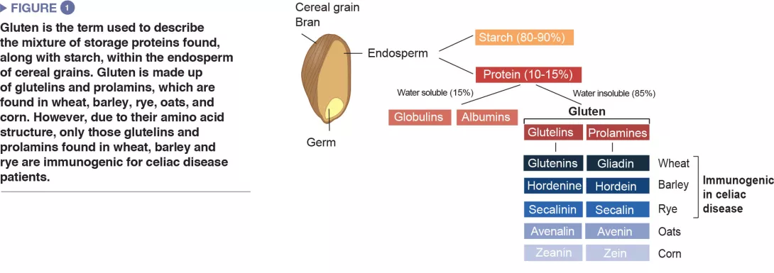
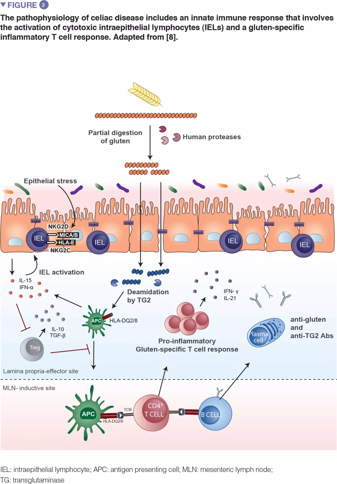
Celiac disease is a common inflammatory and autoimmune reaction that occurs in genetically predisposed individuals after consuming gluten (Figure 1). The characteristic lesion is the destruction of the finger-like projections of small intestinal lining (enteropathy). Clinical manifestations of the disease are varied and include both intestinal or extra-intestinal symptoms. Celiac disease is unique in that it is the only autoimmune disease where the triggering antigen (gluten) is known. The mechanisms that explain HLA genetic risk and the steps triggered by the dietary trigger that ultimately leads to the development of pro-inflammatory gluten specific T-cells and autoantibodies, are well understood (Figure 2). One unsolved question relates to the rapid recent increase in prevalence as well as the fact that celiac disease only develops in a fraction of genetically susceptible individuals, suggesting there must be additional genetic or environmental factors involved in activating the inflammatory cascade. In particular, there has been a growing interest in the role of the microbial factors in celiac disease development [1]. In this review we focus on bacterial alterations and how they could play a role in disease mechanisms as well as how they constitute potential therapeutic targets for celiac disease.

Image

Correlation of dysbiosis and celiac disease: lessons from clinical studies 

One of the first studies to suggest a microbial contribution to celiac disease described the presence of rod-shaped bacteria in duodenal biopsies of children born during a celiac disease “epidemic” in Sweden. These bacteria were not observed in children without celiac disease, or in children born following the epidemic and it was thought that their presence may have contributed to the increase in the disease incidence observed in Sweden.[2] However, the mechanisms underlying this association remain unknown.

Image

A number of studies have since been published that analyzed the fecal and small intestinal microbiota composition in celiac disease patients compared to healthy controls. Some relatively consistent findings across studies include increases in proportions of Bacteroides and members of Proteobacteria, and decreases in Lactobacillus and Bifidobacteria in celiac patients compared to controls.[1] Increased abundance of Proteobacteria was also found in patients who suffered from persistent symptoms, despite adhering to a gluten-free diet.[3] More recently, children at a high genetic risk for developing celiac disease were shown to have a different microbiota composition compared to children who were at a low genetic risk.[4-6] Finally, at-risk children who went on to develop celiac disease were suggested to have higher basal microbial diversity that did not further diversify over time, suggestive of a “premature maturation” of the gut microbiota.[7] While the results suggest that an altered early trajectory of the microbiota could predispose to celiac disease, larger trials with increased sample sizes are needed to confirm the findings. Despite the findings of an altered composition of the microbiota in celiac disease or in at-risk children, no celiac “microbial signature” has been established. Differences in the locations of study population, status of control subjects, fecal vs. small intestinal samples, and methodology may contribute to inconsistencies between studies. Inconsistent findings have also been reported regarding associations between events that can alter the development of the microbiota and celiac disease develop- ment. While early studies suggested anti- biotic use and delivery by C-section could increase celiac disease risk, more recent larger clinical studies did not confirm these associations.[8]

The long-term follow-up of at-risk infants may provide insight into the factors that may contribute to disease onset. Despite no evidence for causation, these clinical associations have stimulated the study of basic causative mechanisms in reductionist systems and animal models.

Mechanisms by which microbiota alterations can influence celiac disease: lessons from basic research 

How microbes could contribute to the pathogenesis or development of celiac disease can be better understood by studying the function of the microbial community in celiac disease patients vs. healthy subjects. Isolation of bacteria from the human small intestine allows for translation into reductionist models. For instance, strains of Enterobacteriaceae isolated from celiac patients were more virulent than those isolated from healthy controls.[9] Moreover, E. coli strains isolated from celiac children had more in vitro pro-inflammatory capacity compared to strains of Bifidobacterium that were isolated from control children.[10] Microbiota “humanized” mouse models of germ-free mice add a layer of complexity and allow in vivo comparison of phenotypes induced by a microbiota of interest. Moreover, these mice can express features of the human immune system (such as MHC-class II expression) that are critical for celiac disease development. Transgenic mice that express the human celiac risk gene, HLA-DQ8, were protected from gluten-induced pathology when they were minimally colonized with a microbiota that was free from pathogens or opportunistic bacteria. However, if an adherent strain of E. coli, isolated from the celiac gut, was added to the protective bacteria, mice developed gluten-induced pathology. Similarly, treatment of mice harbouring a diverse murine microbiota with the antibiotic vancomycin led to an increase in Proteobacteria, including E. coli, and a worsening of gluten-induced pathology.[11]

Image

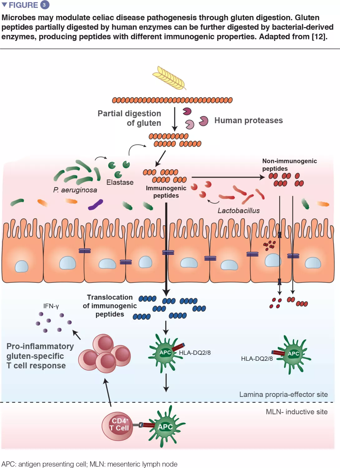
Recent translational work in mice expanded on the observation that bacteria are capable of degrading gluten (Figure 3). The study used gnotobiotic mice that were colonized with either opportunistic pathogens, such as Pseudomonas aeruginosa, or with commensals, such as Lactobacillus. The authors showed that different bacteria can degrade gluten in vivo, but the protein fragments they produce are distinct. The study further demonstrated that enzymes from P. aeruginosa, which was isolated from a celiac disease patient, could degrade gluten. This digestion process produced gluten fragments that stimulated an inflammatory immune response in cells isolated from celiac disease patients and were better able to cross the small intestinal barrier, where interaction with immune cells would occur. Several peptides generated by P. aeruginosa digestion that were subsequently digested with lactobacilli, isolated from a healthy subject and a core member of the healthy microbiome, no longer induced inflammatory immune responses in vitro. This study provided a key mechanism where both opportunistic pathogens and commensals may modify the repertoire and immune properties of gluten peptides in the gut, thereby impacting disease susceptibility.[12]

Can microorganisms be used to treat or prevent celiac disease? 

A diagnosis of celiac disease means strict, life-long avoidance of gluten-containing foods because exposure to small amounts of gluten can trigger a variety symptoms and enteropathy in affected people. Gluten is used ubiquitously in processed foods, which makes strict adherence difficult, and has prompted the search for alternative or adjuvant therapies. Given the key role for microorganisms in regulating immunity and the association between celiac disease and altered composition and function of the microbiota, the therapeutic potential of several probiotics has been tested. A strain of Bifidobacterium longum, previously shown to have anti-inflammatory effects in vitro,[10-13] was tested in children on a gluten-free diet in a double-blind placebo-controlled trial.

Gluten metabolism by bacteria

  • Gluten is highly resistant to breakdown by host digestive enzymes in the small intestine due to its amino acid structure.

  • This leaves large gluten fragments that are capable of inducing immune responses once they cross the epithelial barrier in genetically susceptible individuals.

  • The gastrointestinal tract harbours bacteria that are able to breakdown gluten, and these bacteria may differ between celiac disease patients and healthy subjects.[17]

Administration of the probiotic led to some immune changes, as well as lower levels of potentially harmful bacteria (B. fragilis). However, no changes in symptoms were observed between children that received the probiotic compared to those that received placebo.[14] Because the probiotic was administered together with the gluten-free diet, it is difficult to discern between changes induced by the dietary restriction or the probiotic. Two other studies tested the effects of a strain of Bifidobacterium infantis. The first randomized double-blind placebo-controlled trial demonstrated that patients receiving the probiotic showed significant improvement of symptoms following the 3-week trial, but no differences were found in intestinal permeability.[15] A follow-up trial tested whether the same probiotic could modulate innate immune responses, which could be responsible for the symptomatic improvement previously observed. Administration of a strain of B. infantis led to a decrease in the number of small intestinal Paneth cells, that paralleled a decrease in antimicrobial peptides. These effects of the probiotic were independent of the gluten free diet.[16] Due to the nature of these few studies including only small groups, there is no evidence to date to recommend any probiotic in celiac disease. Moreover, probiotics consumed by patients with celiac disease need to be rigorously certified gluten-free, and this is not the case for every over the counter preparation. Prior to patient consumption, we need a better understanding of mechanisms of action, and those chosen for further testing should be selected due to their involvement in celiac disease pathways. For example, bacteria that aid in the detoxification of gluten could be selected and used to complement the gluten-free diet. However, to date, no single bacterium tested has shown optimal gluten digestion in vitro. Studies have focused on bacterial strains that produce enzymes capable of degrading gluten, but fungal species such as Aspergillus niger, also produce gluten-degrading enzymes, and rational combinations of fungal and bacterial organisms may offer an attractive avenue of therapeutic research in celiac disease.

Conclusion

The role of the intestinal microbiota in celiac disease has become evident. Expanding on clinical associations, reductionist systems and gnotobiotic animal models have provided evidence that specific microbes can modulate key steps in celiac disease pathogenesis. The continued use of these systems to study specific microbe-host and microbe-gluten interactions as well as larger clinical studies where at-risk individuals are followed over time are critical for understanding how microbes could trigger disease. This can allow for microbe targeted preventative strategies or adjuvant therapies to the gluten-free diet.

Sources

1 Verdu EF, Galipeau HJ, Jabri B. Novel players in coeliac disease pathogenesis: role of the gut microbiota. Nat Rev Gastroenterol Hepatol 2015 ; 12 : 497-506.

2 Ou G, Hedberg M, Hörstedt P, et al. Proximal small intestinal microbiota and identification of rod-shaped bacteria associated with childhood celiac disease. Am J Gastroenterol 2009 ; 104 : 3058-67.

3 Wacklin P, Laurikka P, Lindfors K, et al. Altered duodenal microbiota composition in celiac disease patients suffering from persistent symptoms on a long-term gluten-free diet. Am J Gastroenterol 2014 ; 109 : 1933-41.

4 Olivares M, Neef A, Castillejo G, et al. The HLA-DQ2 genotype selects for early intestinal microbiota composition in infants at high risk of developing coeliac disease. Gut 2015 ; 64 : 406-17.

5 Sellitto M, Bai G, Serena G, et al. Proof of concept of microbiome-metabolome analysis and delayed gluten exposure on celiac disease autoimmunity in genetically at-risk infants. PLoS One 2012 ; 7 : e33387.

6 Olivares M, Benítez-Páez A, de Palma G, et al. Increased prevalence of pathogenic bacteria in the gut microbiota of infants at risk of developing celiac disease: The PROFICEL study. Gut Microbes 2018 ; 9 : 551-8.

7 Olivares M, Walker AW, Capilla A, et al. Gut microbiota trajectory in early life may predict development of celiac disease. Microbiome 2018 ; 6 : 36.

8 Tye-Din JA, Galipeau HJ, Agardh D. Celiac disease: a review of current concepts in pathogenesis, prevention, and novel therapies. Frontiers in Pediatrics 2018 ; 6 : 350.

9 Sánchez E, Nadal I, Donat E, et al. Reduced diversity and increased virulence-gene carriage in intestinal enterobacteria of coeliac children. BMC Gastroenterol 2008 ; 8 : 50.

10 De Palma G, Kamanova J, Cinova J, et al. Modulation of phenotypic and functional maturation of dendritic cells by intestinal bacteria and gliadin: relevance for celiac disease. J Leukoc Biol 2012 ; 92 : 1043-54.

11 Galipeau HJ, McCarville JL, Huebener S, et al. Intestinal microbiota modulates gluten-induced immunopathology in humanized mice. Am J Pathol 2015 ; 185 : 2969-82.

12 Caminero A, Galipeau HJ, McCarville JL, et al. Duodenal bacteria from patients with celiac disease and healthy subjects distinctly affect gluten breakdown and immunogenicity. Gastroenterology 2016 ; 151 : 670-83.

13 Laparra JM, Olivares M, Gallina O, et al. Bifidobacterium longum CECT 7347 modulates immune responses in a gliadin-induced enteropathy animal model. PLoS One 2012 ; 7 : e30744.

14 Olivares M, Castillejo G, Varea V, et al. Double-blind, randomised, placebo-controlled intervention trial to evaluate the effects of Bifidobacterium longum CECT 7347 in children with newly diagnosed coeliac disease. Br J Nutr 2014 ; 112 : 30-40.

15 Smecuol E, Hwang HJ, Sugai E, et al. Exploratory, randomized, double-blind, placebo-controlled study on the effects of Bifidobacterium infantis natren life start strain super strain in active celiac disease. J Clin Gastroenterol 2013 ; 47 : 139-47.

16 Pinto-Sánchez MI, Smecuol EC, Temprano MP, et al. Bifidobacterium infantis NLS super strain reduces the expression of α-defensin-5, a marker of innate immunity, in the mucosa of active celiac disease patients. J Clin Gastroenterol 2017 ; 51 : 814-7.

17 Caminero A, Nistal E, Herrán AR, et al. Differences in gluten metabolism among healthy volunteers, coeliac disease patients and first-degree relatives. Br J Nutr 2015 ; 114 : 1157-67.

Summary
Off
Sidebar
Off
Migrated content
Désactivé
Updated content
Désactivé
Hide image
Off
Article

Esophageal microbiome cause or consequence esophageal diseases

Press review

By Pr. Markku Voutilainen
Turku University Faculty of Medicine; Turku University Hospital, Department of Gastroenterology, Turku, Finland

Illustration showing gastroesophageal reflux.

Gastroesophageal reflux disease is common in western world. Barrett’s esophagus (BE) is the complication of reflux disease and the major risk factor of esophageal adenocarcinoma, which has a five-year survival rate less than 20%.

Recent review examined the role of esophageal microbiome in BE and esophageal cancer.[1] Esophagus is exposed to swallowed oral microorganisms and also microbes of the refluxed gastric contents. Esophageal microbiota is not similar to oral nor gastric microbiota. The first bacteria detected in the esophagus were Strectococcus viridans and group D Streptococcus. Later six phyla were observed by broad-range 16S ribosomal DNA gene clone sequencing including Firmicutes, Bacteroidetes, Actinobacteria, Proteobacteria, Fusobacteria, and Streptococcus. BE and high-grade dysplasia were associated with the highest number of bacteria. In patients with esophagitis and BE the number of Streptococcus was diminished while gram-negative anaerobes and microaerophiles were increased.

BE and esophageal adenocarcinoma are associated with increased number of Escherichia coli. Another gram-negative species detected in esophageal cancer patients is Fusobacterium nucleatum. Also oral dysbiosis may be related to the increased risk of esophageal cancer, while gastric Helicobacter pylori seems to protect against esophageal cancer. Gastric dysbiosis such as increase of Clostridiales and Erysipelotrichaceae is associated with esophageal squamous carcinoma. Also fungi, for example Candica albicans and C. glabrata are often detected in esophageal samples from patients with esophaghageal adenocarcinoma. An epidemiologic study showed dose-dependent association between penicillin use and increased risk of esophageal cancer. Also proton pump inhibitors modify gastric and esophageal microbiome.

The present data of esophageal microbiota were obtained from small, selected, and symptomatic patient populations in cross-sectional studies. Thus no conclusions of causality between esophageal microbiota and esophageal diseases can be made. Only a small portion of patients with BE develop adenocarcinoma and further studies are needed to define the role of esophageal dysbiosis in the pathogenesis of cancer. One topic for further research is the impact of proton pump inhibitors on esophageal microbiota and on the risk for esophageal diseases.[1]

Eosinophilic esophagitis (EoE) is an allergic chronic inflammatory disease that is the most common cause of dysphagia in children and young adults in developed countries. EoE has common inflammatory features with other allergic diseases and allergen exposure probably has a central role in EoE pathogenesis. Capucilli and Hill have reviewed EoE epidemiology, pathogenesis and treatment.[2] The esophageal microbiota may be involved in the pathogenesis of EoE. The esophagus is colonized by hundreds of bacterial species and members of Firmicutes and Bacteroidetes phyla are the commonest [2]. In patients with active EoE, the genera Streptococcus and Atopobium are decreased while Neisseria and Corynebacterium are increased. Another study showed that the total amount of esophageal bacteria and Haemophilus genus specifically were increased in EoE. Proton pump inhibitors that are used in the treatment of EoE, cause an enrichment of Proteobacteria phylum. Esophageal bacterial load is increased in EoE patients irrespective of treatment or the severity of esophageal mucosal eosinophilia. Like in other allergic and autoimmune diseases, antibiotic treatment and caesarean delivery are associated with increased risk of EoE.[2]

Conclusion

Studies were cross-sectional and there is no data on the stability of esophageal microbiota over time. More studies are needed to define the role of esophageal microbiota in the pathogenesis and activation of EoE.

Summary
Off
Sidebar
Off
Migrated content
Désactivé
Updated content
Désactivé
Hide image
Off
Press review

The impact of the parents’ microbiome on the descendants health

Press review 

By Pr. Markku Voutilainen
Turku University Faculty of Medicine; Turku University Hospital, Department of Gastroenterology, Turku, Finland

Photo : The impact of the parents’ microbiome on the descendants health

Infant gut is colonized by maternal vaginal and fecal bacteria during vaginal birth. Microbial colonization of the gut starts during fetal life, although its role remains debatable. The hypothesis of Developmental Origins of Health and Disease (DOHaD) suggests that the conditions during fetal life have an impact on the early life of the newborn and also result increased risk for chronic diseases of the offspring.

Professor Friedman has reviewed developmental programming.[1] Maternal obesity, diabetes, and western-style diet have an impact on infant stem cells, immune system and gut microbiota. The gut of the newborn is first colonized by aerobes and facultative anaerobes, which are replaced by strict anaerobes. This modifies innate immune signalling, T helper cell immune responses, and endotoxin tolerance. Maternal obesity may disrupt normal microbial colonization and increase the risk of immunologic and metabolic diseases later in the life. Antibiotic treatment during pregnancy increases the risk of childhood obesity. Children born for obese mothers have lesser abundance of two families of fecal Proteobacteria. Furthermore, maternal high-fat diet causes the loss of key bacteria and decrease in bacterial diversity of the infant fecal microbiota.

Also paternal diet may have impact on the health status of the following generations. Zhang and co-workers examined the impact of unhealthy diet in animal model.[2] They fed male rats in two successive generations (F0 and F1) with with high fat, sucrose and salt diet. The control group was fed with normal diet. The high fat-sucrosesalt diet was associated with increased aspartate aminotransferase levels in the next generation (F2). Unhealthy diet was also associated with higher body weight. In female F2 rats, the Shannon index of gut microbiota indicated significantly higher diversity. Variation in the abundance of bacterial genus was associated with liver function abnormalities. Unhealthy diet in F0 and F1 generations was associated with increased serum cholesterol and lipoprotein levels in male F2 rats.

Conclusion

This data suggest that parent’s unhealthy diet causes dysbiosis of the gut microbiota of the offspring and may increase the risks of overweight and several chronic diseases (T2 diabetes, liver and cardiovascular diseases).

Summary
Off
Sidebar
Off
Migrated content
Désactivé
Updated content
Désactivé
Hide image
Off
Press review

Gut Microbiota for Health World Summit 2019

Congress review

By Pr. Francisco Guarner
Digestive System Research Unit, University Hospital Vall d’Hebron, Barcelona, Spain

IBD

During its 8th edition last March, the GMFH devoted considerable place to diet and the way it interacts with the gut microbiome, preventing or promoting diseases.

Diet and the gut microbiome

Diet is a key element for the symbiotic interactions between gut microbes and the host, and it is considered as one of the main drivers in shaping the gut microbiota across lifetime, as reviewed by Jack A. Gilbert (UC, San Diego), Susan Devkota (Cedars-Sinai, Los Angeles), and Lipping Zhao (Rutgers, New Jersey). Foods deliver numerous substrates for microbial metabolism and the microbiome is a chemical factory that synthesizes metabolites important for human health. Macro- and micronutrients in food influence the structure and functions of the gut microbial ecosystem in such way that diet appears to be the most important determinant of similarity in gut microbial composition across humans.[1]

Self-reported dietary data from the American Gut project [2] suggest that the number of unique plant species that a subject consumes is associated with microbial diversity, rather than self-reported categories such as “vegan” or “omnivore”. Higher microbial diversity and higher abundance of short chain fatty acid (SCFA) producer species was found in individuals eating more than 30 types of plants per week as compared to those eating less than 10 types of plants per week. The faecal metabolome also differed between both groups. In addition, individuals who consume more than 30 types of plants compared to those who consume 10 or fewer plants had significantly lower abundance of antibiotic resistance genes.

Dysbiosis of the gut microbiome is a definable state with mechanistic implications. It is not just a change in microbial diversity but a rupture of the mutualistic balance between microbiota and host, where inadequate diet plays a detrimental role. During homeostasis, colonocyte metabolism is directed towards oxidative phosphorylation, resulting in high epithelial oxygen consumption. The consequent epithelial hypoxia helps maintain a microbial community dominated by obligate anaerobes, which provide benefit by converting fibre into fermentation products (SCFA) absorbed by the host. Conditions that alter metabolism of the epithelium, such as a fibre poor diet, increase epithelial oxygenation, thereby driving an expansion of facultative anaerobes, a hallmark of dysbiosis in the colon.[3] The shift in the colonic microbiota composition from obligate to facultative anaerobes, associated with many chronic human illnesses, might have a common underpinning in colonocyte dysfunction. As highlighted by Susan Devkota, if choosing a strict or extreme dietary regime, consuming mixed fibre types can support the microbiome and prevent nutrient deficiencies.

The "Foundation Guild"

Lipping Zhao pointed out that our ancestors had much higher intake of dietary fibres than current consumption rates. Reduced intake of fibres and diminished prevalence of SCFA-producing bacteria may underlie many chronic diseases such as type 2 diabetes. In a randomised controlled intervention trial with Chinese type 2 diabetes patients,[4] high intake of diverse dietary fibres (WTP diet) selectively promoted abundance of a group of acetic and butyric acid producer strains in the gut. The WTP diet is based on wholegrains, traditional Chinese medicinal foods and prebiotics. The WTP diet improved glucose homeostasis by reducing glycated haemoglobin, fasting blood glucose and meal tolerance test.

Abundance of the SCFA producers in faeces correlated with the metabolic outcomes and the blood levels of glucagon- like peptide-1 and peptide YY, which induce insulin secretion. Moreover, reduction of faecal pH by SCFA production correlated with inhibition of detrimental bacteria that promote inflammation and suppress glucagon-like peptide-1 production. In addition to providing SCFAs to directly benefit the host, this group of SCFA producers played important ecological functions in the gut microbiota. Lipping Zhao suggested that they work as the “foundation guild” for structuring the healthy gut microbiota. To help patients regain a healthy gut microbiota, “this foundation guild must be re-seeded and re-established”, he said.

FODMAPS and IBS

As reviewed by Magnus Simren (University of Gothenburg), the low fermentable oligosaccharides, disaccharides, monosaccharides and polyols (FODMAPs) diet is now being recommended by up to 85% of doctors to treat functional abdominal symptoms. Clinical trials suggest that some patients have a short-term favourable response to a low FODMAP diet, but whether this dietary advice is clearly better than the first line dietary therapy for IBS is uncertain. Of concern, short-term use of the low FODMAP diet has been associated with potentially unfavourable changes in gut microbiota composition, including reduction of fermentative species (Bifidobacterium, Faecalibacterium and Clostridium cluster XIVa) and increased dysbiotic index scores.[5]

A randomized controlled trial compared effects of the low FODMAP diet or the prebiotic GOS on gut microbiota composition.[6] Changes in faecal microbiota differed between both groups after a 4-week treatment period, particularly in relation to bifidobacteria (increase in the prebiotic group and decrease in the low FODMAP group) and Bilophila wadsworthia (the opposite pattern). Despite distinct effects on microbiota, reductions of symptoms were very similar in both groups. Of interest, the decrease in symptoms persisted during the 2-week follow-up after cessation of prebiotic intake, but reappeared immediately after discontinuation the low FODMAP diet. Modulation of gut microbiota as a treatment strategy for IBS seems promising, but long-term safety aspects need to be taken into account. Diets that reduce symptoms but deteriorate gut health (and general health in the long term) should not be the first choice.

Symbiotic trial to prevent newborn sepsis 

Sepsis in early infancy results in one million annual deaths worldwide, most of them in developing countries. Pinaki Panigrahi presented an intervention study to prevent sepsis among infants in rural India.[7] An oral symbiotic preparation (Lactobacillus plantarum plus fructooligosaccharide) significantly reduced sepsis and death in newborns (risk ratio 0.60, 95% confidence interval 0.48–0.74). This finding suggests that a large proportion of neonatal sepsis in developing countries could be effectively prevented using probiotic- prebiotic treatment.

Conclusion

This finding suggests that a large proportion of neonatal sepsis in developing countries could be effectively prevented using probiotic- prebiotic treatment.

Summary
Off
Sidebar
Off
Migrated content
Désactivé
Updated content
Désactivé
Hide image
Off
Congress review

Review of the main contributions related to the intestinal microbiota

Congress review

By Dr Dragos Ciocan
Hepato-gastroenterology and Nutrition, Hôpital Antoine-Béclère, Clamart, France

Bandeau_article5_NL07

The Journées francophones d’hépato- gastroentérologie et d’oncologie digestive (French language Hepatogastroenterology and Digestive Oncology Congress) was held in Paris from 21 to 24 March 2019, with an attendance of more than 5,000 French-speaking doctors and researchers. A number of original studies on the intestinal microbiota (IM) were presented.

Faecal transplantation

Faecal microbiota transplantation (FMT) is a therapeutic strategy which is used in current clinical practice only for recurrent Clostridium difficile infections [1]. Dr. Eymeric Chartrain presented the experience acquired by the Clermont-Ferrand University Hospital reference centre between 2014 and 2018 on the use of FMT in this indication. FMT was effective in 95% of cases with minor side effects occurring in only 16% of patients. Furthermore, patients reported a significant improvement in quality of life at 6 months post-FMT. The total cost of a FMT intervention is approximately 3,100 euros. Despite this high cost, FMT helps lower health care costs by reducing morbidity and mortality in these patients and is a rational and effective option.

The role of FMT is being studied in many diseases involving the IM, including chronic inflammatory bowel diseases (IBDs). Professor Harry Sokol presented the results of a small, randomized, single blind, placebo-controlled pilot trial in 17 patients evaluating the role of FMT in adults with colonic or ileocolonic Crohn’s disease during a flare-up, who were treated with oral corticosteroids. The primary endpoint – donor IM colonization in the recipient at week 6, defined by recipient IM at week 6 more similar to that of the donor (Sorensen similarity index ≥ 0.6) than to that of the patient pre-FMT – was not reached. Nevertheless, among the secondary endpoints, the FMT group had a reduction in endoscopic disease severity whereas the control group had an increase in inflammation. Colonization by donor IM was associated with sustained remission and patients without donor IM colonization had a recurrence earlier. In addition, the IM composition was predictive of steroid-free clinical remission. Despite the small sample size, this study suggests that FMT could be effective after corticosteroid-induced clinical remission in patients with active Crohn’s disease. Several larger studies including one conducted by Professor Sokol’s group are in progress.

Enterobacteriaceae modulate the effects of fungi in colitis 

While the role of the bacterial and fungal IM is known in IBDs, the impact of bacteria- fungal interactions on intestinal inflammation is less clear. Dr. Bruno Sovran presented a study which looked precisely at these interactions in a mouse colitis model. The authors found that administration of a strain of Saccharomyces improved colitis whereas administration of Candida albicans caused it to worsen. However, pre-treatment with colistin, which kills gram-negative bacteria (including proteobacteria) led to a loss of effect of fungi. Administration of colistin-resistant E. coli which restored the enterobacterial population in colistin-treated mice also re-established both the beneficial effects of the strain of Saccharomyces and the deleterious effects of C. albicans on colitis severity. These observations suggest that Enterobacteriaceae are necessary for improved gut colonization by fungi and may explain the effects of some probiotics in colitis.[2]

The gut-brain axis in obesity 

It is now well known that the IM plays a role in the pathophysiology of obesity. The IM can also modulate cognitive and psychological functions via the gut-brain axis.[3] Obesity is a risk factor for cognitive impairment, independently of other comorbidities, but the mechanisms are obscure. The MEMOB study, presented by Dr. Sophie Cambos, investigated memory dysfunctions in obese subjects and their correlation with the IM. In this prospective, longitudinal, monocentric study in obese and normal weight subjects, the obese subjects prior to bariatric surgery had memory dysfunctions in comparison with a control population. Analysis of the microbial profile revealed a link between the abundance of Eggerthellales and memory functions: the greater the Eggerthellales abundance, the worse the memory results. These data suggest that obesity – and therefore the associated microbiota alterations – might accelerate cognitive decline via the gut-brain axis.

Microbiota and the liver 

A Biocodex workshop entitled “Microbiota and the liver, from mechanisms to treatment” took place during the meeting. Professor Gabriel Perlemuter reviewed the latest advances concerning the role of the IM in liver diseases. Among the most prominent recent studies, the IM has been found to play a role in susceptibility to developing alcoholic liver disease and non-alcoholic fatty liver disease when using proton pump inhibitors. These drugs promote overgrowth of Enterococcus in the IM, leading to more translocation to liver where it induces liver inflammation.[4] Several pilot studies also investigated the role of FMT in liver diseases (hepatitis B, hepatic encephalopathy and severe corticoresistant acute alcoholic hepatitis) and reported some efficacy in these indications.

Dr. Anne-Marie Cassard discussed the manipulation of the IM in the case of liver disease. She presented data from her group showing that low levels of Bacteroides are associated with the development of alcohol-induced liver injury. Correcting this IM imbalance by administration of pectin, a soluble fibre, prevented and improved the alcoholic-induced liver lesions.[5] However, not all fibres induce the same changes in the IM, even though they have the same beneficial effect on the host.

Conclusion

Furthermore, among the different strategies studied which target the IM and have shown some effectiveness on liver injury (antibiotics, FMT, probiotics, prebiotics), only antibiotics and FMT can induce long-lasting changes in the IM.

Summary
Off
Sidebar
Off
Migrated content
Désactivé
Updated content
Désactivé
Hide image
Off
Congress review

Treatment of active crohn’s disease with an ordinary foodbased diet that replicates exclusive enteral nutrition

Commented articles - Children's section

By Pr. Emmanuel Mas
Gastroenterology and Nutrition Department, Children’s Hospital, Toulouse, France

Photo : Treatment of active crohn’s disease with an ordinary foodbased diet that replicates exclusive enteral nutrition

Commentary on the original publication by Svolos et al. (Gastroenterology 2019)

Exclusive enteral nutrition (EEN) is the only established dietary treatment in Crohn’s disease (CD), but its acceptability is limited. There is a need for new dietary treatments for CD.

The effects of a personalized diet (CD-TREAT), based on the composition of EEN, were evaluated by analysing the intestinal microbiota, inflammation and clinical response in rats, in healthy adults, and in children with relapsing CD.

Ultimately, it was shown that CD-TREAT replicates EEN changes in the gut microbiota, decreases gut inflammation, is well tolerated and is potentially effective in patients with active CD.[1]

What do we know about this subject?

Exclusive enteral nutrition (EEN) is an effective treatment of Crohn’s disease with ileus involvement, achieving good results (mucosal healing in 80% of cases) that are superior to those obtained with corticotherapy. However, the main obstacle is the acceptability of receiving, for at least 8 weeks, exclusive enteral nutrition. EEN is delivered by nasogastric tube or, as for Modulen IBD®, by the oral route. The mechanism of action of EEN is not fully understood but several studies suggest that it acts by modulating the intestinal microbiota.

What are the main insights from this study? 

This study aimed to determine whether an ordinary diet (CD-TREAT), i.e. oral intake of ordinary foods but based on a nutrient composition similar to that of Modulen IBD®, could be effective in Crohn’s disease. The proportion of carbohydrate was decreased and that of protein increased. A multivitamin tablet provided the micronutrients from EEN. A crossover study was conducted in 25 healthy adult volunteers who received CD-TREAT or EEN for one week each, with a washout period in between. CD-TREAT was easier to follow and more satiating than EEN. Microbiota richness and alpha diversity were not altered by these diets. However, the relative abundance of 58 (49.3%) and 38 (32.3%) bacterial genera changed significantly after EEN and CD-TREAT, respectively, of which 28 changed in the same direction. There were changes in the concentrations of different metabolites (some short chain fatty acids – acetate, propionate and butyrate significantly decreased after EEN and CD-TREAT) and faecal pH increased by about 1 unit. 

5 groups of rats, HLA B27 (inflammatory) and B7 (non-inflammatory): B27-EEN, B27-CD-TREAT, B27-CONTROL, B7-EEN and B7-CONTROL. EEN and CD-TREAT decreased ileal inflammation (Figure 1A), with lower expression of IL-6 in the B27- CD-TREAT group compared to the B27- CONTROL group (p = 0.036). After the 4-week intervention, bacterial diversity was higher in cecum (Figure 1B) and in feces in the B27-CD-TREAT and B27-EEN groups versus B27-CONTROL. Both diets caused changes in faecal concentrations of some short chain fatty acids.

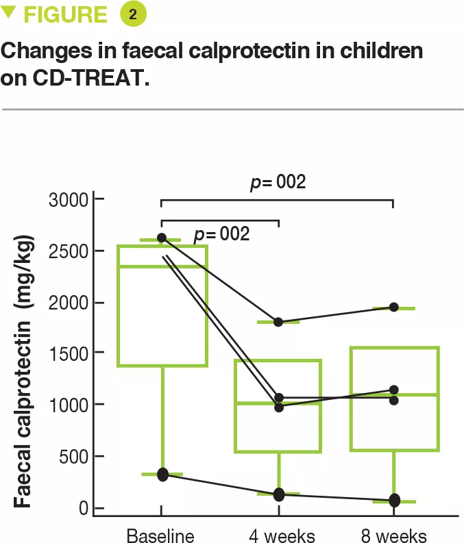
Lastly, 5 children to mild to moderate Crohn’s disease, (wPCDAI score 22.5 to 42.5) were treated with CD-TREAT for 8 weeks. One child discontinued after 9 days because of symptom exacerbation. After 4 weeks, 3 children (60%) had a clinical response (wPCDAI score change > 17.5) and 2 children (40%) were in clinical remission (wPCDAI score < 12.5). After 8 weeks, 80% of children (4 of 5) had a clinical response and 60% (3 of 5) were in clinical remission. The mean baseline level of faecal calprotectin of 1,960 mg/kg decreased by 53% and 55%, respectively, after 4 and 8 weeks (Figure 2). Calprotectin decreased to normal in only one child.

Key points

  • A suitable oral diet could be as effective as exclusive enteral nutrition in Crohn’s disease.

  • Its effect on the intestinal microbiota mimics that of exclusive enteral nutrition.

  • It offers an alternative to exclusive enteral nutrition which is poorly accepted by patients.

Image

What are the consequences in practice? 

This study shows that this diet is more feasible when given orally and that it mimics the effects of EEN with Modulen IBD® on the gut microbiota. CD-TREAT also improves clinical signs and reduces gut inflammation.

Conclusion

This study provides proof-ofconcept that a novel and better tolerated dietary treatment could be effective in Crohn’s disease. These findings need to be confirmed in large, controlled, randomized clinical trials.

Summary
Off
Sidebar
Off
Migrated content
Désactivé
Updated content
Désactivé
Hide image
Off
Commented article