


Focus
Gut microbiota not yet considered “adult” at 5 years old?
A Swedish study has shown that at 5 years the gut microbiota is nearing adult complexity but without yet reaching maturity. The study’s insights on the dynamics of gut microbiota colonization underline the importance of protecting this ecosystem from disturbances throughout childhood.
The gut microbiota Childhood immune system: the benefits of vaginal delivery Potential role of the oropharyngeal microbiota in stunted childhood growth Can fecal transplantation restore the microbiota of Caesarean-born infants?
Summary
Off
Migrated content
Activé
Updated content
Désactivé
Old sources
Old content type
pro_article
Gut microbiota not yet considered “adult” at five years old?
It was once thought that the gut microbiota reaches “adult” complexity at the age of 2 or 3. “Not so”, say the authors of a recent study1: at the age of 5, its composition is still different from that of adults, with certain microorganisms essential to health continuing to develop after this age. Hence the importance of taking good care of the gut microbiota throughout childhood!
The gut microbiota Diet
 Modulating the gut microbiota to better fight child malnutrition
 The 3 key development stages of the gut microbiota in early childhood
Summary
Off
Migrated content
Activé
Updated content
Désactivé
Old sources
Old content type
article
Is the vaginal microbiota to blame for painful periods?
Cramps or discomfort during menstruation is normal but excessive pain that makes you miss work or school is not. This new study looks at the vaginal microbiota’s role in painful periods (dysmenorrhea).
The vaginal microbiota Diet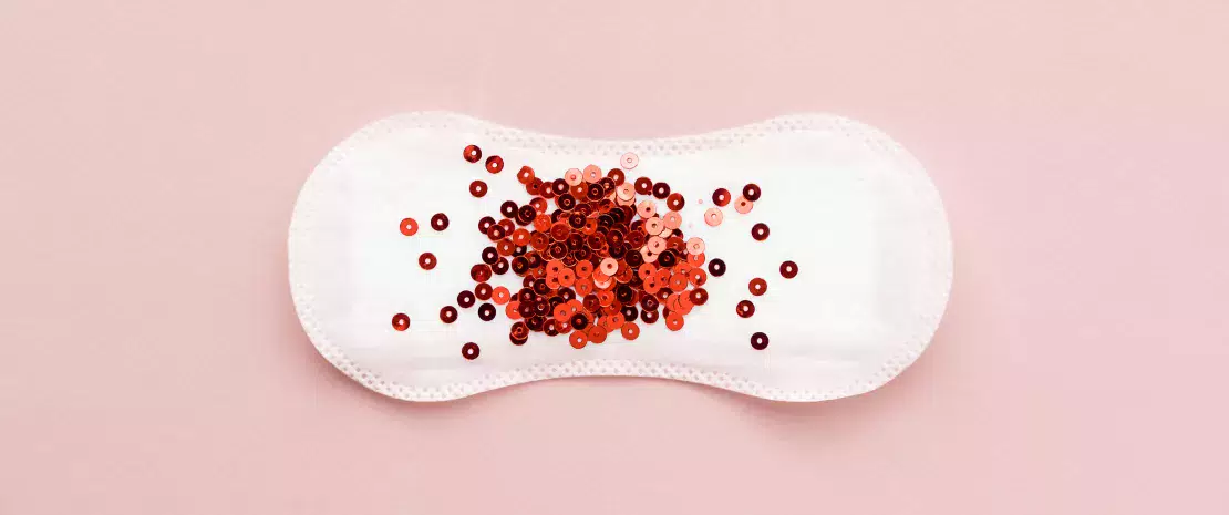
 The prevention of bacterial vaginosis could depend on what you eat
 Can the vaginal microbiota be used as a tool for predicting the severity of endometriosis?
Summary
Off
Migrated content
Activé
Updated content
Désactivé
Old sources
Sources:
Chen CX, Carpenter JS, Gao X, et al. Associations Between Dysmenorrhea Symptom-Based Phenotypes and Vaginal Microbiome: A Pilot Study [published online ahead of print, 2021 Mar 13]. Nurs Res. 2021
Old content type
article
Autism: link between severity of the disorder and changes in the gut microbiota?
A US longitudinal analysis evaluates for the first time links between gut microbiota composition and behavioral changes in children with autism spectrum disorder (ASD).
The gut microbiota Discovery of a new link between autism and gut microbiota Autism: a new fecal microbiota transplant protocol shows promising results What are the long-term effects of antibiotics on the gut microbiota?
Summary
Off
Migrated content
Activé
Updated content
Désactivé
Old content type
pro_article
Gut dysbiosis in SARS-CoV-2 infected monkeys
A French team from the Lille Center for Infection and Immunity, in collaboration with CEA, INRAE, the Pasteur Institute and the Saint-Antoine Hospital in Paris, has shown that SARS-CoV-2, the virus that causes Covid-19, provokes a gut dysbiosis in monkeys which persists even after the virus has been eliminated.
The gut microbiota Covid-19: gut microbiota involved? Gut microbiota and Covid-19: what the experts know and what they suspect How does Covid-19 affect the gut microbiota?
Summary
Off
Migrated content
Activé
Updated content
Désactivé
Old content type
pro_article
Coconut oil for the scalp microbiota!
We’ve all had dandruff on our shirt collar or shoulders. These small, stubborn and unsightly flakes are an extremely common chronic scalp disorder. A recent study has shown that coconut oil can help maintain a healthy scalp by improving the scalp microbiota.
The skin microbiota Diet
 Atopic dermatitis: the skin microbiome has an accomplice!
 Laser and dermatosis: targeting Staphylococcus aureus
Summary
Off
Migrated content
Activé
Updated content
Désactivé
Old sources
Sources:
Saxena, R., Mittal, P., Clavaud, C. et al. Longitudinal study of the scalp microbiome suggests coconut oil to enrich healthy scalp commensals. Sci Rep 11, 7220 (2021)
Old content type
article
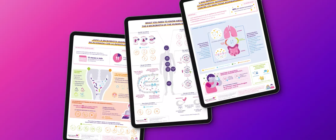
Infographics to share with your patients!
Download original and engaging graphic materials to explain to your patients the essential role of the microbiota in their daily health. These clear and accessible infographics are specifically designed to simplify complex concepts and enhance understanding of the connections between the microbiota and well-being.
Covering a range of topics—from the gut-brain axis to immunity, women’s health, and digestive issues like IBS—they help illustrate how the microbiota influences everything from early development to aging and overall health.
A valuable tool for your consultations and awareness initiatives, they help you educate your patients and strengthen their engagement in their health journey.
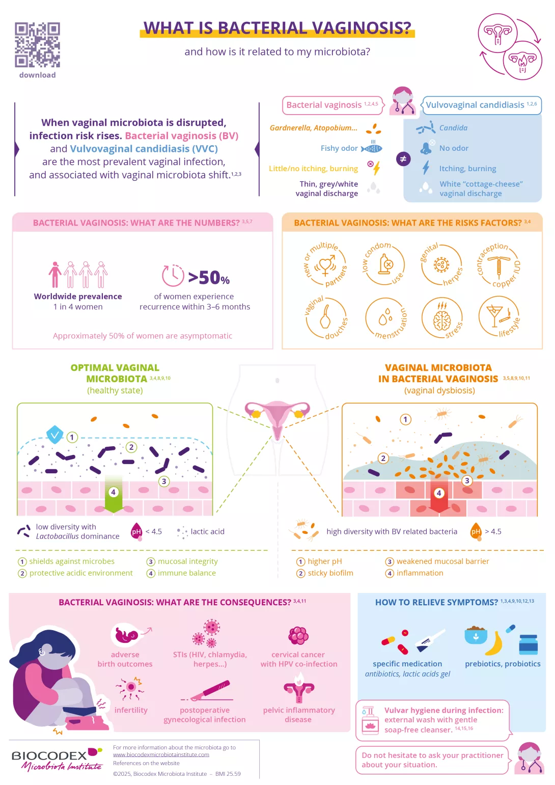
What is bacterial vaginosis?
Bacterial vaginosis is the most common vaginal infection in women of reproductive age. It results from an imbalance in the vaginal microbiota, a delicate ecosystem normally dominated by protective lactobacilli.
This infographic explains what bacterial vaginosis is, how it differs from vulvovaginal candidiasis, its main risk factors and potential consequences, and how symptoms can be relieved. Understanding the role of the vaginal microbiota is key to better prevention and care.
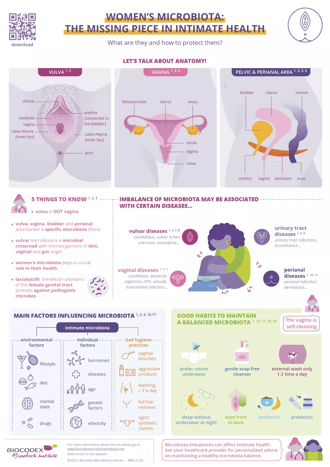
Women’s microbiota: the missing piece in intimate health
This infographic explores the intimate microbiota — an essential yet often overlooked component of women’s health.
It highlights the distinct microbial ecosystems of the vulva, vagina, bladder, and perianal area, and explains how their balance protects against infections and supports overall well-being.
Discover the main factors that influence this delicate microbiota, common signs of imbalance, and practical tips to help patients maintain intimate health through proper hygiene.
Does gut microbiota influence satiety?
This infographic helps healthcare professionals explain to their patients how the gut microbiota influences satiety.
Discover how a balanced microbiota supports appetite regulation and overall health, and why maintaining its balance is essential for well-being.

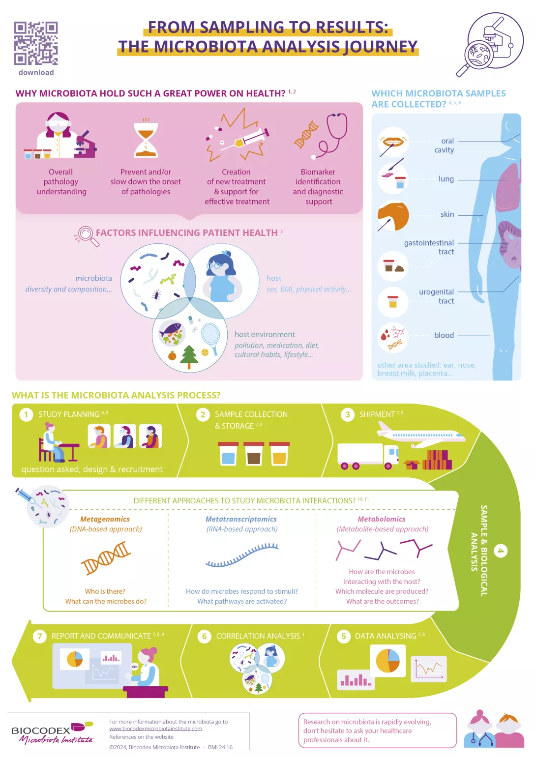
From sampling to results: the microbiota analysis journey
Your patients often ask you about the purpose of microbiota analysis?
This infographic can be a first step in answering them. It outlines the key steps of microbiota analysis, from sample collection to final insights. Simplify complex processes and explore how microbiota data can impact health and personalized care.
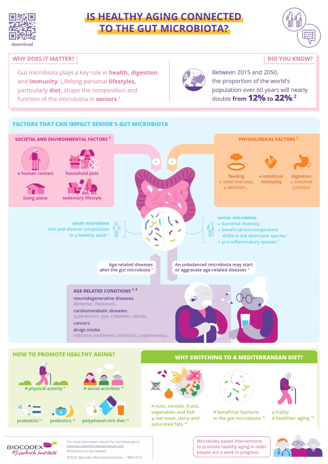
Is healthy aging connected to the gut microbiota?
This infographic explores the link between gut microbiota and aging, highlighting how diet and lifestyle impact digestive health, immunity, and overall well-being in seniors.
Discover key factors influencing microbiota balance and strategies to promote healthy aging.
Only a third of seniors have received information from their healthcare professional on the microbiota, its role and functions
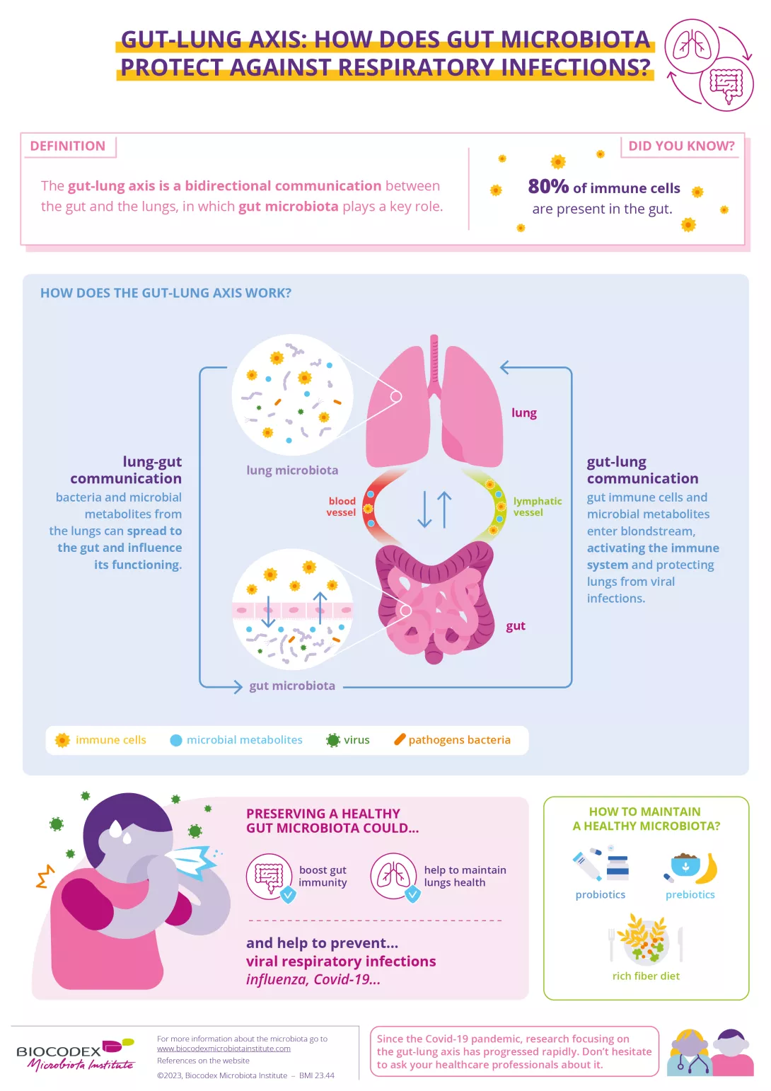
Gut-lung axis: how does gut microbiota protect against respiratory infections?
This infographic illustrates the gut-lung axis, detailing how gut microbiota modulates immune responses to safeguard against respiratory infections. It highlights the bidirectional communication between the gut and lungs.
Discover the mechanisms through which gut bacteria influence lung immunity and learn strategies to support this vital connection.
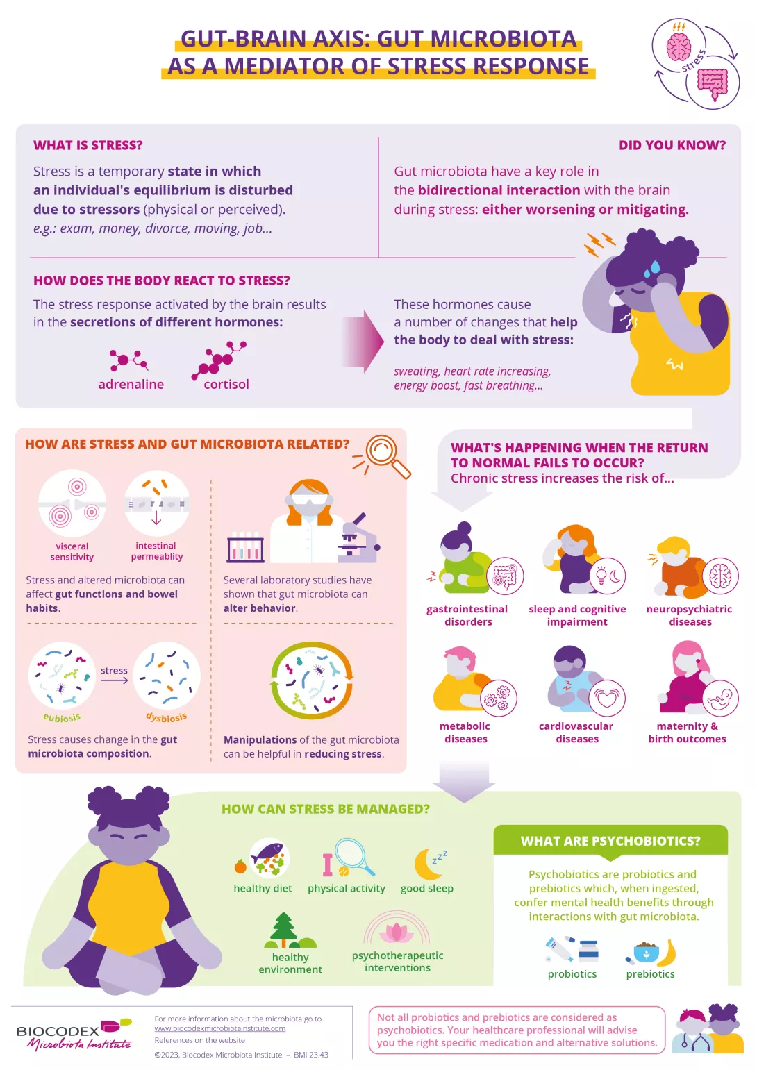
Gut-brain axis: gut microbiota as a mediator of stress response
This infographic explores the connection between gut microbiota and stress, highlighting how the gut-brain axis influences both mental and physical well-being.
It explains how stress impacts gut microbiota, the body's response mechanisms, and strategies to restore balance through lifestyle, diet, and psychobiotics.
Download
Does vaginal microbiota play a role in infertility?
The vaginal microbiota plays a key role in reproductive health, yet its impact on fertility is often overlooked. An imbalance in vaginal flora can reduce pregnancy chances and affect assisted reproduction outcomes.
This infographic highlights the link between microbiota and fertility, emphasizing the importance of maintaining a balanced ecosystem through proper hygiene and patient education.
Download

88% of women would like to have more information about the importance of the vaginal microbiome and its impact on health
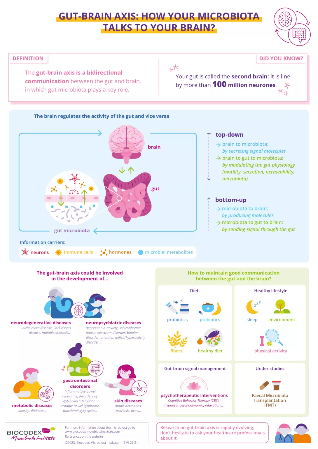
Gut brain axis: how your microbiota talks to your brain?
Your gut and brain are in constant communication, primarily through the gut microbiota. This connection plays a crucial role not only in digestion but also in various diseases, including neurological, psychiatric, and metabolic disorders.
Understanding this link could pave the way for new approaches to maintain our health.
Download
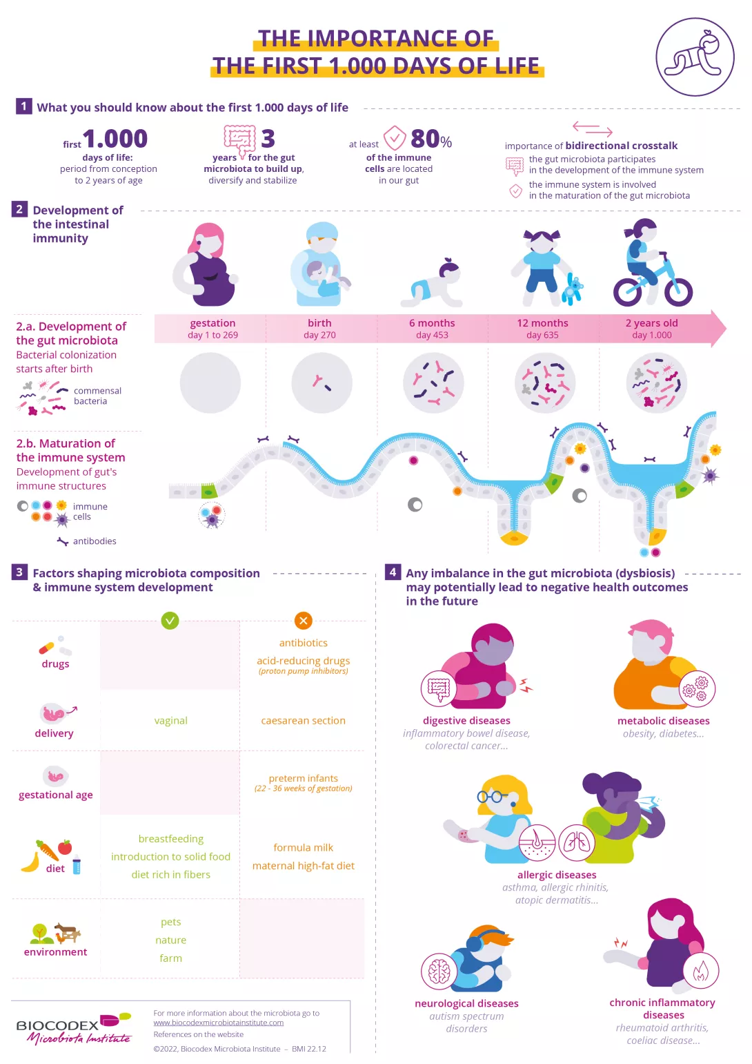
The importance of the first 1000 days of life
This infographic highlights the critical importance of the first 1,000 days of an infant’s life, from conception to toddlerhood, in the development of gut microbiota and intestinal immunity. It explains how early factors, such as diet and environment, influence long-term health.
Learn how these foundational stages shape immune function and disease risk.
Download
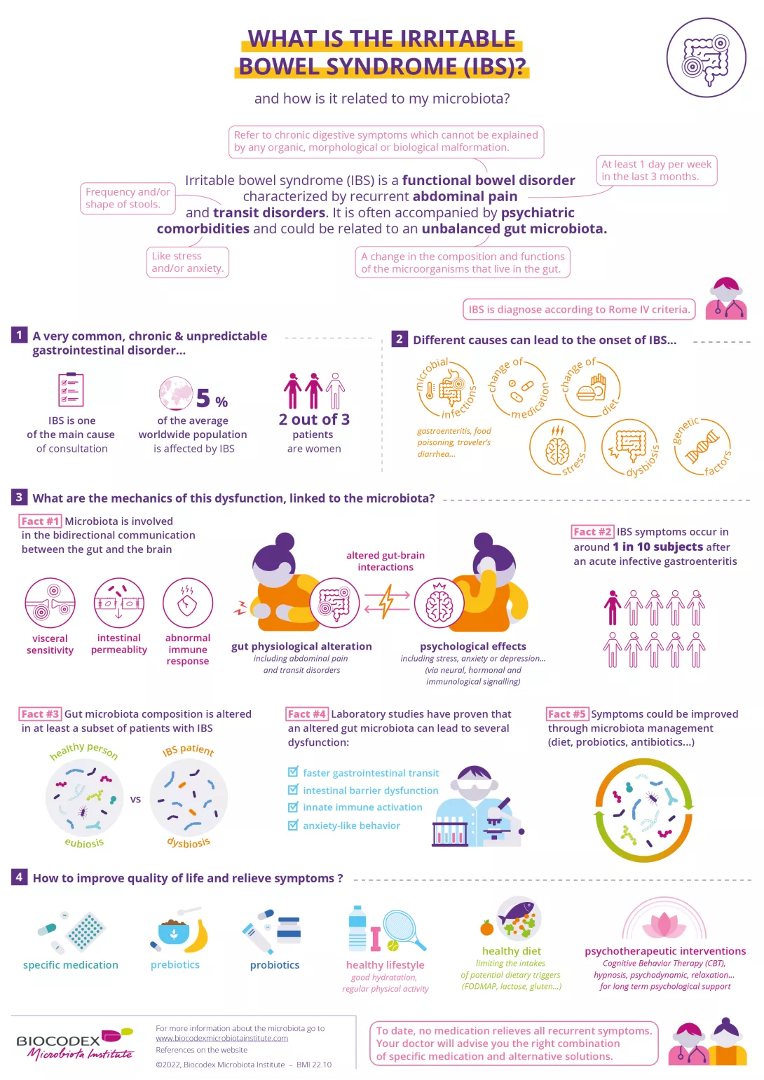
What is the Irritable Bowel Syndrome (IBS)?
This infographic explains Irritable Bowel Syndrome (IBS), a common functional bowel disorder characterized by recurrent abdominal pain and transit issues.
It explores how an imbalanced gut microbiota may contribute to IBS symptoms and highlights the role of diet, lifestyle, and psychological factors in managing the condition.
Discover strategies for improving quality of life and relieving IBS symptoms through microbiota management.
Do you know that an unbalanced microbiota is called a dysbiosis?
This infographic examines dysbiosis and how it disrupts thevarious microbiota of the human body, contributing to conditions like digestive, metabolic, and skin disorders, among others.
It outlines the causes of dysbiosis and offers strategies to restore balance through diet, lifestyle, and medical treatments.

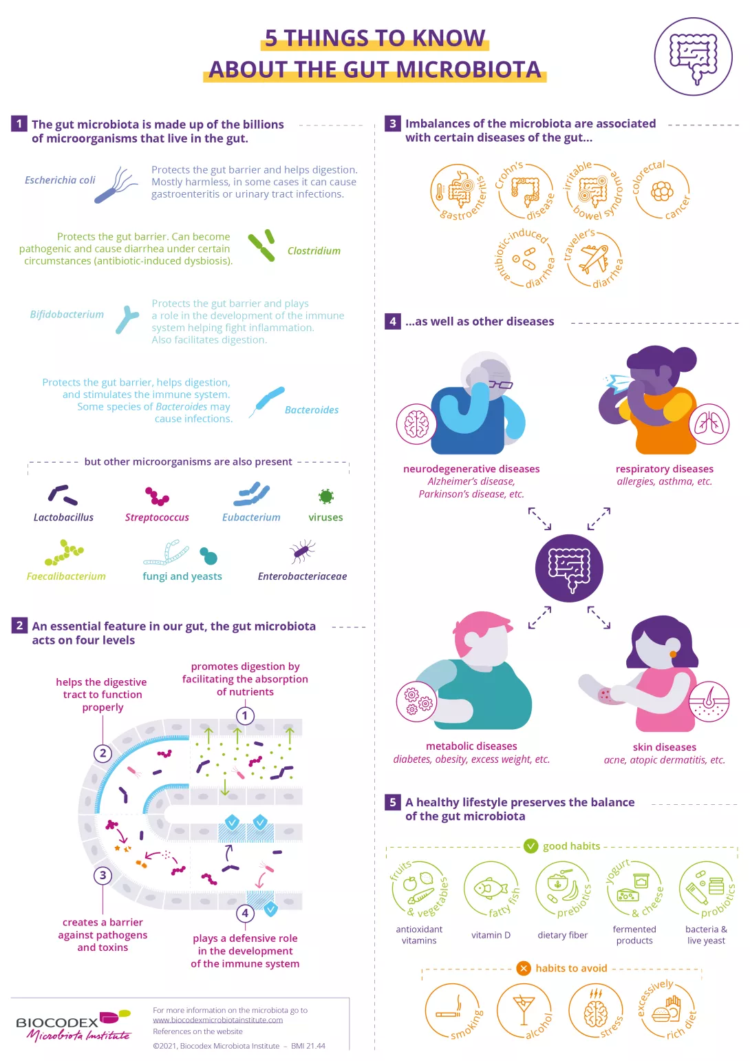
5 things to know about the gut microbiota
This infographic highlights 5 key facts about the gut microbiota, its essential role in digestion, immunity, and overall health. It explains how a balanced microbiota supports gut function and defends against pathogens, while imbalances can be linked to digestive, metabolic, and even neurodegenerative diseases.
Learn how lifestyle choices can influence microbiota health and reduce the risk of disease.
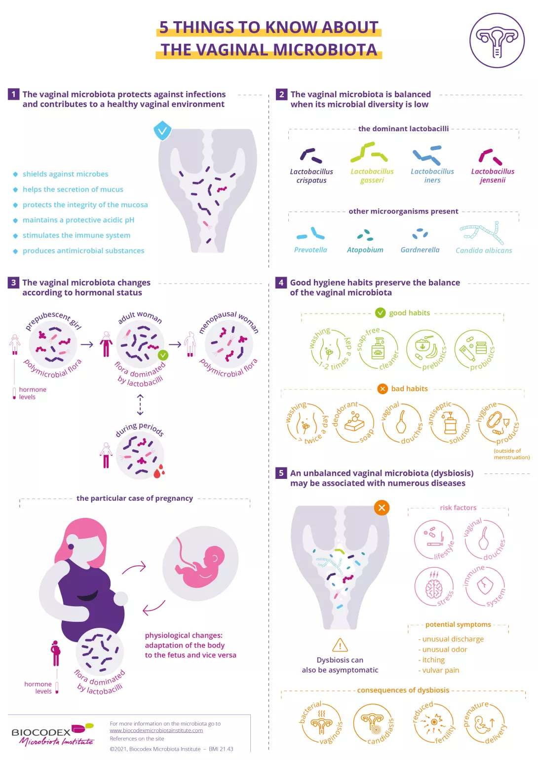
5 things to know about the vaginal microbiota
This infographic outlines 5 important things to know about the vaginal microbiota, including how it protects against infections and how hormonal changes affect its composition.
It also covers the consequences of dysbiosis and offers tips for maintaining a healthy balance through hygiene and lifestyle choices.
What you should know about antibiotics
This infographic covers key facts about antibiotics, explaining how they help fight infections while also affecting the balance of beneficial bacteria in the microbiota.
Overuse or misuse of antibiotics can lead to complications like antibiotic-associated diarrhea and contribute to the rise of antibiotic resistance.
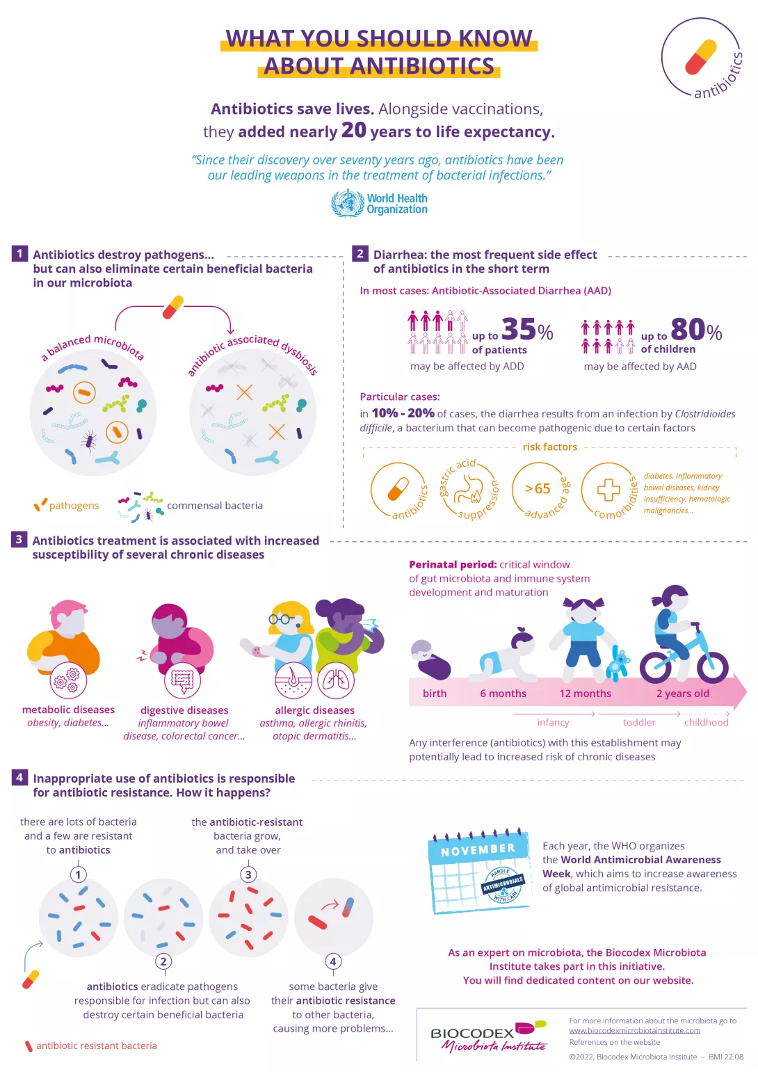
Only 1 in 3 people had been informed by healthcare professionals that taking antibiotics could have negative consequences on the balance of their microbiota
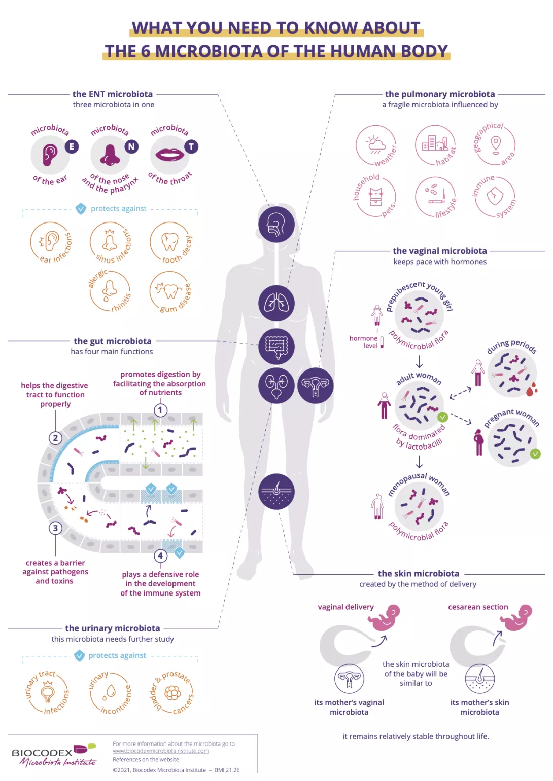
What you need to know about the 6 microbiota of the human body
This infographic introduces the six microbiota of the human body, highlighting the unique functions and characteristics of each one, from the gut to the skin.
It explores how these microbiota are shaped by factors such as hormones, lifestyle, and delivery method, and how they contribute to overall health by protecting against infections and supporting immune function.
Download
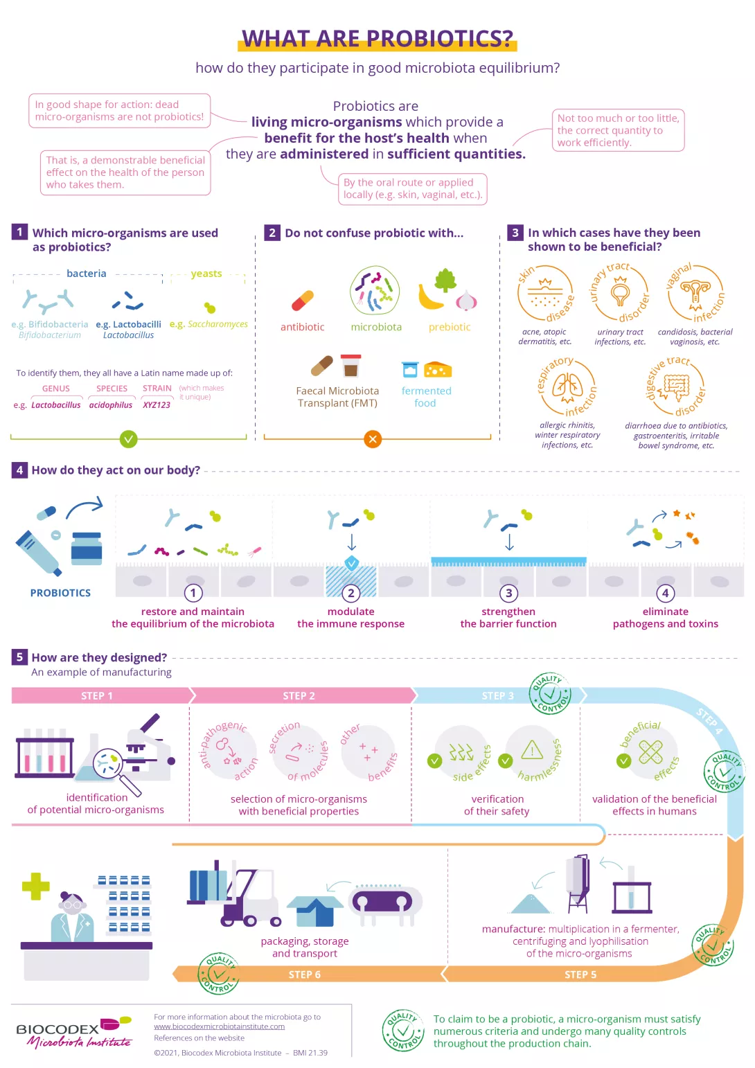
What are probiotics?
This infographic explains what are probiotics and how they help maintain a balanced microbiota, supporting overall health. It explores the types of beneficial microorganisms and the rigorous processes behind their production to ensure their effectiveness in promoting well-being.
Download
Recommended by our community

"Great chart" - Charles Platz (From Biocodex Microbiota Institute on LinkedIn)
"May this institute get more and more successes." -@Mohamma10757688 (From Biocodex Microbiota Institute on X)
"Actually l downloaded these myself. Will follow up on some of the referenced articles. Very interesting stuff" -@HEB2205 (From Biocodex Microbiota Institute on X)
"A great resource for biology teachers." -@OilPaul (From Biocodex Microbiota Institute on X)
Summary
On
Migrated content
Désactivé
Updated content
Désactivé