Clinical and biological predictors of response to standardized pediatric colitis therapy: a prospective multicenter study

Commented articles - Children's section

By Pr. Emmanuel Mas
Gastroenterology and Nutrition Department, Children’s Hospital, Toulouse, France

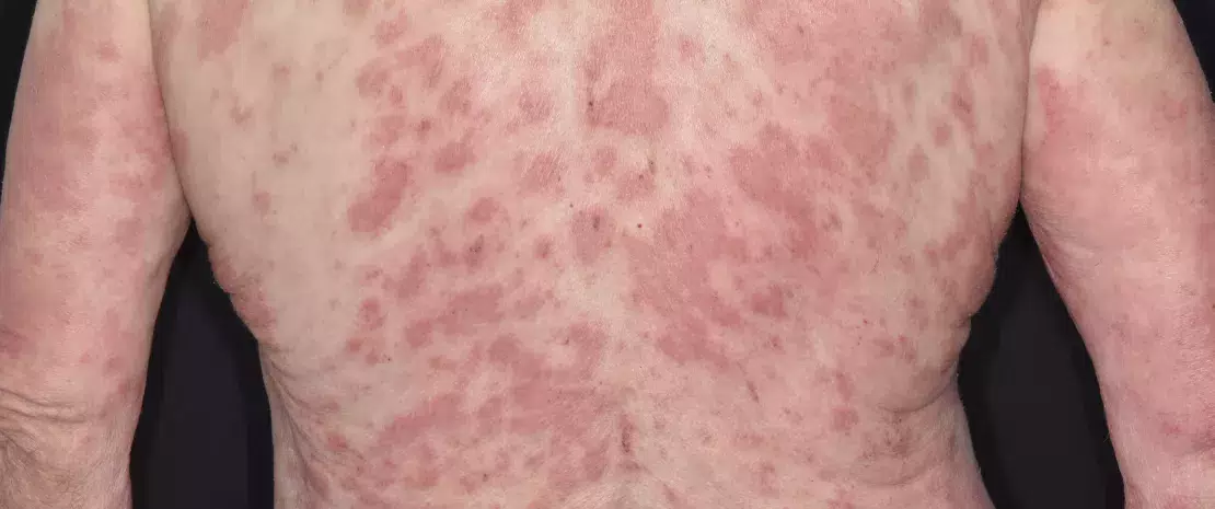
Section of the large intestine with Ulcerative Colitis. Illustration.

Commentary on the original publication by H yams et al. (Lancet 2019) [1]

Due to a lack of evidence-based data on therapeutic efficacy, there is uncertainty in the choice of treatment regimens in children who are newly diagnosed with ulcerative colitis (UC). This is why the authors hypothesized that clinical, transcriptomic, and microbial factors prior to treatment could predict the course of the disease. This inception cohort study recruited patients aged 4 to 17 years with newly diagnosed ulcerative colitis in 29 sites in the United States and Canada. Patients initially received mesalazine or corticosteroids with a pre-established protocol for escalation to immunomodulators (thiopurines) or biologic therapy with anti-TNF. RNA sequencing was used to define rectal gene expression before treatment, and 16S sequencing to characterize rectal and fecal microbiota. The primary outcome was corticosteroid-free remission at week 52 with no therapy other than mesalazine.

What do we already know about this subject?

Clinical, biological and/or endoscopic scores such as the PUCAI (pediatric ulcerative colitis activity index), are used to classify the severity of pediatric UC. Disease severity can be scored as mild (PUCAI 10-30), moderate to severe (35-60) or severe/fulminant (≥ 65). 5-aminosalycilates may be effective in mild forms, while corticosteroids are used in moderate disease, although some children will become steroid-dependent or refractory and require escalation of therapy to immunomodulators or anti-TNFα.

However, there is no prospective study evaluating the response to standardized therapy in newly diagnosed UC.
Image

What are the main insights from this study? 

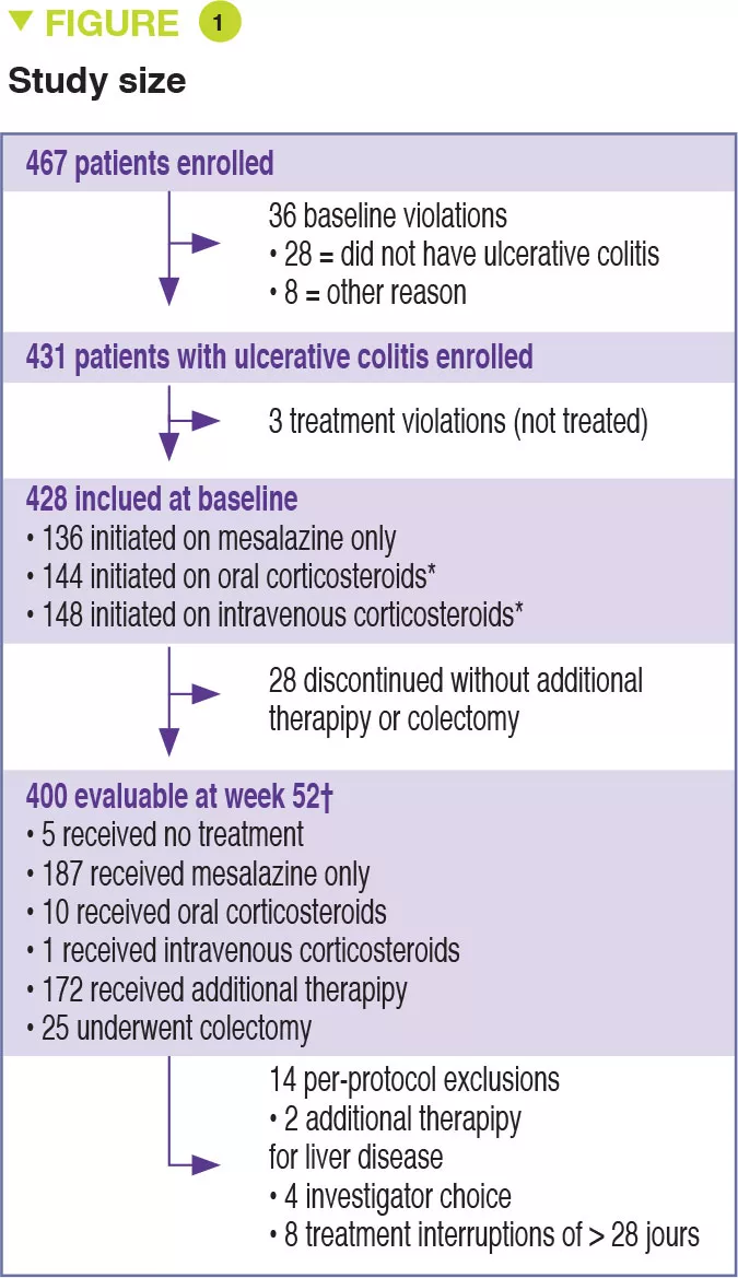
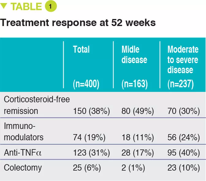
Between 2012 and 2015, 467 children aged 4 to 17 years were recruited into this prospective multicenter study conducted in 29 sites in the United States and Canada (Figure 1). The primary outcome was week 52 remission, defined by a PUCAI score < 10, with no therapy other than mesalazine (no corticosteroids and no colectomy). Baseline severity was defined as mild (therapy with mesalazine or oral corticosteroids with PUCAI < 45), or moderate to severe (oral corticosteroids with PUCAI ≥ 45 or intravenous corticosteroids). In addition to the usual clinical and biological parameters, this study also assessed gene expression from rectal biopsies as well as the rectal and fecal microbiota before treatment. 428 children started therapy: mean age was 12.7 years, 50% female, 42% with mild disease (mean PUCAI 31.9 ± 12.1 SD) and 58% with moderate to severe disease (mean PUCAI 62.9 ± 13.2 SD). At week 52, 150 (38%) of 400 evaluable participants achieved corticosteroid-free remission, of whom 80 (49%) had mild disease and 70 (30%) had moderate to severe disease (Table 1).

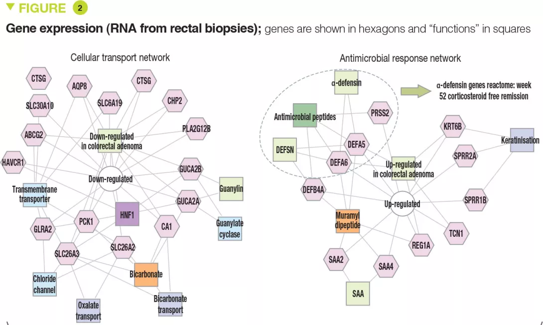
The clinical, biological, and endoscopic parameters that were associated with corticosteroid- free remission were validated in an independent prospective cohort of 307 children. It should be noted that corticosteroid- free remission was achieved by week 16 for moderate to severe disease (after which it can no longer be achieved) whereas it could be obtained up to week 52 in mild forms. Moreover, even patients with very severe disease were able to achieve week 52 corticosteroid-free remission (41/133 [31%] with PUCAI ≥ 65) and, conversely, some patients with mild disease were receiving anti-TNFα at week 52 (13/90 [14%] with PUCAI < 35). The authors identified 33 genes that were differently expressed between patients with moderate to severe disease who achieved corticosteroid-free remission (n = 51) or not (n = 101). Among these genes, 18 genes associated with cellular transport and ion channels were up-regulated and 15 genes involved in the antimicrobial response were down-regulated (Figure 2). The antimicrobial α-defensin signaling pathway showed the strongest negative correlation with week 52 corticosteroid-free remission. However, all 33 genes were negatively associated with the need for therapeutic escalation to anti-TNFα.
Image

Key points

  • The therapeutic decision depends on UC severity (PUCAI, Mayo scores) but also on treatment response by 4 weeks.

  • Other criteria (Hb, rectal eosinophil count, 25OH vitamin D) should also be taken into consideration.

  • Incorporating new parameters (gene expression and microbiota) into treatment decisions could facilitate the development of personalized medicine in the future.
Image

What are the consequences in practice? 

It is important to keep in mind the predictors of corticosteroid-free remission and therapeutic escalation as determined from multivariate analyses.


Predictors of week 52 corticosteroid-free remission were:

  • PUCAI < 45, Hb ≥ 10 g/dL;
  • remission by 4 weeks;
  • low expression of antimicrobial genes;
  • increased relative abundance for Ruminococcaceae and decreased for Sutterella.

Predictors of escalation to anti-TNFα therapy were:

  • total Mayo score ≥ 11;
  • eosinophil count in rectal biopsies < 32 per field at high magnification;
  • 25OH vitamin D < 20 ng/mL; - Hb < 10 g/dL;
  • no remission by 4 weeks;
  • decrease in genes involved in transport and antimicrobial genes;
  • decreased relative abundance of Oscillospira.

Conclusion

This study highlights the parameters that should be taken into consideration to guide the choice of therapy in children who are newly diagnosed with ulcerative colitis. Analysis of rectal gene expression and microbiota could both help predict the response to treatment and identify new therapeutic targets.

Summary
Off
Sidebar
Off
Migrated content
Désactivé
Updated content
Désactivé
Hide image
Off
Commented article

Antibiotic resistance: the lung microbiota pays a heavy price

Broad-spectrum antibiotics used for treating lung infections are regarded as one of the principal contributors to the overall burden of antibiotic resistance.

The pulmonary microbiota

Historically, the lungs of healthy individuals were considered sterile; the description of the LRT microbiota (Lower Respiratory Tract, from larynx to alveoli of the lungs)1 is a recent achievement.2,3 Along with viral and fungal communities, six bacterial phyla dominate a healthy lung microbiota: Firmicutes, Bacteroidetes, Fusobacteria, Proteobacteria, Acidobacteria, and Actinobacteria.1,2,4

“In Western populations, the treatment of lung infections is a primary driver of antibiotic resistance.4

Antibiotics saved millions of lives but their misuse or oversuse now raises serious concerns for health, notably with the further emergence of antimicrobial resistance. Each year, the the World Health Organization (WHO) organizes the World AMR Awareness Week (WAAW) to increase awareness of this public health issue. Let’s take a look at this global threat that required urgent action:

Microbiota at the forefront of antibiotic resistance

The largescale and sometimes inappropriate use of antibiotics is making them in…

A loss of diversity in the lung microbiota

Microbial dysbiosis is observed in a range of respiratory disorders, including lung infection, asthma, chronic obstructive pulmonary disease (COPD) and cystic fibrosis (CF).5,6 But only few studies have explored the direct effects of antibiotics on lung microbiota. Recent investigation has shown that azithromycin treatment decreased bacterial diversity in patients with persistent uncontrolled asthma;1 however clinical benefits are still controversial.1,7,8 In COPD patients, azithromycin treatment lowered alpha diversity;1 in those suffering from CF, antibiotics appear to be the primary drivers of decreased airway microbiota diversity.5

The gut-lung axis

Respiratory diseases, chronic lung disorders and microbial infections are often accompanied by intestinal symptoms.12 Indeed, the intestinal ecosystem undergoes change during the course of several lung diseases.12 While the underling mechanism remains unclear, reciprocal influence between the gut and the lungs could, in part, explain why antibiotic-induced dysbiosis of the gut microbiota in early-life may be a risk factor for subsequent allergic rhinitis and asthma.1,12

The plague of broad-spectrum antibiotics

While the misuse of antibiotics is known to lead to the emergence and selection of resistant bacteria, antibiotic prophylaxis, without a microbial diagnosis, is still widely used to treat lung infections.4 Of the 12 antibiotic-resistant ‘priority pathogens’ listed by the WHO, 4 affect lungs: Pseudomonas aeruginosa, Streptococcus pneumoniae, Haemophilus influenzae and Streptococcus aureus.4,9 There is agreement among the scientific community as a key route to minimize antimicrobial resistance that the disease management of lung infections needs to be improved.4,10,11

Promoting research, raising awareness

  • The Global Alliance Against Respiratory Diseases (GARD), launched by the WHO in 2006 to help combat chronic respiratory diseases, asserts: “Physicians worldwide now face situations in which infected patients cannot be treated adequately because the responsible bacterium is totally resistant to available antibiotics.11

  • At the European level, the ERS (European Respiratory Society) is involved in promoting scientific research, providing access to resources and raising awareness among the public and political decision makers. “Science, education and advocacy are at the core of everything we do.” Its latest monograph, ‘The lung microbiome’,13 reviews the different components of the respiratory microbiome, examines how diseases (asthma, COPD, cancer…) emerge and discusses new developments and therapies.

What is the World AMR Awareness Week?

Each year, since 2015, the WHO organizes the World AMR Awareness Week (WAAW), which aims to increase awareness of global antimicrobial resistance. Held on 18-24 November, this campaign encourages the general public, healthcare professionals and decision-makers to use antimicrobials carefully, to prevent the further emergence of antimicrobial resistance.

Sources

Hufnagl K, Pali-Schöll I, Roth-Walter F, et al. Dysbiosis of the gut and lung microbiome has a role in asthma. Semin Immunopathol. 2020;42(1):75-93.

Barcik W, Boutin RCT, Sokolowska M, et al. The Role of Lung and Gut Microbiota in the Pathology of Asthma. Immunity. 2020;52(2):241-255.

Mathieu E, Escribano-Vazquez U, Descamps D, et al. Paradigms of Lung Microbiota Functions in Health and Disease, Particularly, in Asthma. Front Physiol. 2018;9:1168. Published 2018 Aug 21.

Cookson WOCM, Cox MJ, Moffatt MF. New opportunities for managing acute and chronic lung infections. Nat Rev Microbiol. 2018;16(2):111-120.

Zhao J, Schloss PD, Kalikin LM, et al. Decade-long bacterial community dynamics in cystic fibrosis airways. Proc Natl Acad Sci U S A. 2012;109(15):5809-5814.

Chung KF, Huffnagle GB, Huang YJ. The lung microbiome in obstructive airways disease: potential pathogenetic roles. In: Cox MJ, Ege MJ, von Mutius E, eds. The Lung Microbiome 2019 (ERS Monograph). Sheffield, European.

Gibson PG, Yang IA, Upham JW, et al. Efficacy of azithromycin in severe asthma from the AMAZES randomised trial. ERJ Open Res. 2019;5(4):00056-2019. Published 2019 Dec 23.

Chung KF. Airway microbial dysbiosis in asthmatic patients: A target for prevention and treatment?. J Allergy Clin Immunol. 2017;139(4):1071- 1081.

WHO. Global priority list of antibiotic-resistant bacteria to guide research, discovery, and development of new antibiotics. 25 february 2017

10 Lim WS, Baudouin SV, George RC, et al. BTS guidelines for the management of community acquired pneumonia in adults: update 2009 Thorax 2009;64:iii1-iii55.

11 Forum of International Respiratory Societies. The Global Impact of Respiratory Disease – Second Edition. Sheffield, European Respiratory Society, 2017.

12 Marsland BJ, Trompette A, Gollwitzer ES. The Gut-Lung Axis in Respiratory Disease. Ann Am Thorac Soc. 2015;12 Suppl 2:S150-S156.

13 The Lung Microbiome. Edited by Cox MJ, Ege MJ, and von Mutius E. 2019. Monograph of European Respiratory Society.

Summary
Off
Sidebar
On
Migrated content
Activé
Updated content
Désactivé
Old content type
pro_dossiers_article
Hide image
Off
Dossier detail Pulmonology Gastroenterology Pediatrics

Parkinson’s disease: discovery and inhibition of levodopa metabolism by gut bacteria

Commented articles - Adult's section

By Pr. Harry Sokol
Gastroenterology and Nutrition Department, Saint-Antoine Hospital, Paris, France

Levodopa (L-DOPA) Parkinson's disease drug, molecular model. L-DOPA is a precursor of the catecholamine neurotransmitters dopamine, norepinephrine and epinephrine and is used in the treatment of Parkinson's disease. Atoms are represented as wires. A map of electrostatic potential surrounds the molecule.

Commentary on the original publication by Rekdal et al. (Science 2019)

The human gut microbiota metabolizes the Parkinson’s disease medication Levodopa (L-dopa), potentially reducing drug availability and causing side effects. However, the organisms, genes, and enzymes responsible for this activity in patients and their susceptibility to inhibition by host-targeted drugs are unknown. Here, the authors describe an interspecies pathway for gut bacterial L-dopa metabolism. Conversion of L-dopa to dopamine by a pyridoxal phosphate-dependent tyrosine decarboxylase from Enterococcus faecalis is followed by transformation of dopamine to m-tyramine by a molybdenum-dependent dehydroxylase from Eggerthella lenta. These enzymes predict drug metabolism in complex human gut microbiotas. Although a drug that targets host aromatic amino acid decarboxylase does not prevent gut microbial L-dopa decarboxylation, the authors identified a compound that inhibits this activity in Parkinson’s patients microbiotas and increases L-dopa bioavailability in mice[1].

What do we already know about this subject?

Parkinson’s disease is a debilitating neurological condition affecting more than 1% of the global population aged 60 and above. The primary medication used to treat Parkinson’s disease is levodopa (L-dopa). [2] To be effective, L-dopa must enter the brain and be converted to the neurotransmitter dopamine by the human enzyme aromatic amino acid decarboxylase (AADC). However, the gastrointestinal tract is also a major site for L-dopa decarboxylation, and this metabolism is problematic because dopamine generated in the periphery cannot cross the blood-brain barrier and causes unwanted side effects. Thus, L-dopa is co-administered with drugs that block peripheral metabolism, including the AADC inhibitor carbidopa. Even with these drugs, up to 56% of L-dopa fails to reach the brain. Moreover, the efficacy and side effects of L-dopa treatment are extremely heterogeneous across Parkinson’s patients, and this variability cannot be completely explained by differences in host metabolism. Previous studies in humans and animal models have demonstrated that the gut microbiota can metabolize L-dopa.[3] The major proposed pathway involves an initial decarboxylation of L-dopa to dopamine, followed by conversion of dopamine to m-tyramine by a dehydroxylation reaction.

Although these metabolic activities were shown to occur in complex gut microbiota samples, the specific organisms, genes, and enzymes responsible were unknown. The effects of host-targeted inhibitors such as carbidopa on gut microbial L-dopa metabolism were also unclear. As a first step toward understanding the gut microbiota’s effect on Parkinson’s disease therapy, the authors sought to elucidate the molecular basis for gut microbial L-dopa and dopamine metabolism.

Image

Key points

  • Some gut bacteria can metabolize L-dopa to dopamine and then to m-tyramine, limiting its availability in the brain.

  • The gut microbiota plays a role in the efficacy and toxicity of L-dopa treatment for Parkinson’s disease.

  • Use of a specific inhibitor of bacterial L-dopa metabolism can increase L-dopa bioavailability and thus enhance its efficacy

What are the main insights from this study? 

The authors hypothesized that L-dopa decarboxylation would require a pyridoxal phosphate (PLP)-dependent enzyme. They searched gut bacterial genomes and identified a conserved tyrosine decarboxylase (TyrDC) in Enterococcus faecalis (Figure 1A). Genetic and biochemical experiments revealed that TyrDC simultaneously decarboxylates both L-dopa and its preferred substrate, tyrosine. Next, they used enrichment culturing to isolate a dopamine- dehydroxylating strain of Eggerthella lenta (Figure 1A). Transcriptomics analysis showed that the enzyme responsible for this activity is a molybdenum cofactor-dependent dopamine dehydroxylase (Dadh). Unexpectedly, the presence of this enzyme in gut bacterial genomes did not correlate with dopamine metabolism. Instead, it is a single-nucleotide polymorphism (SNP) in the dadh gene that predicts activity. L-dopa metabolism through this pathway was variable across subjects (Figure 1B). In gut microbiotas from Parkinson’s patients, the abundance of E. faecalis, TyrDC, and the SNPs of dadh correlated with L-dopa and dopamine metabolism, confirming their relevance. The authors then showed that the human AADC inhibitor carbidopa had only a minimal effect on L-dopa decarboxylation by E. faecalis, and was completely ineffective in complex gut microbiotas from patients, suggesting that this drug likely does not prevent microbial L-dopa metabolism in vivo. Given TyrDC’s preference for tyrosine, the authors examined tyrosine “mimics” and identified (S)-α-fluoromethyltyrosine (AFMT) as a selective inhibitor of gut bacterial L-dopa decarboxylation. Co-administering AFMT with L-dopa and carbidopa to mice colonized with E. faecalis increased the serum concentration of L-dopa.

What are the consequences in practice?

These findings show that the gut microbiota can metabolize L-dopa and thereby influence the effectiveness and side effects of this drug. This study paves the way towards the discovery of predictive biomarkers for L-dopa efficacy and toxicity. Furthermore, since the underlying molecular mechanisms are known, the use of specific inhibitors of gut microbial L-dopa metabolism may be possible in patients whose microbiota contains bacteria with deleterious activities.

Conclusion

The gut microbiota in some Parkinson’s patients can metabolize L-dopa. This may underlie the heterogeneous efficacy and side effects of this treatment in Parkinson’s disease. The use of inhibitors of this bacterial metabolism could offer a solution. More generally, this study provides new evidence for the role of the gut microbiota in drug pharmacokinetics and pharmacodynamics. It opens up promising prospects for a field that could be called “pharmacomicrobiomics”.

Summary
Off
Sidebar
Off
Migrated content
Désactivé
Updated content
Désactivé
Hide image
Off
Commented article

Ears, Nose and Throat microbiota: when antibiotics challenge our first line of defense

By disrupting the microbiota in the ears, nose and throat (ENT), antibiotics may leave the door open to opportunistic pathogens implicated in ear and respiratory infections. Their effects could be particularly counterproductive in cases of acute otitis media.

The ENT microbiota A new generation of antibacterial agents? A plasmid capable of killing pathogenic bacteria Can statins combat intestinal dysbiosis? Antibiotic exposure during first six years of life disrupts gut microbiota and impairs child growth

What is commonly referred to as the “ears, nose and throat (ENT) microbiota” is in fact comprised not of one but rather of several microbiota. Antibiotics are likely to act individually on these different microbiota, ranging over the oral cavity through to the pharynx, including inside the sinuses and even the middle ear. This chapter is mainly devoted to the effects of antibiotics on the Upper Respiratory Tract (URT) microbiota, which is an excellent textbook case: the URT microbiota appears to be one of the safeguards of auricular health, yet it is threatened by antibiotics prescribed for this purpose, notably in cases of acute otitis media.

“Within 7 days of antibiotics being administered for URT infections, the incidence of acute otitis media has been shown to increase by a factor of 2.6.”

Pr. Teissier, MD, PhD

The URT microbiota, an ally of aricular health?

The URT microbiota is colonized directly after birth by a variety of commensals (Dolosigranulum Corynebacterium, Staphylococcus, Moraxella, Streptococcus). Mounting evidence suggests that a higher relative abundance of commensal species (Dolosigranulum spp. and Corynebacterium spp.) as well as a greater diversity in the nasopharyngeal microbiota1 are associated with a lower incidence of URT colonization by Streptococcus pneumoniae, Haemophilus influenzae and Moraxella catarrhalis,2,3 three otopathogens implicated in acute otitis media (AOM) .


Antibiotic treatment: much risk for little benefit

Exposure to antibiotics impacts the URT microbiota by decreasing the abundance of protective species and by increasing the abundance of Gram-negative bacteria (Burkholderia spp., Enterobacteriaceae, Comamonadaceae, Bradyrhizobiaceae),4,5 as well as S. pneumoniaeH. influenzae and M. catarrhalis.5 As a result of acquiring antimicrobial resistance, these bacteria, which could not otherwise successfully compete in this niche, are given the opportunity to multiply during treatment to an extent that they may become pathogenic.6 Furthermore, antibiotics are considered unlikely to confer any benefit in most cases of pediatric AOM (the primary reason for prescribing antibiotics to children7) and other URT infections (sore throats or common colds),7,8 due to the frequently non-bacterial nature of these conditions: from 60% to 90% of children with a AOM recover without antibiotics.9,10 Finally, antibiotics lead to gut microbiota dysbiosis that can translate into side effects such as antibiotic-associated diarrhea3,11 (see gut microbiota section).

Expert opinion

In flora hitherto unexposed to antibiotic treatment, there is a harmonious balance between the various commensal bacteria. Disrupting this balance with antibiotics can promote the proliferation of certain bacteria, likely to become pathogenic. In particular, the repeated intake of antibiotics promotes the selection of multidrugresistant bacteria that can no longer be kept in check by the commensal flora, which leads to the more frequent occurrence of infectious complications. It therefore seems essential to preserve the native flora and its natural balance by limiting the use of antibiotics to situations where they are strictly necessary.

Pr. Natacha TEISSIER, MD, PhD Pediatric ENT Department, Robert Debre Hospital, Paris (France)
Sources

Xu Q, Gill S, Xu L, et al. Comparative Analysis of Microbiome in Nasopharynx and Middle Ear in Young Children With Acute Otitis Media. Front Genet. 2019;10:1176.

Laufer AS, Metlay JP, Gent JF, et al. Microbial communities of the upper respiratory tract and otitis media in children. mBio. 2011;2(1):e00245-10.

Pettigrew MM, Laufer AS, Gent JF, et al. Upper respiratory tract microbial communities, acute otitis media pathogens, and antibiotic use in healthy and sick children. Appl Environ Microbiol. 2012;78(17):6262-6270.

Prevaes SM, de Winter-de Groot KM, Janssens HM, et al. Development of the Nasopharyngeal Microbiota in Infants with Cystic Fibrosis. Am J Respir Crit Care Med. 2016;193(5):504-15.

Teo SM, Mok D, Pham K, et al. The infant nasopharyngeal microbiome impacts severity of lower respiratory infection and risk of asthma development. Cell Host Microbe. 2015;17(5):704-715.

Rogers GB, Shaw D, Marsh RL, et al. Respiratory microbiota: addressing clinical questions, informing clinical practice. Thorax. 2015;70(1):74-81.

Mather MW, Drinnan M, Perry JD et al. A systematic review and meta-analysis of antimicrobial resistance in paediatric acute otitis media. Int J Pediatr Otorhinolaryngol. 2019;123:102-109.

Easton G, Saxena S. Antibiotic prescribing for upper respiratory tract infections in children: how can we improve? London J Prim Care (Abingdon). 2010;3(1):37-41.

Massa HM, Cripps AW, Lehmann D. Otitis media: viruses, bacteria, biofi lms and vaccines. Med J Aust. 2009;191(S9):S44-9.

10 Venekamp RP, Sanders SL, Glasziou PP, et al. Antibiotics for acute otitis media in children. Cochrane Database Syst Rev. 2015;2015(6):CD000219.

11 McFarland LV, Ozen M, Dinleyici EC, et al. Comparison of pediatric and adult antibiotic-associated diarrhea and Clostridium diffi cile infections. World J Gastroenterol. 2016;22(11):3078-3104.

Summary
Off
Sidebar
On
Migrated content
Activé
Updated content
Désactivé
Old sources


Old content type
pro_dossiers_article
Hide image
Off
Dossier detail Otorhinolaryngology

Antibiotics, a double-edged sword when managing skin disease

The effects of antibiotics on the skin microbiota have been studied mainly in the context of acne treatment. They may lead to several adverse outcomes including microbiota disruption, bacterial resistance and a risk of further infections hitting the skin or other body sites.

The skin microbiota Antibiotics and risk of IBD in adults What if manipulating the microbiota could improve the response to immunotherapy? A new generation of antibacterial agents? A plasmid capable of killing pathogenic bacteria

Long regarded mainly as a source of infection, the human skin microbiota is nowadays commonly accepted as an important driver of health and wellbeing.1 By promoting immune responses and defense, it plays a key role in tissue repair and barrier functions by inhibiting colonization or infection by opportunistic pathogens.2


To each skin site, its own microbiota

The skin microbiota harbors millions of bacteria, as well as fungi and viruses in lower relative abundances. Corynebacterium, Cutibacterium (formerly known as Propionibacterium), Staphylococcus, Micrococcus, Actinomyces, Streptococcus and Prevotella are the most common genera of bacteria encountered on the human skin.3 However, the relative abundance of bacterial taxa greatly depends on the local microenvironment of the particular piece of skin being considered, and especially on its physiological characteristics, i.e., whether it is sebaceous, moist or dry. Hence lipophilic Cutibacterium species dominate sebaceous sites while Staphylococcus and Corynebacterium species are particularly abundant in moist areas.4


From physiology to pathology, the ambivalent role of C. acnes

The aerotolerant anaerobe C. acnes is one of the most abundant bacterial species in the skin microbiota. It has been implicated in acne, a chronic inflammatory disorder of the skin with complex pathogenesis.5 In contrast with previous thinking, recent studies indicate that C. acnes hyperproliferation is not the only factor implicated in the development of acne.6 In fact, a loss of balance between the different C. acnes strains, together with a dysbiosis of the skin microbiota will trigger acne.6 Moreover, interactions between S. epidermidis and C. acnes are of critical importance in the regulation of skin homeostasis: S. epidermidis inhibits C. acnes growth and skin inflammation. In turn, C. acnes, by secreting propionic acid which participates, among other things, in maintaining the pilosebaceous follicle acidic pH, inhibits the development of S. epidermidisMalassezia, the most abundant skin fungus is also thought to play a role in refractory acne by recruiting immune cells, though its involvement needs to be further explored.6

Antibiotics in atopic dermatitis: friend or foe?

In atopic dermatitis (AD), patients display skin microbiota dysbiosis characterized by an overgrowth of Staphylococcus aureus, which is thought to play a decisive role in the manifestation of AD.14 Though antibiotic treatments have not demonstrated any efficacy in managing AD15 and though they are liable to induce bacterial resistance and result in a deleterious impact on skin commensals,14,16 they are nevertheless commonly used.

Acne treatment, an important source of antibiotic resistance

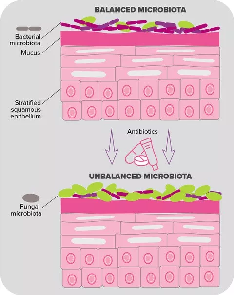
Despite being used routinely to treat acne, topical and oral antibiotics have proved to be problematic in several ways. A first concern expressed by experts is the disruption to the skin microbiota, although precise data on the subject remain scarce. In this vein, a recent longitudinal study compared the cheek microbiota of 20 acne patients before and after six weeks of oral doxycycline therapy. Interestingly, antibiotic exposure was associated with an increase in bacterial diversity; according to the authors, this could be due to a diminished colonization by C. acnes, which would liberate space to allow the growth of other bacteria.7

Dermatologists prescribe more antibiotics than any other specialists. Two thirds of these prescriptions are for acne.8

However, the most significant concern over the use of antibiotics for acne treatment relates to bacterial resistance. First observed in the 1970s, it has been a major worry in dermatology since the 1980s.8 C. acnes resistance is by far the most documented: the latest data point to resistance rates reaching over 50% for erythromycin in some countries, 82-100% for azithromycin and 90% for clindamycin. As for tetracyclines, although still largely effective against the majority of C. acnes strains, their resistance rates are rising, ranging from 2% to 30% in different geographic regions.9 And antibiotic resistance is not limited to C. acnes: while topical antibiotics used by acne patients (especially as monotherapy) have been shown to increase the emergence of resistant skin bacteria such as S. epidermidis, oral antibiotics have been associated with the increased emergence of antibiotic-resistant oropharyngeal S. pyogenes.8,10 In addition, increased rates of upper respiratory tract infection and pharyngitis have been reported as being associated with the antibiotic treatment of acne.11,12

Expert opinion

Antibiotics kill sensitive skin bacteria (Cutibacterium acnes), while concurrently leading to “holes” in the microbiota, which resistant bacteria will fill. This results in cutaneous dysbiosis and the overexpression of multidrug-resistant bacteria. 60% of patients treated for acne have macrolide-resistant C. acnes strains, and 90% of Staphylococcus epidermidis strains are also resistant to macrolides. The use of antibiotics can also have consequences in orthopedic surgery, where many macrolide-resistant strains of C. acnes are similarly observed. During an operation (a hip prosthesis, for example), there is a risk of causing an abscess. This will be all the more difficult to treat, as this bacterium secretes biofilms that adhere to the prosthesis. It is therefore essential, if promoting the selection of resistant bacteria is to be avoided, that the use of topical antibiotics be limited as far as possible (a maximum course of 8 days).

Pr. BRIGITTE DRÉNO, MD, PhD. Chairman of the department of Dermato-Oncology Director of the GMP Unit of Cell & Gene Therapy in University Hospital Nantes (France) Vice Dean of the Faculty of Medicine, Nantes

A call for a limited use of antibiotics in acne

The potential consequences of antibiotic resistance triggered by acne treatment are numerous: failure of the acne treatment itself (see clinical case), infection by opportunistic pathogens (locally or systemically), and the dissemination of resistance among the population.8 Despite this, the levels of antibiotic prescriptions for acne remain high and for longer durations than recommended in the guidelines.13 Against this background of mounting concerns, experts are calling for a more limited use of antibiotics in the treatment of acne.13 In particular, a strategy has been proposed in this regard by the Global Alliance to Improve Outcomes in Acne (see box below).

Strategies from the Global Alliance to Improve Outcomes in Acne to reduce antibiotic resistance in Cutibacterium acnes and other bacteria

First line therapy5

  • Combine topical retinoid with antimicrobial (oral or tropical)

If addition of antibiotic is needed:

  • Limit to short periods; discontinue when only slight or no further improvement

  • Oral antibiotics should ideally be used for 3 months

  • Coprescribe benzyl peroxide-containing product or use as washout

  • Do not use as monotherapy • Avoid concurrent use of oral and topical antibiotics

  • Do not switch antibiotics without adequate justification

Maintenance therapy

  • Use topical retinoids, with benzoyl peroxide added if needed 

  • Avoid antibiotics


Clinical case

by Pr. Brigitte Dreno, MD, PhD

  • A teenager consulted his dermatologist for facial acne (forehead, chin, and cheeks). He received a topical erythromycin-based treatment.

  • 4 to 5 weeks after starting treatment, a new proliferation of papules and pustules appeared on his face. He went back to his doctor, who prescribed oral erythromycin.

  • 1 month later, the patient returned to see his doctor because his acne had extended to his neck (profuse impetigo). The doctor took a sample from one of the pustules for a culture test.

  • The culture test came back positive for Staphylococcus, and the antibiogram indicated a resistance to macrolides. The doctor prescribed benzoyl peroxide, which gave remission within 10 days.

Sources

Egert M, Simmering R, Riedel CU. The Association of the Skin Microbiota With Health, Immunity, and Disease. Clin Pharmacol Ther. 2017;102(1):62-69.

Flowers L, Grice EA. The Skin Microbiota: Balancing Risk and Reward. Cell Host Microbe. 2020;28(2):190-200.

Ederveen THA, Smits JPH, Boekhorst J, et al. Skin microbiota in health and disease: From sequencing to biology. J Dermatol. 2020;47(10):1110-1118.

Byrd AL, Belkaid Y, Segre JA. The human skin microbiome. Nat Rev Microbiol. 2018;16(3):143-155.

Walsh TR, Efthimiou J, Dréno B. Systematic review of antibiotic resistance in acne: an increasing topical and oral threat. Lancet Infect Dis. 2016;16(3):e23-e33.

Dréno B, Dagnelie MA, Khammari A, et al. The Skin Microbiome: A New Actor in Inflammatory Acne. Am J Clin Dermatol. 2020 Sep 10.

Park SY, Kim HS, Lee SH, et al. Characterization and Analysis of the Skin Microbiota in Acne: Impact of Systemic Antibiotics. J Clin Med. 2020;9(1):168.

8. Karadag AS, Aslan Kayıran M, Wu CY, et al. Antibiotic resistance in acne: changes, consequences and concerns. J Eur Acad Dermatol Venereol. 2020;10.1111/jdv.16686.

9. Xu H, Li H. Acne, the Skin Microbiome, and Antibiotic Treatment. Am J Clin Dermatol. 2019;20(3):335-344.

10. Del Rosso JQ, Gallo RL, Thiboutot D, et al. Status Report from the Scientific Panel on Antibiotic Use in Dermatology of the American Acne and Rosacea Society: Part 2: Perspectives on Antibiotic Use and the Microbiome and Review of Microbiologic Effects of Selected Specific Therapeutic Agents Commonly Used by Dermatologists. J Clin Aesthet Dermatol. 2016;9(5):11-17.

11. Margolis DJ, Fanelli M, Kupperman E, et al. Association of pharyngitis with oral antibiotic use for the treatment of acne: a cross-sectional and prospective cohort study. Arch Dermatol. 2012;148(3):326-332.

12. Margolis DJ, Bowe WP, Hoff stad O, et al. Antibiotic treatment of acne may be associated with upper respiratory tract infections. Arch Dermatol. 2005;141(9):1132-1136.

13. Barbieri JS, Spaccarelli N, Margolis DJ, et al. Approaches to limit systemic antibiotic use in acne: Systemic alternatives, emerging topical therapies, dietary modifi cation, and laser and light-based treatments. J Am Acad Dermatol. 2019;80(2):538-549.

14. Wan P, Chen J. A Calm, Dispassionate Look at Skin Microbiota in Atopic Dermatitis: An Integrative Literature Review. Dermatol Ther (Heidelb). 2020;10(1):53-61.

15. George SM, Karanovic S, Harrison DA et al. Interventions to reduce Staphylococcus aureus in the management of eczema. Cochrane Database Syst Rev. 2019 Oct 29;2019(10):CD003871.

16. Seite S, Bieber T. Barrier function and microbiotic dysbiosis in atopic dermatitis. Clin Cosmet Investig Dermatol. 2015;8:479-483.

Summary
Off
Sidebar
On
Migrated content
Activé
Updated content
Désactivé
Old sources


Old content type
pro_dossiers_article
Hide image
Off
Dossier detail

Microbiota and metabolic diseases

Overview

By Pr. Yolanda Sanz
Microbial Ecology, Nutrition & Health Research Unit; Institute of Agrochemistry and Food Technology; National Research Council (IATA-CSIC), Valencia, Spain

Human digestive system microbiota, 3D illustration.

Obesity is one of the greatest public health challenges of the 21st century because of its high prevalence and its role in the development of multiple non-communicable diseases (metabolic syndrome -MetS- and type 2 diabetes mellitus -T2DM). Evidence of the role of gut microbiota alterations, partly due to unhealthy diets, in the mechanisms linking obesity to inflammation and metabolic dysfunction opens new opportunities for a better understanding of the disease aetiology and for designing management strategies. The investigated paths for disease management include faecal microbiota transplants (FMT), dietary ingredients intended to nourish our beneficial microorganisms (like prebiotic fibres) and indigenous bacteria (known as probiotics) to replenish our gut with missing beneficial microorganisms. Evidence is promising but work is still needed to identify constellations of effector intestinal bacteria that help in reprograming and preventing obesity, and to personalize diets to optimize metabolic functions of our gut microbes.

Obesity, metabolic syndrome and diabetes 

We are witnesses of an obesity epidemic worldwide that no country has yet been able to reverse. The overall obesity prevalence has tripled in the US and many EU countries since the 1980s, becoming one of the greatest public health challenges of the 21st century. In fact, obesity shows high comorbidity rates, constituting a major risk factor for the development of multiple non-communicable diseases. Obesity-induced insulin resistance is considered a key causal factor of MetS, which often progress to pancreatic β cell failure that finally triggers T2DM onset.[1]


Metabolic inflammation: the path from obesity to chronic comorbidities

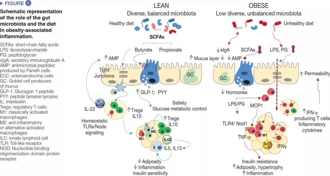
Currently, it is well-documented that the chronic inflammatory state associated with obesity and causally linked to metabolic complications affects the adipose tissue and other organs, including the brain, muscle, liver, pancreas and gut, which show different particularities.[1, 2] Specifically, the involvement of the intestinal immune system and the microbes that expand under the exposure to unhealthy diets have recently emerged as additional drivers of obesity-associated metabolic inflammation and could also represent therapeutic targets.[2, 3]

Image

How is the intestinal microbiota involved? 

The involvement of the gut microbiota in obesity has been partly inferred from observational studies reporting dysbiosis in obese subjects compared to lean ones in cross-sectional assessments. Evidence of gut microbiota changes during dietary, medicinal or surgery interventions intended for weight loss and for improving metabolic complications allowed establishing similar relationships where obesity was associated with reductions in species diversity and increases in bacterial taxa like Proteobacteria (enterobacteria) and Bilophila wadsworthia. By contrast, healthy metabolic phenotypes were often associated with increases in the phyla Bacteroidetes or Bacteroidetes/ Firmicutes ratio or the genera BacteroidesPrevotellaAkkermansiaFaecalibacterium or Christensenella.[4, 5] However, findings were not fully consistent across studies partly due to the heterogeneity of the studies and limitations of their designs. Further meta-analyses have indicated that the only biomarker that could be generalized for obesity was reduced bacterial species diversity.[6] It is also likely that all obese subjects cannot be categorized by the same dysbiotic pattern, particularly considering the high interindividual variability of the microbiota and complexity of the metabolic phenotypes (obesity with and without other complications). More recently, the gut microbiota alterations that precede the development of obesity have been identified as causally involved in the aetiology. Of note it is a recent longitudinal study showing that a reduced bacterial species diversity, linked to unhealthy dietary habits, provides a scenario favouring the overgrowth of Proteobacteria (enterobacteria), which precedes the development of overweight during a 4-year follow-up in children.[7]

More definitive proof of a causal role of the microbiota in defining the metabolic phenotype of the subject has been achieved through FMT, where the dysbiotic microbiota from diseased subjects was transferred to new animal recipients. Most of these experiments showed that FMT was sufficient to replicate the metabolic phenotype of the donor (lean or obese).[8]

The most consistent microbiota based biomarker associated with obesity is a reduced diversity of bacterial species, which may also predispose to the development of obesity, chronic inflammation and metabolic complications.

Microbiota-mediated mechanisms of action 

Gut microbes influence energy metabolism through their capacity to increase the human ability to metabolize nutrients and extract calories from the diet as well as regulating the absorption of sugars and lipids and their deposition in peripheral tissues [8]. Gut microbes and their metabolic products are also involved in the regulation of the enteroendocrine system, for example, via the production of short-chain fatty acids, which induced the synthesis of intestinal hormones (e.g., GLP-1, PYY) that act via endocrine and neural routes regulating appetite, food intake and glucose metabolism [9]. Moreover, the gut microbiota is a major regulator of the gut barrier and the immune system, whose alterations are implicated in obesity-associated low grade inflammation and insulin resistance as detailed below and schematized in (Figure 1).[2,3]

It is likely that a unique pattern of intestinal dysbiosis for obesity cannot be identified and may depend on the underlying metabolic complications and other biological and environmental features of the individual.

Defective mucosal barrier function in obesity 

Unhealthy diets cause defects in the intestinal mucosal barrier affecting its penetrability and favouring the translocation of bacterial components, such as the bacterial lipopolysaccharide (LPS) and the peptidoglycan or even whole microorganisms, which may activate innate immunity in metabolically active organs. The defective intestinal mucosal barrier has been attributed to local inflammation caused by diets rich in saturated fat and the diet-induced dysbiosis as well as to associated disturbances in the mucus layer [10] and in antimicrobial peptides production by Paneth cells (Reg3γ, lysozyme 1) [11]. For example, increased LPS plasma levels (known as “metabolic endotoxemia”) was shown to cause obesity and metabolic dysfunction in animal models and to be associated with an elevated body mass index, HF feeding, postprandial inflammation and risk of T2DM in humans. This could be favoured by the overgrowth of Gramnegative bacteria like enterobacteria, which are a source of LPS under HF feeding. LPS could activate innate immunity in the gut and beyond and induce the recruitment of inflammatory immune cells in metabolic tissues, like macrophages. Diets rich in saturated fat may also promote the growth of other Gram negative bacteria like Bilophila wadsworthia, which generates hydrogen sulphide, a toxic metabolite for enterocytes leading to a leaky gut, inflammation and metabolic dysfunction.[12] Finally, HF diet (HFD) may also increase circulating peptidoglycans, likely through the diet-induced changes in the expression of the antimicrobial peptide lysozyme 1 that hydrolyse components of the bacterial cell-walls. Depending on the peptidoglycan type, these could act as Nod1 ligands of pro-inflammatory macrophages of the adipose tissue or liver causing insulin resistance, while opposite effects seem to occur in pancreas beta cells, possibly as a counterbalance mechanism.[13]

Dysregulation of intestinal immunocompetent cells in obesity

Similar to other metabolic organs, including the adipose tissue and liver, breakdown of immune homeostasis has been observed in the intestine during obesity. In diet-induced obesity diverse subsets of innate and adaptive immune cells within the gut adopt a pro-inflammatory phenotype, primarily demonstrated by increases in proinflammatory macrophages and cytokines (IFNγ).

In parallel, there are reductions in proportions of Treg cells and type 3 ILCs producing IL-22, which help to maintain mucosal integrity and intestinal homeostasis in lean subjects.[2,3] Some of these alterations are reversed through microbiota depletion (e.g., antibiotic treatment) or the administration of, for example, specific bifidobacteria that also restore diet-induced intestinal dysbiosis in obesity, supporting a causal role of the gut microbiota in metabolic inflammation.[3] Also, intestinal IgA+ immune cells act as mucosal mediators of whole-body glucose regulation in HFD-induced obesity. The HFD reduces the number of IgA+ immune cells and secretory IgA.

The reduction of IgA could add another level of destabilization in the bacterial community to that caused by HFD, linked to increases in gut permeability and adipose tissue inflammation.[14]

How microbiota modulation could impact the disease evolution 

FMT

A clinical trial showed that insulin sensitivity could be improved in humans with MetS 6 weeks after being transplanted with gut microbiota from a lean healthy donor.[5] An increase in microbial diversity and abundance of bacteria producing butyrate was also observed. Another trial conducted in subjects with indexes of MetS receiving the microbiota of responders to bariatric surgery reported changes in the expression of dopamine receptors, which could account for a better control of food intake, but did not confirm effects on insulin resistance. Many other trials to evaluate the effects of FMT on obesity have been registered, but the outcomes have not been published yet.[5] Therefore, scientific evidence supporting the use of this strategy to tackle obesity complications is still very limited.

Intestinal dysbiosis sustained by unhealthy diets contributes to the dysregulation of the intestinal immune system, which constitutes an additional driver of obesity-associated metabolic inflammation and also represents a therapeutic target.

Dietary fibres

Consumption of diets with fibre intake above current recommendations (25 g/day for adults) improves weight maintenance and reduces the risk of coronary heart disease and T2DM, according to a large body of evidence from humans. Some of the effects of dietary fibre are due to its physicochemical properties (e.g., indigestibility, viscosity, etc.), which contribute to reducing the glycemic responses and the energy intake, and improving the blood lipid profile. Other effects could be mediated by their impact on the subject’s gut microbiota, which ferments fibres generating metabolites such short chain fatty acids (SCFAs: butyrate, propionate, etc.) with an active role in the host’s metabolism. SCFAs induce the production of enteroendocrine peptides (GLP-1, GLP-2, PYY) that strengthen the gut barrier, induce satiety, improve glucose metabolism and exert anti-inflammatory effects in obesity. Fibre intake also increases gut microbiota diversity which is associated with a healthy metabolic phenotype. Also, beneficial effects of fibres depend not only on the type and amount of fibre, but also on the person’s microbiota structure, its diversity and the presence or absence of specific bacterial species involved in their utilization.[15] Altogether this points to the need of progressing towards more personalized dietary recommendations.[15]

Beneficial bacteria

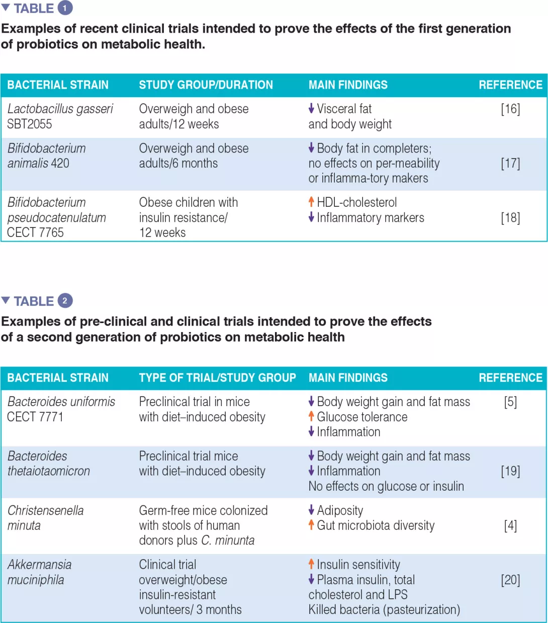
The first and second generation of probiotics: The majority of clinical efficacy trials have been conducted with bacterial strains traditionally used as probiotics for humans, “the first generation of probiotics”, which essentially include Lactobacillus and Bifidobacterium. Table 1 summarizes some examples, although much more can be found in literature, with positive and negative outcomes. A few attempts have also been made to isolate new bacterial species of the human indigenous microbiota, consistently associated with a healthy metabolic phenotype, with a view to create a “second generation of probiotics”. These may help us to enlarge our potential to replenish the gut microbiota with missing microbes. Table 2 summarizes some of the studies conducted in animal models (preclinical trials) and the only one conducted in humans so far.

Image

Conclusion

Scientific evidence confirms the role of gut microbiota alterations, partly due to unhealthy diets, in obesity and metabolic complications. Intestinal inflammation is emerging as a driver of systemic inflammation in obesity. This process is modulated by the gut microbiota and the diet, which altogether represent both a cause and a therapeutic target. Experimental trials provided proof-of-concept that a “healthy microbiota”, specific intestinal bacteria or dietary fibres that potentiate their functions could play a role in obesity management. To make further progresses towards the development of tangible solutions for obesity, further efforts are still needed to identify the effector intestinal bacteria that cooperate in reprogramming obesity and to personalize diets in order to optimize gut microbiota functions in support of our metabolic health.

Sources

1 Johnson AM, Olefsky JM. The origins and drivers of insulin resistance. Cell 2013 ; 152 : 673-84.

2 Winer DA, Luck H, Tsai S, et al. The intestinal immune system in obesity and insulin resistance. Cell Metab 2016 ; 23 : 413-26.

3 Sanz Y, Moya-Pérez A. Microbiota, inflammation and obesity. Adv Exp Med Biol 2014 ; 817 : 291-317.

4 Goodrich JK, Waters JL, Poole AC, et al. Human genetics shape the gut microbiome. Cell 2014 ; 159 : 789-99.

5 Romaní-Pérez M, Agusti A, Sanz Y. Innovation in microbiome-based strategies for promoting metabolic health. Curr Opin Clin Nutr Metab Care 2017 ; 20 : 484-491.

6 Sze MA, Schloss PD. Looking for a Signal in the Noise: Revisiting Obesity and the Microbiome. MBio 2016 ; 7: e01018-16.

7 Rampelli S, Guenther K, Turroni S, et al. Pre-obese children’s dysbiotic gut microbiome and unhealthy diets may predict the development of obesity. Commun Biol 2018 ; 1 : 222.

8 Sonnenburg JL, Bäckhed F. Diet-microbiota interactions as moderators of human metabolism. Nature 2016 ; 535 : 56-64.1.

9 Mulders RJ, de Git KCG, Schéle E, et al.. Microbiota in obesity: interactions with enteroendocrine, immune and central nervous systems. Obes Rev 2018 ; 19 : 435-51.

10 Schroeder BO, Birchenough GMH, Ståhlman M, et al. Bifidobacteria or fiber protects against diet-induced microbiota-mediated colonic mucus deterioration. Cell Host Microbe 2018 ; 23 : 27-40.e7.

11 Everard A, Geurts L, Caesar R, et al. Intestinal epithelial MyD88 is a sensor switching host metabolism towards obesity according to nutritional status. Nat Commun 2014 ; 5 : 5648.

12 David LA, Maurice CF, Carmody RN, et al. Diet rapidly and reproducibly alters the human gut microbiome. Nature 2014 ; 505 : 559-63.

13 Stojanović O, Trajkovski M. Microbiota guides insulin trafficking in beta cells. Cell Res 2019 ; 29 : 603-4.

14 Luck H, Khan S, Kim JH, et al. Gut-associated IgA(+) immune cells regulate obesity-related insulin resistance. Nat Commun 2019 ; 10 : 3650.

15 Sanz Y, Romaní-Perez M, Benítez-Páez A, et al. Towards microbiome-informed dietary recommendations for promoting metabolic and mental health: Opinion papers of the MyNewGut project. Clin Nutr 2018 ; 37(6 Pt A) : 2191-7.

16 Kadooka Y, Sato M, Imaizumi K, et al. Regulation of abdominal adiposity by probiotics (Lactobacillus gasseri SBT2055) in adults with obese tendencies in a randomized controlled trial. Eur J Clin Nutr 2010 ; 64 : 636-43.

17 Stenman LK, Lehtinen MJ, Meland N, et al. Probiotic with or without fiber controls body fat mass, associated with serum zonulin, in overweight and obese adults-randomized controlled Trial. EBioMedicine 2016 ; 13 : 190-200.

18 Sanchis- Chordà J, Del Pulgar EMG, Carrasco-Luna J, et alBifidobacterium pseudocatenulatum CECT 7765 supplementation improves inflammatory status in insulin-resistant obese children. Eur J Nutr. 2019 Oct;58(7):2789-2800. 

19 Liu R, Hong J, Xu X, et al. Gut microbiome and serum metabolome alterations in obesity and after weight-loss intervention. Nat Med 2017 ; 23 : 859-68.

20 Depommier C, Everard A, Druart C, et al. Supplementation with Akkermansia muciniphila in overweight and obese human volunteers: a proof-of-concept exploratory study. Nat Med 2019 ; 25 : 1096-103.

Summary
Off
Sidebar
Off
Migrated content
Désactivé
Updated content
Désactivé
Hide image
Off
Article

Urogenital microbiota: the spectrum of mycosis or urinary tract infections after each antibiotic treatment

A vicious circle. Vaginal tract infections such as vulvovaginal candidiasis often appear after antibiotic therapy, and sometimes following the administration of antibiotics commonly used to treat those same infections. The situation is no better for urinary tract infections: antibiotics typically used to treat them have become a risk factor for their occurrence.

The urinary microbiota What are the long-term effects of antibiotics on the gut microbiota? Role of antibiotics and microbiota in parkinson's disease Is fecal transplant a solution to prevent antibiotic resistance in immunocompromised patients?

Historically, until recent scientific work, urine was regarded as sterile. Compared to other microbiota, this ecosystem has low biomass.1 While a consensus regarding the precise composition has yet to be reached, around 100 species have been identified from 4 principal phyla (Proteobacteria, Firmicutes, Actinobacteria, and Bacteroidetes).2 And though the role of the urinary microbiota is currently a matter of debate, it is well understood that diminished diversity seems to be a risk factor for urinary tract infections.

10 to 30 %

After antibiotic treatment, 10 to 30 % of women develop vulvovaginal candidiasis5

Yet, the vaginal microbiota gains on the other hand from having low diversity and being largely dominated by lactobacilli.3 Despite considerable variability among women, 5 community state types (CST) have been described in the vaginal flora: 4 dominated by one or several species of the Lactobacillus genus (L. crispatus, L. gasseri, L. iners or L. jensenii) and one polymicrobial.4 In both cases, dysbiosis following antibiotic treatment may increase the risk of infection.5

A spectrum of fungus at each antibiotic treatment 

This is what many women being treated with antibiotics dread: developing post-antibiotic vulvovaginal candidiasis. This anxiety is more than justified: antibacterial therapy, whether systemic or applied locally to the vagina, is thought to be one of the main factors leading to vulvovaginal candidiasis.5 This infection may be associated with vaginal microbiota disruption together with Candida yeast proliferation (C. albicans in the majority of cases ; Figure 3). The most common clinical signs of this infection are vulvar pruritus, a burning sensation accompanied by vaginal pain or irritation that may lead to dyspareunia or dysuria.6


FIGURE 3. Yeast proliferation induced by antibiotics exposure

Image

Urinary tract infections: what to prescribe?

According to the 2017 update of the German clinical guidelines on managing uncomplicated urinary tract infections in adult patients:9

  • “For the treatment of acute uncomplicated cystitis (AUC), fosfomycin-trometamol, nitrofurantoin, nitroxoline, pivmecillinam, and trimethoprim (depending on the local rate of resistance) are all equally recommended. Cotrimoxazole, fluoroquinolones, and cephalosporins are not recommended as antibiotics of first choice, due to concerns over the possibility of an unfavorable impact on the microbiome.

  • For AUC with mild to moderate symptoms, instead of antibiotics, symptomatic treatment alone may be considered depending on the patient’s preference after discussing possible adverse events and outcomes.

  • Primarily non-antibiotic options are recommended for prophylaxis of recurrent urinary tract infections.”

The vicious circle of bacterial vaginosis

Though the etiology of bacterial vaginosis (BV), the main form of vaginal infection, remains unclear, it is believed that antibiotic-induced dysbiosis could be partly responsible for its development: dominant lactobacilli are supplanted by polymicrobial flora derived from numerous bacterial genera (Gardnerella, Atopobium, Prevotella, etc.). A vicious circle could be initiated: though antibiotics can be used to treat BV, they are also, alongside sexual history, vaginal douching, contraceptive use, age, stage in the menstrual cycle, tobacco use, etc., among the numerous risk factors associated with this type of infection.7

Urinary microbiota: a textbook case of antibiotic resistance 

Urinary tract infections (UTI) affect millions of men (a 3% annual incidence rate in the US) and women (10%) every year.8 Recurrent UTIs contribute greatly to this incidence: notwithstanding their receiving appropriate antibiotic therapy, more than 30% of women will experience a subsequent infection within the following 12 months.8 UTIs are becoming increasingly difficult to treat because of the rapid spread of drug resistance among Gram-Negative organisms, notably UPEC (uropathogenic Escherichia coli) which cause approximately 80% of UTIs.8

Paradoxically, broadspectrum antibiotics used to treat both community acquired and hospital-associated UTIs have become a risk factor for their occurrence.8 Mechanisms involving both gut and vaginal microbiota are suspected: in the gut, the ultimate reservoir for UPEC, antibiotic exposure increases inflammation and promotes the proliferation of E. coli; in the vagina, they diminish colonization by Lactobacillus species that suppress vaginal UPEC invasion and subsequent bacterial ascension from the vagina into the urinary tract. This is the reason why, experts nowadays recommend that they should be used with caution and that microbiota-sparing treatments should be developed.8

Expert opinion 

Urinary tract infections are closely linked to imbalances in any one of three microbiota: the urinary microbiota, since urine is not sterile; the vaginal microbiota, with which the urinary microbiota shares many similarities; and the gut microbiota, from where the pathogens involved in urinary tract infections originate (e.g. E. coli, which passes from the anus to the vulvar vestibule and then to the bladder).

Dr. JEAN-MARC BOHBOT, MD, PhD. Andrologist and Infectious Diseases Specialist, Institut Alfred Fournier, Paris (France)

Clinical case

by Dr. Jean-Marc Bohbot, MD, PhD

  • 18-year-old Solène consults for recurrent vulvo-vaginal candidiasis. For about 3 months, she suffers from recurrent candidiasis (2 episodes per month) with abundant white leucorrhea and intense vulvo-vaginal pruritus. These episodes have a very negative impact on her daily life, not to mention her sex life.

  • A vaginal sample confirmed the presence of Candida albicans with an intermediate vaginal microbiota (Nugent score 6). Solène has a regular partner who experiences no symptoms. She is not diabetic. The candidiasis appeared a few weeks after starting a daily antibiotic treatment (cyclines) for acne. These antibiotics promote vaginal dysbiosis and facilitate the development of fungi.

  • After consultation with the dermatologist, oral cyclines were replaced by a local treatment; the candidiasis disappeared within 2 weeks.

In cases of acne, the use of antibiotics should be limited or should be accompanied by probiotic cures to preserve the balance of the vaginal microbiota.

Sources

1 Neugent ML, Hulyalkar NV, Nguyen VH, et al. Advances in Understanding the Human Urinary Microbiome and Its Potential Role in Urinary Tract Infection. mBio. 2020 Apr 28;11(2):e00218-20.

2 Morand A, Cornu F, Dufour JC, et al. Human Bacterial Repertoire of the Urinary Tract: a Potential Paradigm Shift. J Clin Microbiol. 2019 Feb 27;57(3). pii: e00675-18.

3 Gupta S, Kakkar V, Bhushan I. et al. Crosstalk between Vaginal Microbiome and Female Health: A review. Microb Pathog. 2019 Aug 23;136:103696.

4 Greenbaum S, Greenbaum G, Moran-Gilad J, et al. Ecological dynamics of the vaginal microbiome in relation to health and disease. Am J Obstet Gynecol. 2019;220(4):324-335.

5 Shukla A, Sobel JD. Vulvovaginitis Caused by Candida Species Following Antibiotic Exposure. Curr Infect Dis Rep. 2019 Nov 9;21(11):44.

6 Gonçalves B, Ferreira C, Alves CT, et al. Vulvovaginal candidiasis: Epidemiology, microbiology and risk factors. Crit Rev Microbiol. 2016 Nov;42(6):905-27.

7 Coudray MS, Madhivanan P. Bacterial vaginosis-A brief synopsis of the literature. Eur J Obstet Gynecol Reprod Biol. 2019 Dec 24;245:143-148.

8 Klein RD, Hultgren SJ. Urinary tract infections: microbial pathogenesis, host-pathogen interactions and new treatment strategies. Nat Rev Microbiol. 2020;18(4):211-226.

9 Kranz J, Schmidt S, Lebert C, et al. The 2017 Update of the German Clinical Guideline on Epidemiology, Diagnostics, Therapy, Prevention, and Management of Uncomplicated Urinary Tract Infections in Adult Patients. Part II: Therapy and Prevention. Urol Int. 2018;100(3):271-278.

Summary
Off
Sidebar
On
Migrated content
Activé
Updated content
Désactivé
Old sources


Old content type
pro_dossiers_article
Hide image
Off
Dossier detail Urology

From diarrhea to chronic diseases: the well-documented consequences of antibiotic-related gut microbiota dysbiosis

Antibiotic treatment may sometimes take place without any obvious short-term side effects. Nevertheless, the dysbiosis triggers diarrhea for up to 35% of patients; in the long term, antibiotic-induced microbiota alterations may represent a risk factor for allergic, autoimmune or metabolic diseases.

The gut microbiota What are the long-term effects of antibiotics on the gut microbiota? Role of antibiotics and microbiota in parkinson's disease Is fecal transplant a solution to prevent antibiotic resistance in immunocompromised patients?
Clostridium difficile spores surrounding a long Cl. difficile bacterium.

Antibiotics are a powerful tool in the fight against bacterial infections. However, research has also documented detrimental effects on the trillions of commensal bacteria that live in the intestinal tract. This resultant dysbiosis renders the gut microbiota less able to fulfil its protective functions. In the short term, dysbiosis leaves the door open for opportunistic pathogens and the selection of multi-resistant bacteria. In the long term, the gut microbiota, despite having a certain degree of resilience, can sometimes fail to fully restore itself;1,2 this is understood to pave the wave to a range of diseases. Recent research has shown that antibiotics may alter the bacterial diversity and abundance of the normal microbiome and that this impact may be prolonged (typically 8-12 weeks after antibiotics have been discontinued).3,4

35%

Diarrhea occurs in up to 35% of patients who receive antibiotics3, 5, 6

Diarrhea, the most common adverse effect of antibiotics

As the main short-term consequence, some patients treated with antibiotics experience a change in their intestinal transit, most often resulting in diarrhea. The incidence of antibiotic-associated diarrhea (AAD) depends on several factors (age, setting, type of antibiotic, etc.) and may range between 5 and 35% of patients taking antibiotics.3,5,6

Expert opinion

Antibiotics disrupt the protective intestinal microbiota, which can lead to unintended consequences including antibiotic-associated diarrhea (in up to 35% of patients) and the development of antibiotic resistant strains of pathogens that are of global concern in regards to increased healthcare costs and mortality.

Lynne Mc Farland, PhD. Clinical Epidemiologist Public Health Reserve Corps University of Washington, Seattle

Among children this percentage can reach up to 80%.3 Most of the time, the diarrhea is purely functional, caused by the antibioticinduced dysbiosis. It is usually of mild intensity and is self-limiting, lasting 1-5 days. Antibiotics displaying a broader spectrum of antimicrobial activity like clindamycin, cephalosporins, and ampicillin/amoxicillin are associated with higher rates of diarrhea.6

Antibiotics are an extraordinary scientific discovery that saves millions of lives but their excessive and inappropriate use has now raised serious concerns for health, notably with antibiotic resistance and microbiota dysbiosis. Let’s take a look at this dedicated page:

The ambivalent role of antibiotics

By destroying the bacteria responsible for infection, antibiotics can also lead…

The particular case of C. difficile 

In 10 to 20% of cases, diarrhea results from infection with Clostridioides difficile (formerly known as Clostridium difficile) colonizing the microbiota.6 This bacterium, which persists in the environment via spores, is a gram-positive, spore-forming, obligate anaerobe. Infection occurs via spores ingestion. Under specific circumstances (e.g., antibiotic-induced dysbiosis), the spores may germinate and vegetative bacterial cells of this opportunistic pathogen may colonize the intestines. In the infective phase, C. difficile produces 2 toxins that damage the colonocytes and trigger an inflammatory response with a variety of clinical outlooks, ranging from moderate diarrhea to pseudomembranous colitis, toxic megacolon and/or death.


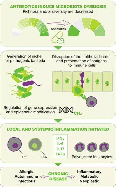
FIGURE 2. Downstream effects of antibiotic-induced gut dysbiosis. (source : adapted from Queen et al., 202010)

Image

1/3

Nearly 1/3 of AAD cases are due to C. difficile3

Most recognized common risk factors for C. difficile infection (CDI) include age > 65 years, use of proton pump inhibitors, comorbidities and of course antibiotic use. The latest is the most relevant modifiable risk factor for CDI. The association of antibiotics with CDI has been established in hospitals and more recently in community settings,7 where the risk of infection varies from intermediate for people exposed to penicillins, high for these exposed to fluoroquinolones and highest for those receiving clindamycin. As for tetracyclines, they trigger no increased risk.8 In a hospital setting, the highest risk of developing CDI was observed for cephalosporins (from 2nd to 4th generations), clindamycin, carbapenems, trimethoprim sulfonamide, fluoroquinolones and penicillin combinations.9

When the gut microbiota becomes a reservoir of antibiotic resistance 

When exposed to antibiotics, microbial communities respond in the short term not only by changing their composition, but also by evolving, optimizing and disseminating antibiotic resistant genes. The human gut microbiota overly exposed to antibiotics is now considered a significant reservoir of resistance genes, in adults as well as in children.2 By contributing to the growing difficulty to combat bacterial infections, antibiotic resistance has become a major public health concern.

An open door to non-communicable diseases

Disruption of the gut microbiota resulting from antibiotic exposure is also suspected of increasing the risk of several chronic diseases by elevating inflammatory responses locally and systemically, thereby leading to a deregulated metabolism and compromised immune homeostasis10 (Figure 2, page 4). The perinatal period, characterized by the development of the immune system along with the maturation of the gut microbiota, has been shown to be a particularly sensitive time, one during which antibiotic-driven dysbiosis translates into long-lasting health effects, i.e. a higher risk of diseases later in life, including inflammatory bowel diseases (e.g., Crohn’s disease), atopic diseases (e.g., asthma) and metabolic disorders (e.g., type 2 diabetes, obesity).

Clinical case

by Lynne V. McFarland, PhD

  • 53-year old woman consulted her physician with a 3-day history of respiratory tract symptoms (cough, sore throat and runny nose) with fever and fatigue. No co-morbidities and was otherwise healthy. Her physician prescribed a sputum sample and a 10-day course of oral cefaclor (500 mg, b.i.d). The sputum cultures came back negative for pathogens.

  • She was admitted at hospital on the 3rd day of the antibiotics because she developed acute diarrhea (with six watery stools per day and abdominal cramping) and unresolved respiratory symptoms. Laboratory cultures (sputum and stool) were negative for pathogens. She was asked to discontinue her antibiotics, but the diarrhea continued for the next two days.

  • Her physician prescribed erythromycin (500 mg, t.i.d.) and a probiotic for one week. Her respiratory symptoms and diarrhea resolved within four days and she was discharged one day later with no complications.

What is the World AMR Awareness Week?

Each year, since 2015, the WHO organizes the World AMR Awareness Week (WAAW), which aims to increase awareness of global antimicrobial resistance.
Held on 18-24 November, this campaign encourages the general public, healthcare professionals and decision-makers to use antimicrobials carefully, to prevent the further emergence of antimicrobial resistance.

Sources

1 Dethlefsen L, Relman DA. Incomplete recovery and individualized responses of the human distal gut microbiota to repeated antibiotic perturbation. Proc Natl Acad Sci U S A. 2011;108 Suppl 1(Suppl 1):4554-4561.

2 Francino MP. Antibiotics and the Human Gut Microbiome: Dysbioses and Accumulation of Resistances. Front Microbiol. 2016;6:1543.

3 McFarland LV, Ozen M, Dinleyici EC et al. Comparison of pediatric and adult antibiotic-associated diarrhea and Clostridium difficile infections. World J Gastroenterol. 2016;22(11):3078-3104.

4 Kabbani TA, Pallav K, Dowd SE et al. Prospective randomized controlled study on the effects of Saccharomyces boulardii CNCM I-745 and amoxicillin-clavulanate or the combination on the gut microbiota of healthy volunteers. Gut Microbes. 2017;8(1):17-32.

5 Bartlett JG. Clinical practice. Antibiotic-associated diarrhea. N Engl J Med 2002;346:334-9.

6 Theriot CM, Young VB. Interactions Between the Gastrointestinal Microbiome and Clostridium difficile. Annu Rev Microbiol. 2015;69:445-461.

7 Kuntz JL, Chrischilles EA, Pendergast JF et al. Incidence of and risk factors for community-associated Clostridium difficile infection: a nested casecontrol study. BMC Infect Dis. 2011;11:194.

8 Brown KA, Khanafer N, Daneman N et al. Meta-analysis of antibiotics and the risk of community-associated Clostridium difficile infection. Antimicrob Agents Chemother. 2013;57(5):2326-2332.

9 Slimings C, Riley TV. Antibiotics and hospital-acquired Clostridium difficile infection: update of systematic review and meta-analysis. J Antimicrob Chemother. 2014;69(4):881-891.

10 Queen J, Zhang J, Sears CL. Oral antibiotic use and chronic disease: long-term health impact beyond antimicrobial resistance and Clostridioides difficile. Gut Microbes. 2020;11(4):1092-1103.


Summary
Off
Sidebar
On
Migrated content
Activé
Updated content
Désactivé
Old content type
pro_dossiers_article
Hide image
Off
Dossier detail Pediatrics Gastroenterology

Modulating gut microbiota in metabolism disorders and alcoholic hepatitis

Literature selection

By Pr Markku Voutilainen
Turku University Faculty of Medicine; Turku University Hospital, Department of Gastroenterology, Turku, Finland

Alcoholic hepatitis. Light micrograph of a section through a liver affected by hepatitis (inflammation of the liver) caused by excessive alcohol intake.

Gut microbiome fermentation determines the efficacy of exercise for diabetes prevention

Liu Y, Wang Y, Ni Y, et al. Gut microbiome fermentation determines the efficacy of exercise for diabetes prevention. Cell Metabolism. 2020 Jan 7;31(1):77-91.e5. 

The impact of exercise on gut microbiota was examined in prediabetic men. Exercise responders had a decrease in fasting insulin and insulin resistance (HOMA-IR), whereas in non-responders they remained unchanged or even deteriorated. Exercise caused increased abundance of FirmicutesBacteroides, and Proteobacteria. Alterations of the gut microbiota correlated with the reduction of HOMA-IR. DNA synthesis, amino acid (AA) metabolism, and short chain fatty acid (SCFA) synthesis enhanced in responders. In non-responders, AA fermentation was shifted to production of colonic gases and detrimental compounds, which associate with increased insulin resistance. Increased serum short chain fatty acids, but decreased branched chain amino (BCAA) and aromatic amino acids were detected only in responders. SCFAs have a beneficial role in energy and glucose metabolism, whereas increased BCAAs associate with insulin resistance.

In conclusion, exercise responders’ gut microbiome had enhanced capacity to produce short chain fatty acids but increased breakdown of BCAAs, whereas the microbiome of non-responders produced metabolically detrimental compounds.

Bacteriophage targeting of gut bacterium attenuates alcoholic liver disease

Duan Y, Llorente C, Lang S, et al. Bacteriophage targeting of gut bacterium attenuates alcoholic liver disease. Nature. 2019 Nov;575(7783):505-511.

Alcoholic hepatitis (AH) patients have increased fecal Enterococcus faecalis (EF), 80% of AH patients are positive for EF. Germ-free mice on ethanol diet were colonized with cytolysin-positive (CL) EF feces of AH patients. Those infected with CL + feces developed a more severe ethanol-induced liver damage. Mice having overgrowth of intestinal enterococci and on ethanol diet were given bacteriophages lysing CL + EF. They developed less severe liver damage. Thus phage therapy may attenuate ethanol-related liver disease caused by CL + EF and improve prognosis in severe AH

Summary
Off
Sidebar
Off
Migrated content
Désactivé
Updated content
Désactivé
Hide image
Off
Press review

Skin microbiota : which role in atopic dermatitis and acne?

Literature selection

By Pr Markku Voutilainen
Turku University Faculty of Medicine; Turku University Hospital, Department of Gastroenterology, Turku, Finland

Atopic dermatitis on the upper body of an 83 year old male patient. Atopic dermatitis, also called atopic eczema, is an inflammatory skin reaction that occurs without any obvious external cause.

Microbe-host interplay in atopic dermatitis and psoriasis

Fyhrqvist N, Muihead G, Prat-Nielsen S, et al. Microbe-host interplay in atopic dermatitis and psoriasis. Nat Commun. 2019 Oct 16;10(1):4703. 

The authors compared atopic dermatitis (AD) and psoriasis (PSO) microbiota with that of healthy volunteers. The authors detected 26 and 24 microbes typical for AD and PSO, respectively. The most discriminative taxa for AD were genus Staphylococcus, and most discriminating microbes for PSO were Corynebacterium simulansNeisseriaceae g. spp., C. kroppenstedtii, Lactobacillus spp. and L. iners.

AD is characterized by S. aureus abundance. In PSO, many different bacteria such as Corynebacterium may be involved. The depletion of Lactobacillus is typical for both diseases. In AD, loss of strictly anaerobic bacteria is typical with diminished production of lactic and short chain fatty acids leading to increased skin pH. Microbe-host interactions are important both in skin homeostasis and disease pathogenesis.

Staphylococcus epidermidis: a potential new player in the physiopathology of acne?

Claudel JP, Affret N, Leccia MT, et alStaphylococcus epidermidis: a potential new player in the physiopathology of acne? Dermatology; 2019;235(4):287-294. 

The interplay between skin and cutaneous microbiota is essential to differentiate between commensal and pathogenic bacteria. During puberty, over-colonization of skin pilosebaceous units (PU) by Cutibacterium acnes (CUA) may cause acne. Some strains of S. epidermidis modulate host innate immune reactions, and some isolates have antimicrobial activity against CUA. Conversely, some CUA strains have antimicrobial activity against S. epidermidis which may also control CUA via succinic acid. The use of topical antibiotics may result in the development of antibiotic-resistant strains of CUA and S. epidermidis. Eliminating only CUA may lead to proliferation of S. aureus and S. epidermidis increasing the risk of infections. Lactobacillus may be efficient in acne and other inflammatory skin diseases. The authors suggest that regular oral or topical supplementation of skin microbiota could be treatment option in acne.

Summary
Off
Sidebar
Off
Migrated content
Désactivé
Updated content
Désactivé
Hide image
Off
Press review