Metagenomics of the faecal virome: cumulative effect of the quantity of gluten and enteroviruses risk of coeliac disease autoimmunity in predisposed children – the TEDDY study

Commented articles - Children's section 

By Pr. Emmanuel Mas
Gastroenterology and Nutrition Department, Children’s Hospital, Toulouse, France
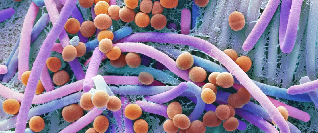
Faecal bacteria. Scanning electron micrograph (SEM) of bacteria cultured from a sample of human faeces.

Commentary on the original article of Lindfors et al. (Gut 2019) [1]

A higher gluten intake, frequent gastrointestinal infections and adenovirus, enterovirus, rotavirus and reovirus have all been proposed as environmental triggers of coeliac disease (CD). However it is not known whether an interaction exists between the quantity of gluten ingested and exposure to viruses in the development of CD. This study sought to determine whether distinct viral exposures, alone or associated with gluten, increased the risk of CD autoimmunity in genetically predisposed children. It was concluded that frequent exposure to enteroviruses between the ages of 1 and 2 years was indeed associated with increased risk of CD autoimmunity, indicating a cumulative effect of the interaction between enteroviruses and higher gluten intake.

What do we already know about this subject?

Coeliac disease is an autoimmune pathology which occurs in genetically predisposed individuals of genotype HLA DQ2 and/ or DQ8-positive. It is characterised by the presence of villous atrophy and lymphocyte infiltration of the epithelium of the small intestine. Gluten present in the diet induces an autoimmune response directed against tissue transglutaminase. The appearance of anti-transglutaminase antibodies (ATA) indicates the presence of coeliac disease autoimmunity

The rise in the incidence of autoimmune diseases has led to suspect that environmental factors may have a role in their pathogenesis. Observational studies suggest that viral infections could cause a loss of oral tolerance to gluten and the development of coeliac disease.

What are the main insights from this study?

It is a nested case-control study in the TEDDY (The Environmental Determinants of Diabetes in the Young) birth cohort which included 8,676 children before the age of 4 and a half months followed to the age of 15 years. The principal objective of this cohort was to identify the genetic and environmental factors associated with type 1 diabetes and coeliac disease. After forming pairs matched for family history of type 1 diabetes, sex and study inclusion site, 83 pairs (child with predisposition (case) and control) were retained in the final analysis for whom faecal virome data were available after introduction of gluten. Of these pairs, 16 had a family history of type 1 diabetes. During follow-up, 28 of the coeliac disease autoimmunity cases developed coeliac disease.

Stool samples were collected every month from the age of 3 months to 2 years; tests for enterovirus, adenovirus, astrovirus, norovirus, reovirus and rotavirus were performed. Every 3 months, a food questionnaire was used to collect information on breast-feeding and the age of introduction of gluten-containing foods. A 3-day record of food intake enabled a calculation of the quantities of gluten ingested at 6, 9, 12, 18 and 24 months.

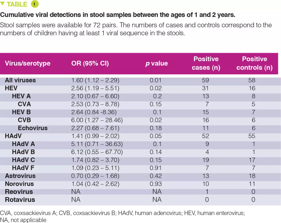
The percentage of stool samples positive for any virus fluctuated from 22 to 50%, without any age-related peak and for the enteroviruses this ranged from 0 to 21% after 6 months. Between 1 and 2 years, enteroviruses were detected in 31 cases versus 16 controls (Table 1). The cumulative number of stool samples positive for any virus was associated with an increased risk for coeliac disease autoimmunity (OR 1.60; p = 0.01), with a stronger association conferred by the enteroviruses (OR 2.56; p = 0.02).

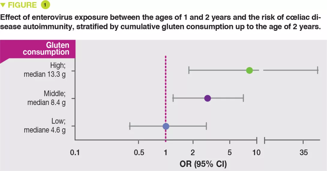
The risk of coeliac disease autoimmunity was not increased by viral infections occurring after the age of gluten introduction while breast-feeding was still continuing. In contrast, after weaning, in stool samples collected between the ages of 1 and 2 years after gluten introduction, both the cumulative number of viruses detected (OR 1.41; p = 0.05) and also the numbers of enteroviruses (OR 2.47; p = 0.03) were associated with the risk of coeliac disease autoimmunity. There was a significant interaction between the presence of enterovirus detected between 1 and 2 years and the quantity of gluten ingested up to the age of 2 years in the risk of coeliac disease autoimmunity (p = 0.03). It is suggested that this increases with the amounts of gluten ingested: high (OR 8.3), middle (OR 2.9) and low (OR 1.0) (Figure 1).

Key points

  • Environmental factors play a role in cœliac disease.

  • Enterovirus exposure is a risk factor for the development of anti-transglutaminase antibodies in HLA DQ2 and/or DQ8-positive children.

  • This risk is potentiated by the intake of large amounts of gluten in the diet.
Image
Image

What are the consequences in practice?

The results of this study indicate the value of preventing the appearance of coeliac disease auto-antibodies in children at risk. This could be done by carefully monitoring the amounts of gluten ingested, in particular in case of exposure to enteroviruses, especially when the child is no longer being breast fed.

Conclusion

This study demonstrated an association between gastro-intestinal exposure to enteroviruses and the risk of coeliac disease autoimmunity in genetically at-risk children. This risk increases as greater amounts of gluten are ingested.

Summary
Off
Sidebar
Off
Migrated content
Désactivé
Updated content
Désactivé
Hide image
Off
Commented article

The Mediterranean diet alters the gut microbiota in the elderly, reduces frailty and improves health: study NU-AGE

Commented articles - Adult's section

By Pr. Harry Sokol
Gastroenterology and Nutrition Department, Saint-Antoine Hospital, Paris, France

Bandeau_Article2_NL10

Commentary on the article of Ghosh et al. Gut 2020 [1]

Ageing is accompanied by a deterioration of many bodily functions and inflammation, which collectively contribute to frailty. It has already been shown by the authors and other teams that frailty is associated with cha nges in the gut microbiota, and more especially in the context of a poorly diversified diet. The Mediterranean diet is associated with good health. In this study the authors sought to determine if 12 months of Mediterranean diet, known to be associated with good health, could modify the gut microbiota, reduce frailty and improve cognitive function. The gut microbiota was profiled in non-frail or pre-frail subje cts in five European countries before and after the adoption for 1 year of a Me diterranean diet tailored for elderly subjects (NU-AGE diet). The results showed that it is feasible to improve the usual dietar y regime in order to modulate the gut microbiota, and thus promote healthier ageing.

What do we already know about this subject?

Frailty which accompanies ageing involves the failure of several physiological systems and constant activation of the innate inflammatory immune response. Frailty can include the development of chronic lowgrade inflammation, impaired cognitive function, sarcopenia and the development of chronic diseases such as diabetes and atherosclerosis. The modification of dietary regimens such as the adoption of a Mediterranean diet has been suggested as a key therapeutic strategy to combat frailty.[2] The Mediterranean diet is characterised by the consumption of larger amounts of vegetables, pulses, fruits, nuts, olive oil, fish and the consumption of smaller amounts of red meat, dairy products and saturated fats. The adhesion to this type of diet is associated with reduced mortality and increased anti-oxidant activity, as well as a reduction in the incidence of several diseases and inflammation.

Several studies have shown that the adoption of this diet is related to a reduction in frailty. Beyond the inverse relationship with disease, closer adhesion to a Mediterranean diet was associated with beneficial changes in the composition of the gut microbiota (reduction in proteobacterial abundance, increased production of short chain fatty acids [SCFAs]). As a general rule however, few elderly subjects follow this type of diet and a large number suffer because of a restricted diet associated with a low-diversity gut microbiota. Changing this is a major challenge, in particular concerning persons in care homes.

In previous studies the authors used bioinformatic analysis to identify specific microbial taxa which are gradually lost in the transition from a high-diversity microbiota of healthy subjects to a low-diversity microbiota of frail subjects. In a recent 6-month dietary intervention study in elderly individuals given supplementation with 5 prebiotics (up to 20 g/day), several microbial taxa were modified, but no change was noted in the overall diversity of the microbiota nor in the inflammatory markers. The authors therefore concluded that a more drastic dietary intervention was necessary. The dietary intervention NU-AGE project aimed to study the effect of administration of a personalised Mediterranean diet for 12 months in a large cohort of over 1,200 persons aged 65 to 79 years, distributed across five European countries. A significant relationship was observed between increased adherence to the Mediterranean diet and global cognitive capacity and improved episodic memory.[3]

Moreover, it was shown that greater adherence reduced the rate of bone loss in individuals with osteoporosis and improved innate immune function, blood pressure and arterial stiffness.[4-6] In the study described here, the authors analysed the gut microbiota of a sub-group of study subjects.

What are the main insights from this study? 

A total of 612 subjects were analysed (289 controls: 145 males, 144 females and 323 on a Mediterranean diet: 141 males, 182 females). At baseline, differences in terms of diet and microbiota were observed between the various countries. Relationships between the Mediterranean diet and the gut microbiota were revealed. Among the taxa associated with good adherence with the Mediterranean diet (DietPositive), we find an over-representation of species such as Faecalibacterium prausnitziiEubacterium and Roseburia, a majority of which are associated with good health (including the production of SCFAs and anti-inflammatory effects). Inversely, certain taxa are depleted in case of good adherence to this diet, some of which have been linked to type 2 diabetes, colorectal cancer, cirrhosis or chronic inflammatory bowel disease. Taken together, these results suggest that adherence to a Mediterranean diet can modulate the microbiota in a direction positively associated with health.

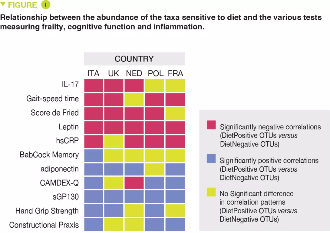
Lastly, the authors observed that the abundance of DietPositive taxa were negatively correlated with some inflammatory markers (high-sensitivity CRP (hsCRP) and IL-17), and with clinical scores associated with increased frailty (Fried scores, gait speed time). In contrast, the abundance of these taxa was positively correlated with the improvement in cognitive function (Constructional Praxis score, Babcock memory score) and reduced frailty (hand grip strength) and two anti-inflammatory markers (adiponectin and sGP130). The opposite trend was observed with DietNegative taxa (Figure 1). Analysis of the inferred microbial metabolite profiles indicated that the diet-modulated change in microbiota was associated with an increase in production of short/branched chain fatty acids and a lower production of secondary bile acids, p-cresols, ethanol and carbon dioxide.

Key points

  • Ageing is associated with a deterioration in numerous functions and inflammation which lead to frailty.

  • A Mediterranean diet induces an increase in the abundance of bacterial taxa which are associated with a reduction in frailty.

  • The role of an increase in the production of some bacterial metabolites, including SCFAs, is suggested in the positive effects of the diet.

What are the consequences in practice? 

These results confirm that dietary intervention is an effective means of improving health, at least partially, via a modulation of the gut microbiota. Of course we can recommend that elderly subjects adopt a Mediterranean diet, but the feasibility of this type of dietary intervention is questionable in the long-term. As this study has identified bacteria associated with the beneficial effects of the Mediterranean diet, it lays the groundwork for their use in the form of next-generation probiotics. This type of approach based on bacteria from the gut microbiota should be tested in this indication.

Image

Conclusion

This study highlights the complex interactions between diet, the gut microbiota and health. It suggests that the beneficial effects of a Mediterranean diet on the health of elderly subjects is due, at least in part, to a modulation of the gut microbiota.

Recommended by our community

"Good Science - worth a read..."  -@ethicos2013 (From Biocodex Microbiota Institute on X)

Summary
Off
Sidebar
Off
Migrated content
Désactivé
Updated content
Désactivé
Hide image
Off
Commented article

Gut microbiota and Clostridioides difficile

Overview

By Pr. Gianluca Ianiro
Digestive Disease Center, Fondazione Policlinico “A. Gemelli” IRCCS, Rome, Italy

 Illustration of the bacteria Clostridioides difficile.

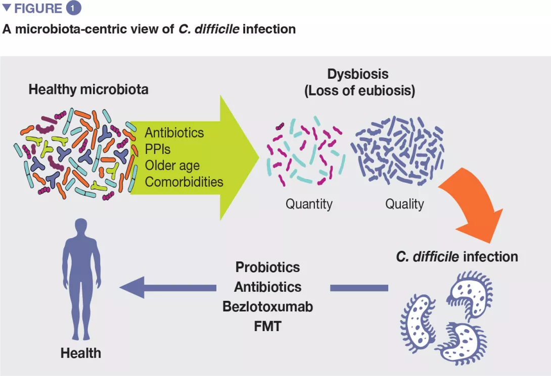
C. difficile infection (CDI) has become in recent years a clinical and socio economical burden worldwide, due to its increase in morbidity, severity, mortality, and likelihood to recur. There is a considerable involvement of gut microbiota in CDI, for many reasons. First, most risk factors associated with the development of CDI, including the overuse of broad-spectrum antibiotics or proton pump inhibitors, are associated with an imbalance of gut microbiota. Moreover, specific microbiota modulators are involved in the prevention (specific probiotics) or treatment (fecal microbiota transplantation) of CDI. In this paper, we will review epidemiology, risk factors, and approved therapies of CDI, with a microbiota-centric view.

Clostridioides difficile (C. difficile, previously called Clostridium difficile) is a gram-positive, spore-forming, obligate anaerobe. Spores allow C. difficile to persist in environments, and to be spread from infected subjects. Under specific circumstances (e.g., antibiotic-driven dysbiosis), spores are driven to germination in the large bowel, and present in a vegetative form that leads to clinical infection (Clostridium difficile infection [CDI]). In the infection phase, C. difficile produces two toxins, enterotoxin A and cytotoxin B that both cause damage to colonocytes and trigger the inflammatory response, leading to a variety of clinical pictures, from mild colitis to pseudomembranous colitis and toxic megacolon. [1]

In recent years, CDI has become a considerable healthcare and economical burden in most countries. Studies from the United States report an incidence of nearly 453,000 cases and of nearly 29,000 CDIrelated deaths in 2011, while the incidence in Europe is 124,000 cases/year, with nearly 3,700 deaths/year. Increased morbidity, hospitalization length and mortality, contribute to the considerable economic burden of CDI, which accounted for nearly $ 5 billions in the US in 2011, and for nearly € 3.7 billions in Europe in 2013. [2, 3] These figures show that the CDI incidence has risen worldwide, for several reasons. First, the increased use of antibiotics, which are a known as risk factors for CDI development. Furthermore, the spreading of specific ribotypes (mainly the virulent ribotype 027, but also the 017 in Asia, the 018 in Italy, the 17,621 in Eastern European countries, 24,422 in Oceania) has let CDI clusters develop. Additionally, there was also an increased number of diagnoses, due to the development of highly sensitive diagnostic tests (e.g., PCR), and the risen awareness of CDI among healthcare professionals. Overall, the main cause of the overall increase in CDI incidence appears to be the increased rate of recurrences. From 2001 to 2012, the annual incidence of recurrent CDI has increased by nearly 189%, while the increase in overall CDI incidence in the same time period was nearly 43%. [2] As recurrent infection is less likely than first episode to be cured by antibiotics, it is associated with longer hospitalization, increased morbidity and mortality too.

CDI is widely known to be the main cause of healthcare associated infectious diarrhoea, but recent evidence suggests that its diffusion in the community settings is growing. To date, nearly 25%-35% of CDI cases are acquired in community, probably due to several fecal-oral transmission pathways (e.g., zoonosis and food).

Despite this increase in diagnoses, the misdiagnosis/underdiagnosis of CDI is still relevant, as observed in the EUCLID study.
This finding suggests that a considerable number of patients with CDI is still not diagnosed, increasing the risk of disease diffusion.

Nosocomial CDI, a community-acquired CDI, appear to differ for several characteristics. First, nosocomial patients are more likely to present with a severe clinical picture, while community patients can even be asymptomatic carriers, increasing the risk of CDI spreading. Moreover, community- based CDI is known to spread also among patients without standard risk factors.

Risk factors for C. difficile infection

Although the exact pathogenic pathways of CDI are not yet clarified, several risk factors have been identified over time. [4] Their knowledge is relevant as the management of modifiable risk factors is a prevention measure against CDI. Most relevant risk factors include older age, use of antibiotics, proton pump inhibitors, and others (Figure 1).

Image
Antibiotics

If antibiotics remain today essential molecules in the therapeutic arsenal, it is also necessary to take into account their undesirable effects on the gut microbiota, as a considerable body of evidence supports the association between their use and many dysbiosis-associated diseases, including CDI.[5]

First, antibiotics may kill commensal bacteria that may have a direct action against C. difficile (by secreting a number of bacteriocins) and also compete with the pathogen for nutrients (e.g., sialic acid and succinate). Moreover, there is also an indirect protective role of commensal bacteria through the regulation of bile acids.

Recently, Clostridium scindens was associated with resistance to C. difficile colonisation. It has a bile acid inducible operon which is able to encode dehydroxylating enzymes that convert primary bile acids into secondary bile acids. Primary bile acids promote the germination of C. difficile spores, while secondary bile acids are able to inhibit this process.[6]

The use of systemic antibiotics is the most relevant modifiable risk factor for the development of CDI. Healthy gut microbiota can determine the successful colonisation of the large bowel by C. difficile or not, by direct and indirect pathways. In principle, the imbalance of healthy gut microbiota by broad-spectrum antibiotics may bring several consequences that drive to CDI.

As a corollary of this evidence, patients with recurrent CDI are known to have an imbalanced microbial profile, with higher relative abundance of detrimental bacterial families as Enterobacteriaceae and Veillonellaceae and lower relative abundance of beneficial families, including Ruminococcaceae, Bacteroidaceae and Lachnospiraceae.

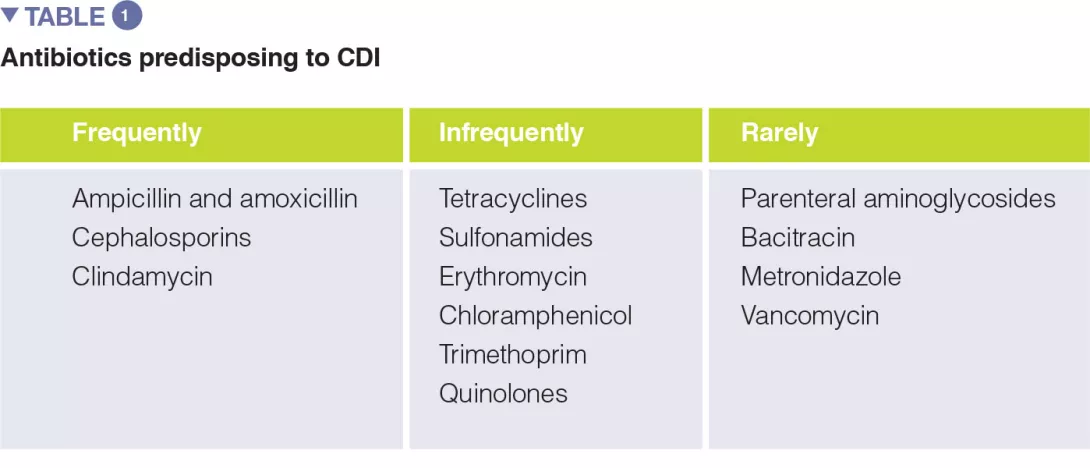
A number of systematic reviews, alone or with meta-analysis, have assessed the relevance of different antibiotic classes in CDI development. In the earliest meta-analysis (1998), antibiotics use was associated with a 6-fold increase in the risk of developing CDI, and the highest risk was observed for fluoroquinolones, clindamycin, cephalosporins. Moreover, the use of antibiotics was found to be an independent predictor of CDI recurrence (relative risk 1.76). One of the key factors to prevent CDI is represented by the antibiotic stewardship approach, so the knowledge of the CDI risk for different antibiotic classes is of paramount importance (Table 1).

The use of the following antibiotics is associated with a 2-fold higher risk of CDI among inpatients: clindamycin, cephalosporins, carbapenems, fluoroquinolones, trimethoprim, sulphonamides. In the community setting, respectively, antibiotics were found to have different risk levels for CDI development or recurrence, including: clindamycin (risk increased of 8 to 20 times), cephalosporins and fluoroquinolones (3-5 times increase), macrolides (2-3 times increase).[5]

Image
Gastric acid suppression
  • Proton pump inhibitors (PPIs) are largely used worldwide for several upper gastrointestinal disorders, including gastroesophageal reflux disease, hiatal ernia, gastritis, H. pylori infection (together with antibiotic eradication therapy), peptic ulcer disease.

  • Overall PPIs are considered safe drugs. However, a large body of evidence shows that the use of PPIs is significantly associated with the development of CDI.

  • In principle, PPIs can increase the risk of C. difficile colonization by several pathways, including reduced acid production that can lead to small intestinal bacterial overgrowth and dysbiosis, and increase of bile salts that can promote the germination of C. difficile spores. Finally, there is no clear evidence if increased gastric pH is a safer environment for spores. [6]

  • The clinical evidence of a significant association between PPIs and CDI comes from several systematic reviews and meta-analyses, with odds ratios ranging from 1.26 to 2.34, based on different reports (from 3 to 67, according to different meta-analyses).

  • Most evidence is heterogeneous and comes from observational cohorts, so potentially confounding factors, including other drugs and co-morbidities, could reduce the quality of this finding. However, the association between PPIs and CDI kept significant even after stratification for antibiotic use, both in cohort studies and in case-control reports.

  • The detrimental role of PPI was found to be stronger toward community-associated CDI, suggesting that there is a chronic overuse in communities rather then in hospitals.

  • Specifically, PPIs have been associated not only with CDI overall, but also with recurrent CDI by several meta-analyses (including from 3 to 16 studies), with odd ratios ranging from 1.52 to 2.51, although definitions of recurrence varied significantly among studies.

Advanced age 
  • Advanced age is one of the best known risk factors for primary CDI and recurrent CDI.

  • Established evidence shows that CDI rates are much higher in adults over 65 years than in younger population. In a meta-analysis of 33 studies, age older than 65 years was identified as an independent predictor of recurrent CDI (relative risk 1.63).

  • However, age is a considerable confounder, as the use of several drugs that promote CDI, such as antibiotics or PPIs, is more common in older age. Increasing evidence suggests that the microbiota of elderly patients is less healthy (in terms of reduced microbial diversity and increase of opportunistic species) than normal, supporting again the role for microbiota imbalance in CDI.[7]

Other disorders

The association between CDI and selected comorbidities has also been explored systematically. In a systematic review, significantly higher risk of CDI was found for inflammatory bowel disease (OR 3.72), kidney insufficiency (OR 2.64), hematologic malignancies (OR 1.75), and diabetes mellitus (OR 1.15). This was especially true for community-acquired CDI.[7]

Therapeutic management of CDI 

Conventional treatment of CDI

Traditionally, metronidazole and vancomycin have been the most common treatment options for CDI, being used both as first line options, while only vancomycin was recommended, as tapered or pulsed regimen, to treat recurrent disease.[8]

However, in recent years CDI has become more cumbersome to treat. In particular metronidazole was shown to achieve lower cure rates than vancomycin, so that vancomycin has been preferred to metronidazole also in primary infection. Overall, also vancomycin is losing its efficacy, and the rates of recurrent disease have grown. Morover, hypervirulent strains of C. difficile have emerged, specifically the ribotype 027, which is less responsive to standard antibiotic therapy and is associated with more severe clinical pictures [8]

In recent years fidaxomicin, a narrow spectrum antibiotic, was shown to be superior than vancomycin in treating CDI recurrences. However, its high costs and the recent evidence of its inferiority compared with fecal microbiota transplantation (FMT) in treating recurrent CDI are potential limitations to its widespread use.[9]

Antibiotics are an extraordinary scientific discovery that saves millions of lives but their excessive and inappropriate use has now raised serious concerns for health, notably with antibiotic resistance and microbiota dysbiosis. Let’s take a look at this dedicated page:

The ambivalent role of antibiotics

By destroying the bacteria responsible for infection, antibiotics can also lead…

What is the World AMR Awareness Week?

Each year, since 2015, the WHO organizes the World AMR Awareness Week (WAAW), which aims to increase awareness of global antimicrobial resistance.
Held on 18-24 November, this campaign encourages the general public, healthcare professionals and decision-makers to use antimicrobials carefully, to prevent the further emergence of antimicrobial resistance.

Therapeutic microbiota modulators: probiotics and fecal microbiota transplantation 

Generally, probiotics are considered a reliable option to restore healthy gut microbiota after a dysbiotic event, e.g., antibiotic treatments. Overall, some probiotics are known to be effective against antibiotic-associated diarrhea (AAD), which is a common adverse event of antibiotic regimens.[10-12] In a metanalysis of 21 randomized trials, Saccharomyces boulardii decreased significantly the risk of AAD (risk ratio: 0.47).[11]

As CDI is basically a subgroup of AAD, the efficacy of probiotics in preventing CDI was then investigated. Recently, a Cochrane review has shown, in a meta-analysis of 23 trials, that probiotics are both safe and effective for preventing CDI.[13] However, only specific probiotics, including Saccharomyces boulardii, Lactobacillus casei, a mixture of L. acidophilus and Bifidobacterium bifidum, and a mixture of L. acidophilus, L. casei and L. rhamnosus, have been found to be effective in preventing primary CDI after antibiotic therapies. In particular, S. boulardii was effective in preventing CDI in a cohort of elderly hospitalized patients, with likely saving of money. Indeed, a Canadian study showed that the use of preventative probiotics was able to save $ 518/patient than usual care, and to reduce the risk of CDI.[11] However, further, larger studies are needed to confirm the role of specific probiotics in CDI prevention.

Based on this outstanding evidence, scientific societies have included FMT among the treatment options for recurrent CDI.[14, 15] FMT is also known to increase overall survival and decrease hospitalization length in patients with recurrent CDI.[16]
Although FMT has been increasingly standardized over years, is still underdiffused worldwide. Future microbiota-based approaches that will guarantee a widespread diffusion of FMT include capsulized FMT and microbiota-based drugs.

FMT is the infusion of stools from healthy donors in the gut of a recipient to cure a dysbiosis related disorders. To date, several systematic review and meta-analyses have shown that FMT is highly effetive in curing recurrent CDI (up to 90% cure rates).

Conclusion

CDI is a burdensome disease that occurs mainly in patients with several risk factors, most of which are associated with gut microbiota imbalance, including antibiotic overuse, proton pump inhibitors, and older age. Also from a microbiological point of view, the microbial profile of patients with CDI is characterized by a deep imbalance of gut microbiota. Therapeutic microbiota modulators have been shown to be effective in preventing (specific probiotics, some Lactobacillus strains and S. boulardii) or curing (FMT) recurrent CDI, paving the way for a microbiota-based approach for the management of this disorder.

Sources

1 Guery B, Galperine T, Barbut F. Clostridioides difficile: diagnosis and treatments. BMJ 2019 ; 366 : l4609.

2 Ma GK, Brensinger CM, Wu Q, et al. Increasing incidence of multiply recurrent Clostridium difficile infection in the United States. A cohort study. Ann Intern Med 2017 ; 167 : 152-8.

3 Paredes-Sabja D, Shen A, Sorg JA. Clostridium difficile spore biology: sporulation, germination, and spore structural proteins. Trends Microbiol 2014 ; 22 : 406-16.

4 De Roo AC, Regenbogen SE. Clostridium difficile Infection: An Epidemiology Update. Clin Colon Rectal Surg 2020 ; 33 : 49-57.

5 Ianiro G, Tilg H, Gasbarrini A. Antibiotics as deep modulators of gut microbiota: between good and evil. Gut 2016 ; 65 : 1906-15.

6 Abt MC, McKenney PT, Pamer EG. Clostridium difficile colitis: pathogenesis and host defence. Nat Rev Microbiol 2016 ; 14 : 609-20.

7 McDonald LC, Gerding DN, Johnson S, et al. Clinical Practice Guidelines for Clostridium difficile Infection in Adults and Children: 2017 Update by the Infectious Diseases Society of America (IDSA) and Society for Healthcare Epidemiology of America (SHEA). Clin Infect Dis 2018 ; 66 : 987-94.

8 Furuya-Kanamori L, Stone JC, et al. Comorbidities, exposure to medications, and the risk of community-acquired Clostridium difficile infection: a systematic review and meta-analysis. Infect Control Hosp Epidemiol 2015 ; 36 : 132-41.

9 Hvas CL, Dahl Jørgensen SM, et al. Fecal microbiota transplantation is superior to fidaxomicin for treatment of recurrent Clostridium difficile infection. Gastroenterology 2019 ; 156 : 1324-32.

10 Ianiro G, Murri R, Sciumè GD, et al. Incidence of bloodstream infections, length of hospital stay, and survival in patients with recurrent clostridioides difficile infection treated with fecal microbiota transplantation or antibiotics: a prospective cohort study. Ann Intern Med 2019 ; 171 : 695-702.

11 Cammarota G, Ianiro G, Kelly CR, et al. International consensus conference on stool banking for faecal microbiota transplantation in clinical practice. Gut 2019 ; 68 : 2111-21.

12 Cammarota G, Ianiro G, Tilg H, et al. European consensus conference on faecal microbiota transplantation in clinical practice. Gut 2017 ; 66 : 569-80.

13 Goldenberg JZ, Lytvyn L, Steurich J, Parkin P, Mahant S, Johnston BC. Probiotics for the prevention of pediatric antibiotic-associated diarrhea. Cochrane Database Syst Rev 2015; (12) : CD004827.

14 Carstensen JW, Chehri M, Schønning K, et al. Use of prophylactic Saccharomyces boulardii to prevent Clostridium difficile infection in hospitalized patients: a controlled prospective intervention study. Eur J Clin Microbiol Infect Dis 2018 ; 37 : 1431-9.

15 McFarland LV. Probiotics for the primary and secondary prevention of C. difficile infections: a meta-analysis and systematic review. Antibiotics 2015 ; 4 : 160-78.

16 Szajewska H, Kołodziej M. Systematic review with meta-analysis: Saccharomyces boulardii in the prevention of antibiotic-associated diarrhoea. Aliment Pharmacol Ther 2015 ; 42 : 793-801.

Summary
Off
Sidebar
Off
Migrated content
Désactivé
Updated content
Désactivé
Hide image
Off
Article Gastroenterology

Gut Microbiota #11

By Prof. Markku Voutilainen
Turku University Faculty of Medicine; Turku University Hospital, Department of Gastroenterology, Turku, Finland

 Coloured scanning electron micrograph (SEM) of Mycobacterium tuberculosis.

TUBERCULOSIS AND GUT MICROBIOTA

Eribo OA, du Plessis N, Ozturk M, et al. The gut microbiome in tuberculosis susceptibility and treatment response: guilty or not guilty? Cell Mol Life Sci 2020 ; 77 : 1497-509.

5-10% of persons infected worldwide with Mycobacterium tuberculosis (MT) will progress to active TB. Recent research highlighted that gut dysbiosis induced by treatment could be involved in the disease development by compromising immune protection against MT. This review summarizes how the gut microbiota, lung immunity could be linked during the disease; and how the gut microbiota dysbiosis induced by the protracted anti-TB antibiotics treatment is involved to an increased susceptibility to MT re-infection or TB recrudescence after successful treatment cure. The authors also indicate that the gut microbiota biosignature might help recognizing healthy from active TB patients.

KETOGENIC DIET, GUT MICROBIOTA AND IMMUNE RESPONSES

Ang QY, Alexander M, Newman JC, et al. Ketogenic diets alter the gut microbiome resulting in decreased intestinal Th17 cells. Cell 2020 ; 181 : 1263-75.

Very low-carbohydrate, high-fat ketogenic diet (KD) is used in refractory pediatric epilepsy, and some evidence supports KD use in diabetes and obesity but their metabolic and immune consequences remain unclear. The authors examined the impact of KD on human and mice gut microbiota via metagenomics and metabolomics and compared with high-fat diets impact: several bifidobacterial species were reduced, and an increase of Firmicutes/Bacteroidetes ratio induced by high-fat diet reversed. Increased plasma β-hydroxybutyrate levels inhibit bifidobacterial growth. KD reduced proinflammatory Th17 cell accumulation in mice adipose tissue and inhibited induction of intestinal Th17 cells.

Summary
Off
Sidebar
Off
Migrated content
Désactivé
Updated content
Désactivé
Hide image
Off
Press review

Skin Microbiota #11

By Prof. Markku Voutilainen
Turku University Faculty of Medicine; Turku University Hospital, Department of Gastroenterology, Turku, Finland

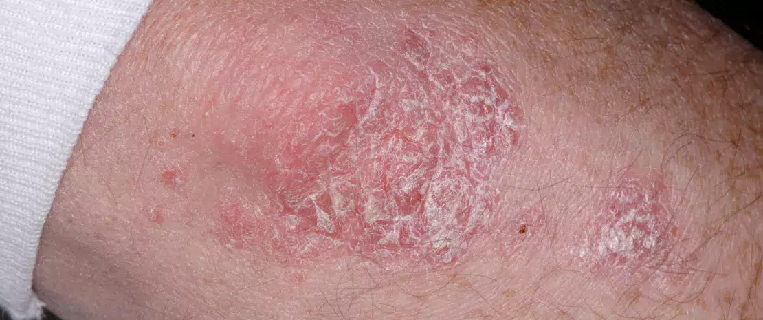
Close-up of plaque psoriasis (Psoriasis vulgaris) on an elbow.

THE ROLE OF SKIN MICROBIOTA IN ITCH

Kim HS, Yosipovitch G. The Skin Microbiota and Itch: Is There a Link? J Clin Med 2020 ; 9 : 1190.

The authors discuss the role of skin microbiota in the pathogenesis of itch. Itch sensation is mediated via epidermal nerve fibres (pruriceptors) driving by chemical mediators that originate from a complex interaction between keratinocytes (KC), inflammatory cells, nerve endings and the skin microbiota, relaying itch signals to the brain. Skin dysbiosis is characterized by production of proteases, pathogen-associated molecular patterns, and toxins, leading to skin barrier damage. Mast cell degranulation induced by delta-toxin prompt inflammation and itching. Skin microbiota and brain communicate via neurochemicals (acetylcholine, histamine, catecholamines, corticotropin) originate from skin microbiota. Stress intensifies itch via the skin-brain axis, where the amygdala seems to modulate itching sensation via microbial signals. Chronic stress increases cortisol production, directly activates skin bacteria by increasing the virulence of skin pathogens, leading to a weakening of the skin barrier and to an aggravation of the itch sensation. The authors conclude that cosmetics/transdermal drugs that modulate skin microbiota might have the potential to ameliorate itch.

SKIN MICROBIOTA IN PSORIASIS

Quan C, Chen X-Y, Li X, et al. Psoriatic lesions are characterized by higher bacterial load and imbalance between Cutibacterium and Corynebacterium. J Am Acad Dermatol 2020 ; 82 : 955-61.

The authors examined the microbiota in psoriatic lesions and unaffected skin in psoriasis vulgaris (PS) patients and healthy controls by quantitative PCR and 16S rRNA sequencing. Higher bacterial load and lower diversity was observed in PS lesions than patients unaffected and controls’ skin. Cutibacterium (Cu) was reduced in lesions, whereas Corynebacterium (Cr) was increased. Compared with patients’ unaffected skin, Cr/Cu + Cr ratio was higher in the lesions. These findings indicate that PS was the major cause for the imbalance between Cu and Cr between lesions and unaffected skin or controls. Cr load correlated with the severity of PS lesions, whereas Cu load showed correlation with the abnormity of skin capacitance. The present study suggests that skin microbiota might play a significant role in the pathogenesis of PS.

Summary
Off
Sidebar
Off
Migrated content
Désactivé
Updated content
Désactivé
Hide image
Off
Press review

Vaginal Microbiota #11

By Prof. Markku Voutilainen
Turku University Faculty of Medicine; Turku University Hospital, Department of Gastroenterology, Turku, Finland

Bandeau-Microbiota_NL11_FR.jpg

VAGINAL DYSBIOSIS AND RECURRENT IMPLANTATION FAILURE

Fu M, Zhang X, Liang Y, et al. Alterations in vaginal microbiota and associated metabolome in women with recurrent implantation failure. mBio 2020 ; 11 : e03242-19.

Recurrent implantation failure (RIF) is defined as a failure to achieve a clinical pregnancy after transfer of at least four good-quality embryos in a woman under the age of 40 years. Embryonal and uterine factors or maternal systemic diseases may cause RIF, but some women do not have recognizable etiology. The authors focused on the vaginal microbiota and metabolome of women with RIF. They found that RIF patients suffered vaginal dysbiosis, having a more diverse and abundant bacteria with an increase of many anaerobic and aerobic bacteria which could be linked to bacterial vaginosis and aerobic vaginitis or urinary tract infection, respectively. Conversely, at genus level their vaginal microbiota was decreased in Lactobacillus (LB); at species level L. iners was reduced and L. crispatus was the most abundant species in the RIF group. Increased vaginal bacterial diversity, LB depletion and related metabolic changes could serve as biomarkers capable of predicting the risk of RIF.

THE ROLE OF VAGINAL MICROBIOTA IN URINARY TRACT INFECTIONS

Lewis A, Gilbert NM. Roles of the vagina and the vaginal microbiota in urinary tract infection: evidence from clinical correlations and experimental models. GMS Infect Dis 2020 ; 8 : Doc02.

This review summarizes the role played by vaginal microbiota in urinary tract infections (UTI) as mounting evidence indicates that vagina may serve as a reservoir for uropathogens and increase susceptibility to UTI. Escherichia coli is the commonest cause of UTI and can colonize the vagina, which can be increased if the vaginal Lactobacillus (LB) colonization is reduced. Some vaginal bacteria are frequently detected in the urine but are underappreciated as uropathogens, because they are difficult to detect in routine clinical practice. For example, bacterial vaginosis (BV) is characterized by Gram-negative anaerobes, species belonging to Actinobacteria and Firmicutes phyla while LB is reduced and BV patients have a higher UTI risk. Gardnerella vaginalis is detected in BV and can cause acute or recurrent UTI. Group B Streptococcus may cause both aerobic vaginitis and UTI. Finally, some vaginal bacteria may enter the urinary tract and can transit briefly, cause immunomodulation or injury and unbalanced the host-pathogen interactions to influence the outcomes of uropathogenesis.

Summary
Off
Sidebar
Off
Migrated content
Désactivé
Updated content
Désactivé
Hide image
Off
Press review

EJFHOD 2020

Congress review

By Dr Dragos Ciocan
CCA Hepato-gastroenterology and nutrition department Antoine-Béclère Hospital, Clamart, France

IBD
Article5_Bandeau-Microbiota_NL11_FR.jpg

Despite the Covid-19 pandemic, this year we were able to benefit from an on-line version of the Journees francophones d’hépato-gastroenterologie (French-speaking hepato-gastroenterology congress) with the advantage of being able to access presentations recorded between 3 and 20 July 2020. These eJFHOD reached a total of 7,924 users, and 172,937 pages were viewed. As each year, original studies on the intestinal microbiota (IM) were presented at this congress.

Microbiota and colorectal cancer

Third most frequent cancer in humans, sporadic colorectal cancer (CRC) develops following interactions between the host and its environment, and the IM is thought to be implicated [1]. Professor Sobhani presented the results of a study which investigated the links between epigenetic mechanisms promoted by bacteria of the IM and the onset of CRC [2]. Mice transplanted with faecal samples from patients with CRC developed precancerous colonic lesions, associated with an increase in hypermethylated genes. Donors with CRC exhibited methylation anomalies of several gene promoters associated with intestinal dysbiosis. Using the identified microbial and epigenetic signatures, a pilot study (n = 266) was conducted in humans in order to develop a blood test for the diagnosis of CRC. A cumulative methylation index (CMI, measuring the hypermethylation level of 3 genes) was identified as a predictive factor in the onset of CRC. These results were validated in a prospective cohort of 1,000 patients. Intestinal dysbiosis in patients with a positive CMI was characterised by an increase in pro-methylating bacterial species. This work indicates that intestinal dysbiosis associated with CRC could promote colon carcinogenesis via deregulation of the methylation of certain genes. The cumulative hypermethylation index (CMI) and/or pro-methylating bacteria are thus potential biomarkers for CRC diagnosis, or be used in the evaluation of the effects of treatments modulating the IM in patients with CRC.

A new dysbiosis marker in Crohn’s disease

In a study coordinated by Professor Seksik, the authors studied the role of MAM (microbial anti-inflammatory molecule, produced by Faecalibacterium prausnitzii and reduced in patients with Crohn’s disease, CD [3]) as a biomarker of intestinal dysbiosis and diagnostic aid in CD. The authors showed that loss of MAM is associated with the diagnosis of CD. This preliminary study in a small number of patients (24 patients in relapse, 24 in remission and 12 healthy controls) paves the way to the diagnosis of CD based on the IM, but these preliminary results require validation in independent cohorts.

A new therapeutic perspective in IBD

It is known that bacteria detect and respond to environmental signals (an ability called Quorum Sensing). Of the molecules which are part of this system, 3-oxo-C12:2 is low in patients with chronic inflammatory bowel disease (IBD), this reduction appears to be correlated with the observed intestinal dysbiosis [4]. In a study presented by D. Aguanno, the authors studied the impact of this molecule on the epithelial cells of the intestine and showed that this did not modify paracellular permeability but attenuated the deleterious effects on the tight junctions induced by pro-inflammatory cytokines. In a second study, Coquant et al. showed that 3-oxo-C12:2 exerted an anti-inflammatory effect on immunoreactive cells, partly mediated by the T2R138 receptor. This molecule may therefore have protective effects on the intestinal barrier, modulate the inflammatory response and thus represent a novel therapeutic perspective in IBD.

Summary
Off
Sidebar
Off
Migrated content
Désactivé
Updated content
Désactivé
Hide image
Off
Congress review

Gut mucosal and faeca l microbiota profiling combined to intestinal immune system in neonate s affected by intestinal ischemic injuries

Commented articles - Children's section

By Pr Emmanuel Mas
Gastroenterology and Nutrition Department, Children’s Hospital, Toulouse, France
 

NEC
Article3_Bandeau-Microbiota_NL11_FR.jpg

Comments on the original article of Romani et al. (Frontiers in Cellular and Infection Microbiology 2020 [1])

In the first weeks of life the microbiota plays a crucial role i n health by acting as a barrier against the invasion of pathogens and by maintaining intestinal immune homoeostasis. Changes in the ecology of the faecal microbiota (FM) have been reported in neonates with intestinal ischaemia. The aim of this study was to describe the FM, the mucosal microbiota (MM) and mucosal immunity in these patients.

Fourteen neonates underwent intestinal resection due to intestinal ischaemia. Two groups were identified on the basis of lesion severity: extensive (EII) and localised intestinal ischaemia (LII). This study showed the variations in FM and MM in the EII and LII groups.

What do we already know about this subject?

The intestinal microbiota of neonates is characterised by lower bacterial diversity and a higher proportion of pathogenic bacteria. Moreover, their intestinal immune system is immature. These two factors modify the intestinal epithelial barrier and enhance the production of pro-inflammatory mediators.

Necrotising enterocolitis (NEC) is an ischemic and inflammatory disorder of the gastro- intestinal tract which affects premature neonates. The physiopathology of NEC remains poorly understood but intestinal dysbiosis is present, and also an inflammatory process. The use of antibiotics and antacids promote dysbiosis and also increases the risk of onset of NEC.

Neonates can also suffer from other ischaemic and inflammatory disorders, such as small bowel volvulus and localised gastro- intestinal perforations.

What are the main insights from this study?

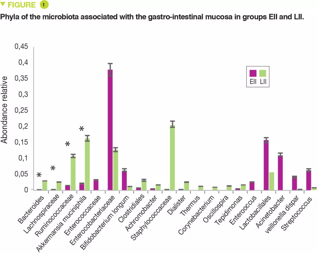
This single centre pilot study profiled the composition of the FM and MM and also the mononuclear cells of the lamina propria and the pro-inflammatory cytokines of two groups of neonates: full-term or premature. Seven infants had extensive intestinal ischaemia (EII) (5 NEC, 1 small bowel volvulus and 1 total colonic ischaemia) and 7 infants had localised intestinal ischaemia (LII) (4 isolated perforations and 3 cases of intestinal atresia). The FM of 9 full-term infants was used as control. The MM of neonates with EII, compared with those with LII, contained: more Proteobacteria (p = 0.049) and fewer Bacteroidetes (p = 0.007) and Verrucomicrobia (p = 0.01) (Figure 1); fewer Bacteroides, Lachnospiracee, Ruminococcaceae and Akkermansia muciniphila (p < 0.05).

The FM was less diverse (Shannon index) in EII than in LII (p = 0.01). The relative abundance for the MM was similar between EII and LII for Proteobacteria and Firmicutes (p < 0.05). Similarly, a bacterial distribution with more Enterobacteriaceae in EII and more RuminococcaceaeBacteroidesLachnospiracee and Staphylococcaceae in cases of LII.

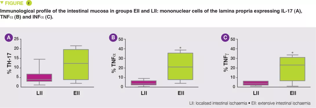
The EII group exhibited increased numbers of lymphocytes B, T and NK (Natural Killer), in particular T CD3+ lymphocytes, TH17 (Figure 2A) and reduced numbers of T lymphocyte regulators (Tregs), with increased numbers of cells expressing TNFa (Figure 2B) and INFg (Figure 2C).

Image

Key points

  • Necrotising enterocolitis is a severe gastro-intestinal pathology which affects premature neonates

  • Anomalies of the faecal microbiota and the gastrointestinal-associated microbiota may be involved in the inflammatory and ischaemic of processes NEC

Image

What are the consequences in practice?

This pilot study confirms that lack of bacterial diversity and the predominance of Enterobacteriaceae are risk factors for NEC, as is a reduction in numbers of Akkermansia muciniphila. Correction of this dysbiosis could modify the TH17/Tregs imbalance and reduce the production of mediators of inflammation (TNFa and INFg).

Conclusion

The faecal microbiota and the gastro-intestinal mucosal microbiota have specific characteristics in premature neonates with ischemic lesions. Additional studies are needed to determine the role of these bacteria in the inflammatory and ischaemic process of NEC.

Summary
Off
Sidebar
Off
Migrated content
Désactivé
Updated content
Désactivé
Hide image
Off
Commented article

Cholesterol metabolism by uncultured human gut bacteria influences host cholesterol level

Commented articles - Adults' section

By Pr. Harry Sokol
Gastroenterology and Nutrition Department, Saint-Antoine Hospital, Paris, France


Article2_Bandeau-Microbiota_NL11_FR.jpg

Comments on the original article of Kenny et al. (Cell Host & Microbe 2020)

The human microbiome possesses extensive metabolic capabilities but our understanding of the mechanisms linking gut microbes to human metabolism remains limited. In this article, the authors focused on the conversion of cholesterol to the poorly absorbed sterol coprostanol by the gut microbiota to develop a framework for the identification of functional enzymes and microbes. By integrating paired metagenomics and metabolomics data from e xisting cohorts with biochemical knowledge and experimentation, the authors predicted and validated a group of microbial cholesterol dehydrogenases that contribute to coprostanol formation. These enzymes are encoded by ismA genes in a group of uncultured microbes, which are prevalent in geographically diverse human cohorts. Individuals harbouring coprostanol-forming microbes have significantly lower faecal cholesterol levels and lower total serum cholesterol with effects comparable to those attributed to variations in lipid homoeostasis genes. Thus, cholesterol metabolism by these microbes may play important roles in reducing intestinal and serum cholesterol concentrations, directly impacting human health. [1]

What do we already know about this subject?

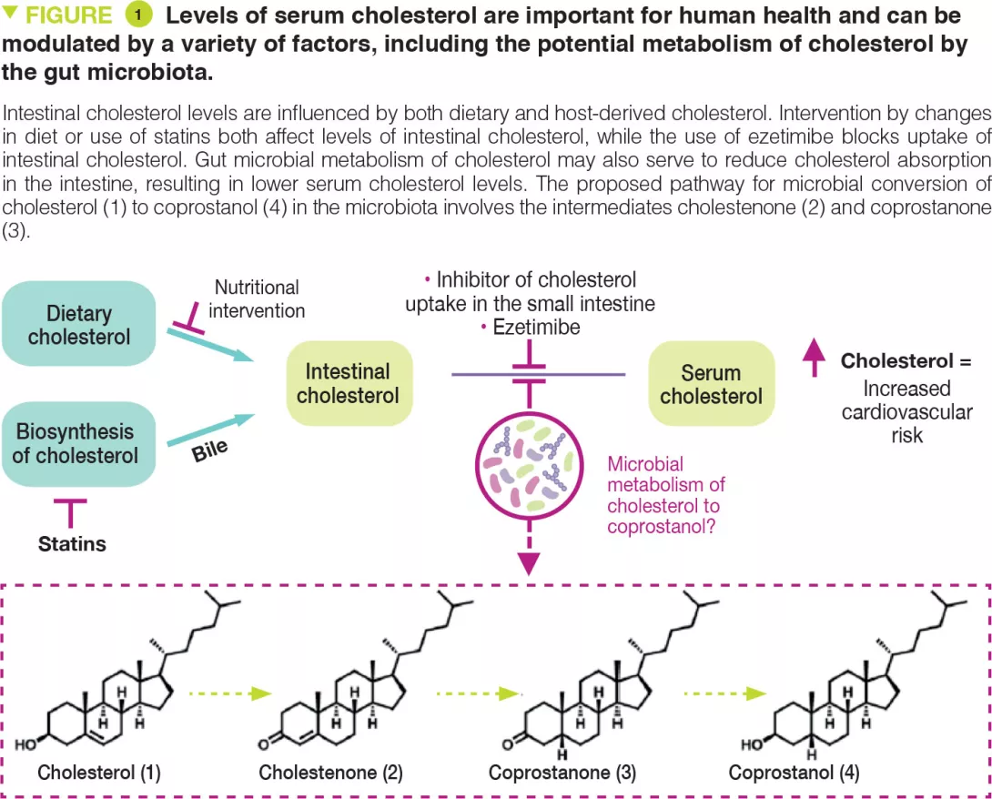
Cholesterol is a key biological molecule that functions as a structural component of all animal cell membranes and is a pre-cursor of steroid hormones, vitamin D, and bile acids. Two main sources of cholesterol are thought to influence concentrations of this metabolite in serum: endogenous cholesterol synthesised in the liver and exogenous cholesterol derived from dietary components of animal origin (Figure 1). The cholesterol synthesised in hepatocytes is transported to the gallbladder and is then secreted into the small intestine along with other bile salts. In the intestine, biliary cholesterol (~1–2 g/day) mixes with dietary cholesterol (~0.2–0.4 g/day in the average American diet), and both sources are eventually transported into enterocytes for packaging into lipoprotein particles and secretion into the plasma. Hypercholesterolaemia is a risk factor for cardiovascular disease (CVD), which is the cause of one-fourth of all deaths in industrialised countries.

Reducing cholesterol transport in the intestine is a clinically validated strategy for lowering serum cholesterol levels. A range of gut microbes metabolise and modify dietary and host-derived molecules in the small intestine. Because both sources of cholesterol pass through this environment, the gut microbiota may influence serum cholesterol levels. Indeed, microbiota transfer from human donors with elevated serum cholesterol levels can impart this hypercholesterolaemia phenotype to mice. [2, 3] Other studies have reported that administering specific bacterial species can have cholesterol-lowering effects. [4] However, the precise mechanisms underlying these observations are currently unknown. The gut microbiota may exert cholesterol-lowering effects by metabolising intestinal cholesterol to coprostanol (Figure 1), which would reduce the amount of cholesterol absorbed from the intestine.

This microbiota-dependent transformation has been known to occur in humans since the early 1900s. Several coprostanol-generating gut bacteria with similar physical and biochemical characteristics have been reported from a variety of different sources including rats, baboons, and humans. However, most of these strains are not currently available and were never sequenced. Early work showed that coprostanol formation by this group of gut bacteria proceeds through an indirect reduction pathway involving the initial oxidation of cholesterol (1) to cholestenone (2), followed by reduction of the D4,5 double bond to form coprostanone (3), and subsequent re-reduction of ketone to generate coprostanol (4) (Figure 1). The bacterial enzymes responsible for this metabolism were never identified. More recently, other reports have implicated additional phylogenetically diverse gut bacteria in coprostanol formation. [5] While efforts to elucidate how gut microbial metabolism of cholesterol affects human serum cholesterol levels span over 100 years, mechanistic support for this connection has remained elusive due to a limited understanding of the gut microbes, genes, and enzymes responsible for coprostanol formation.

Image

Key points

  • Some bacteria found in the human intestinal microbiota possess enzymes belonging to the ismA family capable of breaking down cholesterol.

  • The presence of ismA+ species in gut microbiota is associated with decreased faecal and serum cholesterol in humans.

  • The effect of ismA+ species on serum cholesterol is on par with human genetics.

What are the main insights from this study?

The authors used a multi-disciplinary strategy to discover gut bacterial enzymes. This strategy, based on correlations between metagenomics and metabolomics data from existing human cohorts, identified and characterised an extensive family of cholesterol dehydrogenase enzymes from a clade of uncultured intestinal bacteria implicated in the metabolism of cholesterol to coprostanol. Firstly, the enzyme responsible for the first step in cholesterol transformation, called ismA, was identified in Eubacterium coprostanoligenes, a bacteria already known for this function. Analysis of sequencing data from human cohorts then identified homologous enzymes in a group of uncultured anaerobic bacteria.

The presence of these ismA genes in the microbiome was associated with the presence of coprostanol in stools and lower faecal cholesterol levels. Finally, to demonstrate the potential for these cholesterol- metabolising bacteria to influence human health, the authors showed that presence of ismA genes in human metagenomes is associated with a decrease in total cholesterol concentrations in serum that is on par with the effects observed from variants in human genes involved in lipid homoeostasis

What are the consequences in practice?

Overall, these findings confirm the role of gut-bacterial metabolism in modulating host cholesterol levels in the intestine and also, more importantly, on a systemic level. This work paves the way for the use of the gut microbiota as a predictive biomarker of high cholesterol and establishes the foundations for microbiota-targeted therapeutic interventions.

Conclusion

This study highlights the role of the gut microbiota in breaking down cholesterol with an effect on serum cholesterol levels. Gut microbiota could soon become the target of cholesterol lowering therapies.

Summary
Off
Sidebar
Off
Migrated content
Désactivé
Updated content
Désactivé
Hide image
Off
Commented article

The gut-lung axis during viral respiratory infections

Overview

By Dr François Trottein
Centre d’Infection et d’Immunité de Lille, Inserm U1019, CNRS UMR 9017, University of Lille, Lille Hospital, Institut Pasteur de Lille, France

The novel coronavirus (SARS-CoV-2) disease 19 (COVID-19) reminds us that interactions between the gut microbiota and the immune system are essential during viral respiratory tract infections. Respiratory viruses can trigger gastrointestinal symptoms emphasizing the role of the lung-gut axis in disease. Clinical studies and experimental models indicate that acute vi ral respiratory infections alter the composition and functions of the gut microbiota, an essential component of human health. Here, we review these major changes and discuss the potential causes of intestinal dysbiosis. We also present the consequences of gut dysbiosis that develops during infection on secondary disease outcomes. Lastly, we suggest interventional strategies that might be used to target the gut microbiota in order to reduce the viral respiratory disease severity.

THE ONGOING THREAT OF RESPIRATORY VIRAL INFECTIONS

Viral respiratory tract infections are still a major concern worldwide as they lead to considerable socio-economic and health issues. Despite vaccine campaigns and antiviral medications, influenza - the commonly called “flu” - remains the most impacting in terms of infected individuals (5-10% of the global population) and deaths (> 600,000 per year). Along with annual epidemics, influenza is also responsible for pandemics every 10-20 years, the most famous being the 1918-1919 Spanish flu (50 million deaths) and the most recent, the 2009 H1N1 influenza pandemic (400,000 excess deaths) [1]. Pandemics are not restricted to influenza, as exemplified by the COVID-19 [2]. Of great concern is the increasing frequency of pandemics occurring over the last few decades, a phenomenon explained in part by climate changes and human practises, in general.

THE GUT MICROBIOTA IN HEALTH AND DISEASES

The gut microbiota has a crucial role in the maintenance of human health and is critical in the control of (respiratory) infections [3, 4]. Many factors can alter the diversity and composition of the gut microbiota, leading to dysbiosis. Among them, disease situations, such as infections and chronic inflammatory or metabolic disorders, can trigger profound alteration of the gut microbiota’s composition and function. Changes in intestinal bacterial communities can influence disease outcomes even in distant organs (including the lungs) [3, 4], as demonstrated by transfer experiments with dysbiotic microbiota. Below, we summarize the effects of acute viral respiratory infections on the gut microbiota.

VIRAL RESPIRATORY TRACT INFECTIONS LEAD TO GUT DYSBIOSIS

Clinical evidences suggest a gut microbiota dysbiosis during influenza infection. The relative abundance of ActinobacteriaErysipelotricheaClostridia and beneficial butyrate producers (Lachnospiraceae and Ruminococcaceae families) is decreased in patients H1N1. On the other hand, opportunistic pathogens such as Escherichia- Shigella and Prevotella develop [5]. In experimental (mouse) models, transient gut dysbiosis also occurs with a peak at 5-7 days post-infection [6-9]. Changes occurred at taxonomic levels with no change in alpha diversity. Infection blunts the growth of health-promoting bacteria such as Lactobacilli, Bifidobacteria, and segmented filamentous bacteria. Many species capable of fermenting dietary fibres into short-chain fatty acids (SCFAs) are affected. In line, the production of SCFAs dramatically drops during influenza infection [9]. The reduced levels of beneficial commensals associate with the overgrowth of deleterious bacteria including Gammaproteobacteria (Escherichia coli) and mucus-degrading bacteria such as Verrucomicrobia (Akkermansia genus) and Ruminococcus. SARS-CoV-2 infection also triggers gut microbiota alterations in patients, including lower abundance of butyrate producers such as several genera from the Ruminococcaceae and Lachnospiraceae (Roseburia) families [5, 10]. On the other hand, a significantly higher relative abundance of opportunistic bacterial pathogens including Streptococcus (class Bacilli), Rothia and Actinomyces was observed. Of note, overgrowth of opportunistic fungal pathogens (Aspergillus and Candida spps) was also described in COVID-19 patients [11]. Collectively, viral respiratory infections lead to depletion of beneficial commensals and enrichment of opportunistic harmful pathogens. Putative changes in the gut microbiota’s structure, composition and functional activity might be biomarkers of disease severity.

MECHANISMS LEADING TO GUT DYSBIOSIS

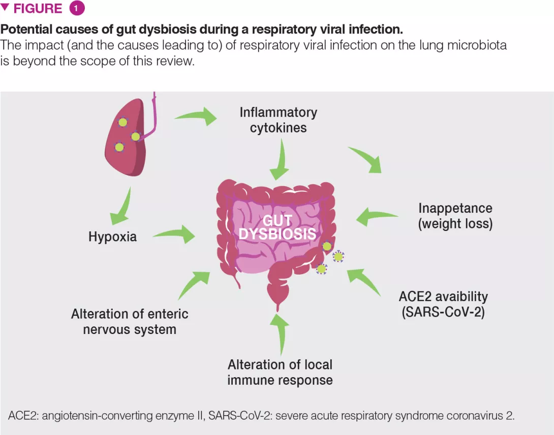
There are probably several causes of gut dysbiosis during viral respiratory infections; these may include the release of inflammatory cytokines and the reduced food intake (Figure 1). Infection induces substantial weight loss due to a loss of appetite. Pair-feeding experiments in mice clearly indicate that a rapid fall in food intake mimics the changes in the gut microbiota observed during influenza infection [8]. Recent evidence suggests a role for TNFa in inappetence-associated dysbiosis during viral respiratory infection [12]. Type I and II interferons, which are essential for the host antiviral response, also play a part in gut dysbiosis [5,6].

Image

Hypoxia (a feature of acute viral respiratory infection), alterations of the enteric nervous system and dysregulated local immune response are also likely to participate in gut dysbiosis [13] (Figure 1). In the case of COVID-19, along with these mechanisms, local viral replication is likely to play a role in gut dysbiosis. Angiotensin- converting enzyme II (ACE2), the receptor of SARS-CoV-2, is instrumental to maintain the gut’s microbial ecology. Considering the lack of available ACE2 during SARS-CoV-2 infection, one expects that this might influence the composition and functions of the gut microbiota [13].

CONSEQUENCES OF GUT DYSBIOSIS ON SECONDARY OUTCOMES

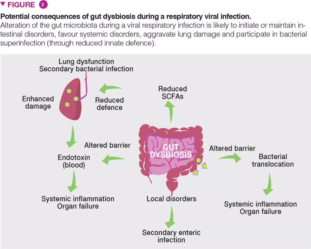
Gut dysbiosis during viral respiratory infection has local and distal consequences and might be an important contributor of disease severity and fatal outcomes (Figure 2). Patients experiencing viral respiratory infection can develop gastroenteritis- like symptoms such as abdominal pain, nausea, vomiting and diarrhoea. Alterations of the gut microbiota may explain these disorders. It is also likely that altered gut microbiota including the emergence of pathobionts and mucus degrading bacteria play a role in intestinal inflammation and disruption of the gut barrier integrity [6]. In turn, intestinal barrier leakage might increase endotoxin concentrations in the blood, ultimately triggering inflammation, cytokine overproduction and lung dysfunctions [14]. Acute viral respiratory infections can lead to secondary enteric infections and sepsis. Gut dysbiosis (and drop of SCFAs) might be important in this setting. Indeed, SCFAs play a key role in intestinal homeostasis, barrier integrity and control of enteric pathogens [15]. Along with local disorders, our recent data show that gut dysbiosis can remotely hamper host defense in the lungs [9] (Figure 2). In healthy conditions, the gut microbiota remotely arms the lungs against bacterial infection, in part by reinforcing the bactericidal activity of pulmonary macrophages [16].

Image

During influenza, this axis is altered and opportunistic bacteria invade the lungs leading to bacterial superinfection, a major cause of death during influenza epidemics and pandemics [1]. We have shown that the reduced production of acetate (the major SCFAs) by the gut microbiota is partly responsible for this effect [9]. Collectively, dysbiosis might influence the gastrointestinal and pulmonary signs and symptoms (and overall mortality) of viral respiratory infections. Can we use the gut-lung pathway as a basis to better control the severity and mortality rate of viral respiratory infections?

INTERVENTIONAL PERSPECTIVES

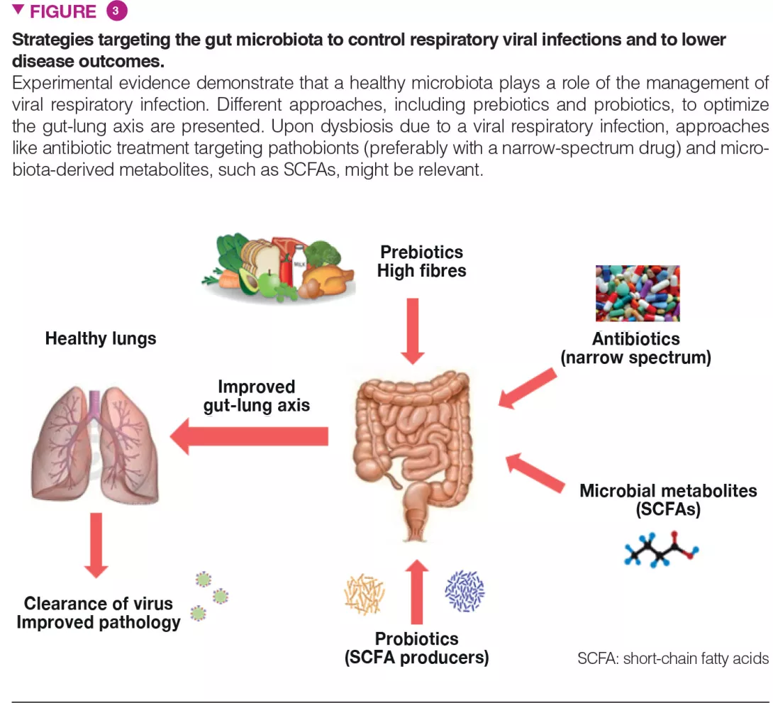
The gut microbiota is vital in the lung’s defences against respiratory infection and interventional strategies that target intestinal commensals to preventively arm the lungs against viral pathogens and to protect the microbiota against the perturbations associated with viral infections are of major interest (Figure 3). This is particularly true in individuals with a general imbalance of the gut microbiota such as the elderly and individuals with co-morbidities, well known to be more susceptible to infections.

Image

Approaches like (i) dietary ingredients intended to nourish our beneficial microorganisms (like prebiotic fibres) and (ii) indigenous bacteria (known as probiotics) to replenish our gut with missing beneficial microorganisms and to optimize their metabolic functions, might be relevant. These strategies, especially personalized (i.e. based on the analysis of the gut microbiota in the “at risk” population) might improve clinical outcomes and accelerate the recovery of patients that experience acute viral respiratory tract infections. Probiotics, including some Bifidobacteria and Lactobacillus spp, can decrease the severity of influenza infection, through still undetermined mechanisms [17]. Of highly topical concern, a recent study showed that oral bacteriotherapy in addition to the standard drug therapy showed promising clues in the management of COVID-19 patients [18].

Conclusion

The gut-lung axis is important during viral respiratory tract infections. Alterations of the gut microbiota plays a part in secondary outcomes. The gut microbiota therefore represents a target to limit disease severity. Solutions for disease management include dietary ingredients (like prebiotic fibres) and probiotics.

Sources

1 McCullers JA. The co-pathogenesis of influenza viruses with bacteria in the lung. Nat Rev Microbiol 2014. 12: 252-62.

2 Wang C, Horby PW, Hayden FG, et al. A novel coronavirus outbreak of global health concern. Lancet 2020; 395: 470-3.

3 McAleer JP, Kolls, JK. Contributions of the intestinal microbiome in lung immunity. Eur J Immunol 2018; 48: 39-49.

4 Budden KF, Gellatly SL, Wood DLA, et al. Emerging pathogenic links between microbiota and the gut-lung axis. Nat Rev Microbiol 2017; 15: 55-63.

5 Gu S, Chen Y, Wu Z, et al. Alterations of the gut microbiota in patients with COVID-19 or H1N1 Influenza. Clin Infect Dis. 2020 Dec 17;71(10):2669-2678. 

6 Wang J, Li F, Wei H, et al. Respiratory influenza virus infection induces intestinal immune injury via microbiota-mediated Th17 cell-dependent inflammation. J Exp Med 2014; 211: 2397-410.

7 Deriu E, Boxx GM, He X, et al. Influenza Virus Affects Intestinal microbiota and secondary Salmonella infection in the gut through type I interferons. PLoS Pathog 2016; 12: e1005572.

8 Groves HT, Cuthbertson L, James P, et al. Respiratory disease following viral lung infection alters the murine gut microbiota. Front Immunol 2018; 9: 182-93.

9 Sencio V, Barthelemy A, Tavares LP, et al. Gut dysbiosis during influenza contributes to pulmonary pneumococcal superinfection through altered short-chain fatty acid production. Cell Rep 2020; 30: 2934-47.

10 Zuo T, Zhang F, Lui GCY, et al. Alterations in gut microbiota of patients with COVID-19 during time of hospitalization. Gastroenterology 2020 Sep;159(3):944-955.e8.

11 Zuo T, Zhan H, Zhang F, et al. Alterations in fecal fungal microbiome of patients with COVID-19 during time of hospitalization until discharge. Gastroenterology. 2020 pii: S0016-5085(20)34852-6.

12 Groves HT, Higham SL, Moffatt MF, et al. Respiratory viral infection alters the gut microbiota by inducing inappetence. mBio 2020; 11: e03236-19.

13 Trottein F, Sokol H. Potential causes and consequences of gastrointestinal disorders during a SARS-CoV-2 Infection. Cell Rep 2020; 32: 107915.

14 Openshaw PJ. Crossing barriers: infections of the lung and the gut. Muc Immunol 2009; 2: 100-2.

15 Koh A, De Vadder F, Kovatcheva-Datchary P, et al. From dietary fiber to host physiology: short-chain fatty Acids as key bacterial metabolites. Cell 2016; 165: 1332-5.

16 Brown RL, Sequeira RP and Clarke TB. The microbiota protects against respiratory infection via GM-CSF signaling. Nat Commun 2017; 8: 1512-22.

17 Zelaya H, Alvarez S, Kitazawa H, et al. Respiratory antiviral immunity and immunobiotics: beneficial effects on inflammation-coagulation interaction during influenza virus infection. Front Immunol 2016; 7: 633.

18 d’Ettorre G, Ceccarelli G, Marazzato M, et al. Challenges in the management of SARS-CoV2 infection: the role of oral bacteriotherapy as complementary therapeutic strategy to avoid the progression of COVID-19. Front Medecine 2020; 7: 389-96.

Summary
Off
Sidebar
Off
Migrated content
Désactivé
Updated content
Désactivé
Hide image
Off
Article