Microbiota viruses may help monitor progression of HIV infection
Immunocompromised HIV-positive individuals have an abundance of viruses in their gut microbiota. Some of these viruses could be used to predict the effectiveness of HIV treatment and monitor the recovery of immunity. 1
The gut microbiota
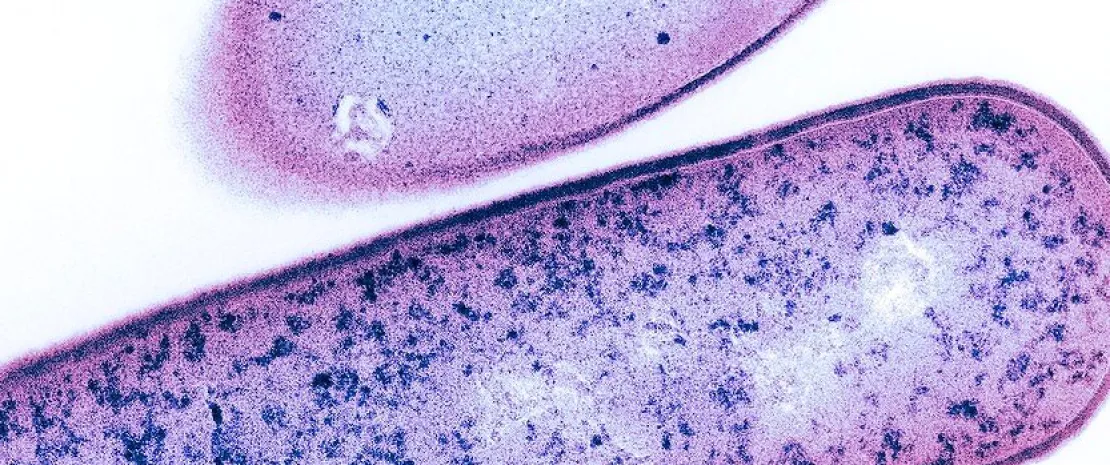
The digestive tract is the main replication site of the human immunodeficiency virus (HIV), which is the cause of AIDS. The presence of HIV is associated with inflammation of the gut mucosa and an imbalance in the bacteria of the microbiota ( (sidenote: Dysbiosis Generally defined as an alteration in the composition and function of the microbiota caused by a combination of environmental and individual-specific factors. Levy M, Kolodziejczyk AA, Thaiss CA, et al. Dysbiosis and the immune system. Nat Rev Immunol. 2017;17(4):219-232. ) ), which could influence progression of the disease.
39 million people worldwide living with HIV in 2022. ²
2/3 of those affected by HIV live in Africa (25.6 million). ²
Not only bacteria in the microbiota...
But what about the viruses which are present alongside bacteria, fungi, and archaea in the gut microbiota? Are they, like bacteria, involved in disease?
To answer this question, a team of Mexican researchers analyzed the ‘virome’ (viral component of the microbiota) in the feces of 92 HIV-positive individuals at different stages of infection but not receiving treatment and compared it with that of 53 healthy individuals. 1
1.3 million people contracted the virus in 2022. ²
630,000 people died of AIDS in 2022. ²
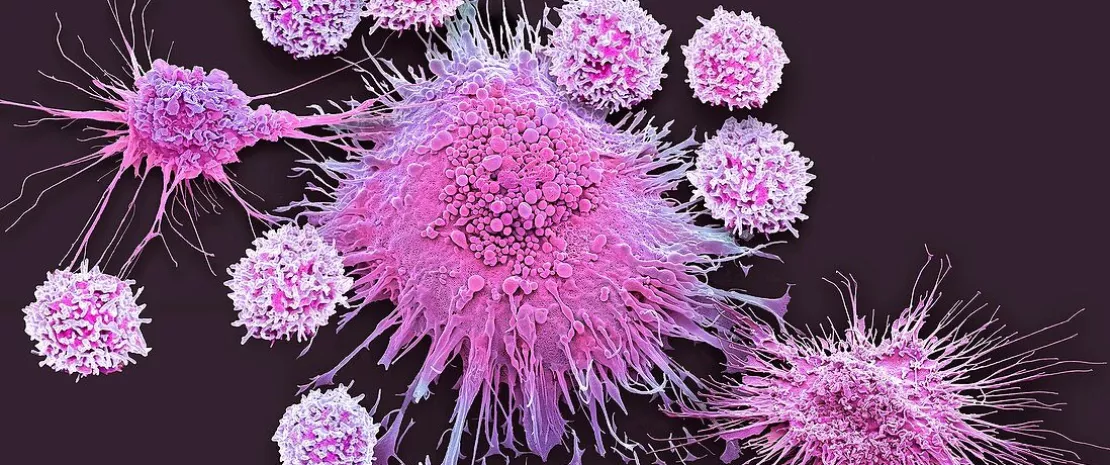
From the HIV-positive individuals, they then selected 14 people suffering from immunodeficiency, i.e. a low level of CD4 T cells, the cells in which HIV multiplies.
They took stool and blood samples from them before antiretroviral treatment and at four timepoints during antiretroviral treatment. Their aim was to study changes in immunity and the gut microbiota during the first two years of therapy.
A striking expansion of certain virus species
They found that in those most affected by the disease, i.e. those suffering from severe immunodeficiency (CD4 T cell count < 350), three species of virus are present in great abundance in the microbiota: Anelloviridae (anelloviruses), Adenoviridae, and Papillomaviridae. Anelloviruses appear to be particularly affected by antiretroviral treatment, their markers decreasing significantly after 24 months of treatment.

One notable finding was that the presence of anelloviruses at the start of treatment is associated with a poorer recovery of immunity and a lower CD4 T cell count, and therefore less effective treatment.
For the scientists, this study represents an important step forward. Not only does it provide a better understanding of the virome, a component of the gut microbiota little studied and less well understood than the bacterial component; it also provides a better understanding of the involvement of microbiota viruses in HIV infection.
Towards better monitoring of patients
These results open up the prospect of one day being able to use anelloviruses as a marker to predict the effectiveness of treatment and monitor the immune recovery of those affected by HIV. This is good news, since the fight against AIDS remains a major public health concern.