Scams, (fake) science, and publishing

The publication of scientific articles is essential to research but today this process faces two evils: predatory journals and doctored articles.
Details below. 

They might look and read like scientific articles and journals, but they are not worthy of the name. Two evils are currently plaguing the world of scientific publication: “bogus” journals and “fake” articles.

Predatory journals

The principle of predatory journals is simple: they publish articles because they are paid by the authors and not because of the articles’ quality. This allows authors to publish lackluster findings unworthy of publication. It also allows interest groups to promote studies they know to be biased or falsified in order to promote the product or the sector they represent (e.g. a drug). The problem is that these predatory journals closely resemble legitimate journals, at times even partially plagiarizing their names, making it difficult to recognize them. The resemblance is so close that good scientists are being fooled into publishing legitimate articles in these journals, or are being influenced by the bad articles they read in them. Fortunately, lists of these predatory journals (more than 14,000 titles as of the beginning of 2021) can be found online, published by researchers and associations (such as predatoryjournals.com) constantly on the lookout for them.

Paper mills

In addition to fake journals, there are also fake articles. Such articles are written by paper mills, whose role is to provide authors in need of inspiration or career advancement with ready-made articles in return for a few dollars. The problem is that these articles contain fabricated scientific results and error-filled data. One can occasionally read articles on cancer, for example, whose data are pure science fiction, and this even in highly respected journals, whose editors have been duped.

14,000 More than 14,000 titles as of the beginning of 2021

400 Over 400 fake research papers published

Elisabeth Bik, a Dutch microbiologist specializing in scientific integrity (and in microbiota!) has identified over 400 fake research papers published in China by just one such paper mill1. Even with researchers constantly on the lookout for these black sheep of the scientific publishing world, caution is still required, as anyone can be deceived. Aware of such scams, the Biocodex Microbiota Institute takes the greatest possible care in selecting the articles featured on its website.

Elizabeth Bik has created the blogs Integrity Digest (on scientific integrity) and Microbiome Digest (on microbiota research).

Summary
Off
Sidebar
Off
Migrated content
Désactivé
Updated content
Désactivé
Hide image
Off
News

Scientific publications: Dr Science and Mr Fraud

The world of scientific publishing is essential to the sharing of research findings, but today it is plagued by two evils: predatory journals and paper mills.
Let’s take a closer look at this world without law.

The publication of scientific articles allows researchers to share their findings. A peer review system guarantees the quality of published work. Every article in a scientific journal worthy of the name has been previously reviewed by other experts in the field. Among other things, these experts can request improvements from the author or even refuse publication if they consider the article to contain errors or to be of little interest.

Predatory journals

However, some scientific journals are scientific in name only. Known as predatory journals, their principle is that the author pays to be published, whatever the quality of their article. The problem is that the researchers are at times fooled themselves, either as authors (some non-predatory journals ask for a contribution to publication costs) or as readers of such articles, which they believe to be correctly reviewed by peers. American library scientist, Jeffrey Beall, first denounced such journals1,2 in 2012. He proposed a number of criteria to identify them (e.g. articles accepted too quickly) and drew up a list of culprits3. Since then, several groups have taken up the torch, among which predatoryjournals.com. To reveal the extent of such practices, some authors have gone so far as to submit bogus articles. For example, in exchange for $55, a team of Franco-Swiss authors was able to publish in one such journal a nonsense article co-signed by fictitious authors from non-existent institutes (Institute for Quick and Dirty Science), with absurd methodologies, wild conclusions (enriching table salt with hydroxychloroquine to reduce scooter accidents), and a bizarre bibliography.

Paper mills

As if bogus journals were not enough, a second scourge is also eating away at scientific publishing: paper mills. Paper mills provide authors lacking inspiration, time or ethical values, but who wish to boost their careers in return for money, with ready-made articles containing data produced from thin air. Since these articles may be published in mainstream (rather than predatory) journals, it is sometimes difficult to identify them. The scam may have reached industrial scale, involving thousands or tens of thousands of articles.4

Fightback underway

With the help of volunteer scientific investigators, such as microbiologist Elisabeth Bik5, the scientific community is preparing to fight back against this second scourge. Searching for images too similar to be true, in 2020, Bik’s blog6 identified more than 400 articles that were likely produced by a single Chinese paper mill. Iran and Russia were also singled out for blame. As a result, the publishers are reviewing the articles flagged, with many already retracted and others marked with “expressions of concern”7. Serious publishers are also becoming more careful about new submissions, not hesitating to ask authors for raw data to ensure the veracity of the studies performed.

400 More than 400 articles that were likely produced by a single Chinese paper mill

Due to the extent of these two phenomena, caution and critical thinking are required, since anyone can be deceived. Aware of such scams, the Biocodex Microbiota Institute takes the greatest possible care in selecting the articles featured on its site

Summary
Off
Sidebar
Off
Migrated content
Désactivé
Updated content
Désactivé
Hide image
Off
News Gastroenterology Pediatrics

What to take away? Skin immunity

skin microbiota

Skin and gut dysbiosis, as well as immune dysregulation, have been observed in the development of atopic dermatitis, a complex disease with multifactorial causes.8

2 Atopic dermatitis is characterized by the activation of type 2 lymphocytes resulting in an overactive and exaggerated immune response.(9)

INFLAMMATION

Atopic dermatitis is a chronic inflammatory skin disease that appears in periodic flare-ups.9 The inflammation observed is the result of an immune dysregulation.9

Summary
Off
Sidebar
Off
Migrated content
Désactivé
Updated content
Désactivé
Hide image
Off
Dossier detail

What to take away? Respiratory immunity

Pulmonary Tract Microbiota

The bidirectional communication axis between the gut and lungs, called the “gut–lung axis” influences the immune status of both organs.

Lung and gut microbiota influence each other and may have an impact on respiratory diseases.4

1 1 factor known to control vaccine efficacy can be gut microbiota.(5)

DYSBIOSIS

Lung and gut dysbiosis are observed during viral respiratory infections.6 Common definition of dysbiosis describes it as a compositional and functional alteration in the microbiota that is driven by a set of environmental and host-related factors that perturb the microbial ecosystem.7

Summary
Off
Sidebar
Off
Migrated content
Désactivé
Updated content
Désactivé
Hide image
Off
Dossier detail

What to take away? Intestinal Immunity

The microbiota plays critical roles in the development and education of the host’s innate and adaptive immune system components, while the immune system orchestrates the maintenance of key features of host-microbe symbiosis. Maintaining homeostasis between the gut microbiota and the immune system is essential, determinants interfering with neonatal gut establishment (antibiotics...) may potentially lead to negative health outcomes.1

80% At least 80% of the body Ig-producing cells are located in the gut (2)

MUCUS

The intestinal mucus layer is at the crucial interface between host and gut microbiota. Its disruption leads increased penetration or passage of potentially harmful bacteria that can eventually cause inflammation and infection.3

Summary
Off
Sidebar
Off
Migrated content
Désactivé
Updated content
Désactivé
Hide image
Off
Dossier detail

Expert interview: Pr. Brigitte Dréno

Atopic dermatitis (AD) is a chronic inflammatory skin disease that appears in periodic flareups. Like asthma, hay fever or allergic conjunctivitis, it is classified as an allergic disease. The disease causes very poorly defined oozing red lesions to appear in specific locations on the skin, such as in the folds of the elbow or behind the knees, but at times also on the face or the rest of the body. AD usually appears in early childhood, and may persist into adulthood. The causes are multifactorial and complex and include a genetic predisposition (mutation of the skin protein, filaggrin), an alteration of the skin barrier, a dysbiosis of the skin and gut microbiota, and immune dysregulation.

AD affects 15%-20% of children and 10% of adults in “developed” countries. The number of cases has increased significantly in recent decades due to pollution and contact with allergens.1

What factors cause flare-ups ? 


Inflammatory outbreaks can be triggered by multiple factors, including stress, pollution, cold, humidity, certain allergens (pollen), certain medications, woolen clothing, and certain cosmetics containing plants or essential oils.

The causes of atopic dermatitis are multifactorial and complex and include a genetic predisposition, an alteration of the skin barrier, a dysbiosis of the skin and gut microbiota, and immune dysregulation.

Pr. Brigitte Dréno

What do we know about the links between atopic dermatitis, the microbiota and immunity?

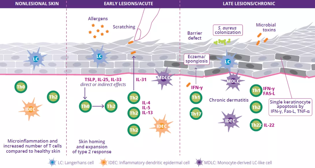
On a pathophysiological level, AD is characterized by an alteration of the skin barrier, a skin and gut dysbiosis, and immune dysregulation with the activation of Th2 lymphocytes. This immune dysregulation leads to a major cytokine surge, which in turn causes the inflammatory reactions.2

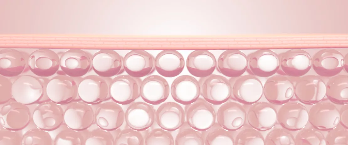
An alteration of the skin barrier is the starting point for a dysbiosis of the skin microbiota characterized by a reduction in bacterial diversity and the proliferation of Staphylococcus aureus. Allergen penetration leads to the activation of keratinocytes and the production of interleukin (IL-33, IL-25, TSLP), resulting in the differentiation of Th2 lymphocytes. These in turn secrete pro-inflammatory cytokines (IL-4, IL-5 and IL-13) characteristic of Type 2 inflammation (Fig 9). These cytokines directly activate the sensory nerves, provoking pruritus.

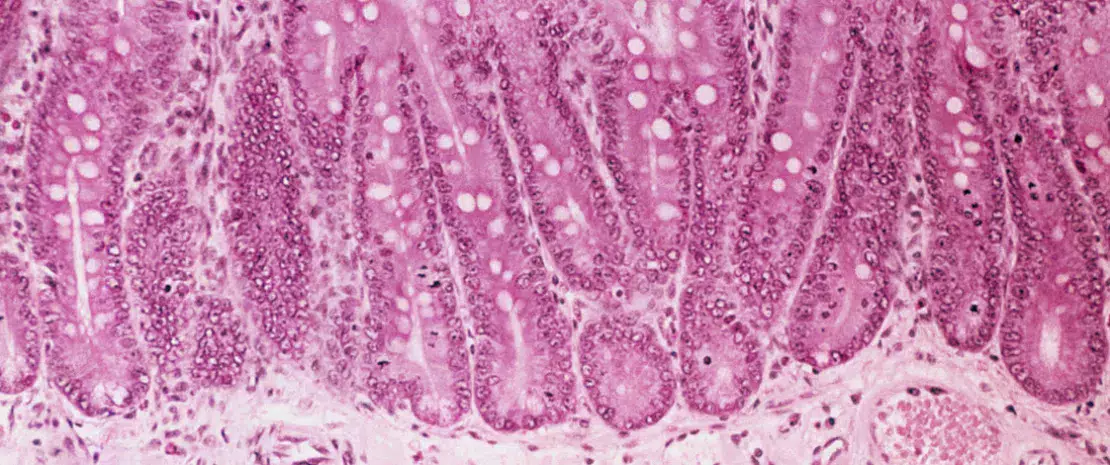
With chronic lesions, the skin barrier repairs itself poorly and becomes thicker, since it is subject to chronic inflammation. There is also a progressive increase in cytokines and Th cells (Th1, Th2, Th22) which secrete cytokines that contribute to the destruction of keratinocytes. Lastly, a gut dysbiosis may play a role in the disease’s pathophysiological mechanism.3

FIGURE 9: Pathogenesis, main mechanisms and pathophysiology of atopic dermatitis.


Adapted from Sugita K et al, 20204

What have recent discoveries about the microbiota taught you? Has your practice changed?

Recent discoveries about the microbiota have led me to better understand the importance of maintaining and repairing the skin barrier to control inflammation. As a systemic treatment, I advise my patients to use a cleansing gel that preserves the skin’s pH (pH ~5, avoid products with a basic pH), as well as a moisturizer and tailored cosmetic products. The findings also help us to better understand the skin’s immunological system and how to respect the skin’s microbiota.

The immune dysregulation in atopic dermatits leads to a major cytokine surge, which in turn causes the inflammatory reactions.2

Pr. Brigitte Dréno

What are your thoughts on the use of probiotics to treat AD or prevent relapse?

There are many ways to rebalance the skin microbiota in case of AD (probiotics, prebiotics, symbiotics, etc.)5 but the postbiotic approach seems to me the most interesting. Postbiotics are preparations of inanimate microorganisms and/or their components that confers a health benefit on the host.6 They can restore the skin barrier via an anti-inflammatory action that allows bacteria to recolonize, therefore having a long-term impact on the microbiota. Oral probiotics or prebiotics are another interesting approach to regulating the intestinal system, which itself plays a general immunomodulatory role in the immune system.7

Summary
Off
Sidebar
On
Migrated content
Désactivé
Updated content
Désactivé
Hide image
Off
Dossier detail

Targeting the gut microbiota to optimize vaccine efficacy?

by Dr Genelle Healey

Photo : Vaccination after antibiotic therapy: effect on immunity and role of the microbiota

Since the COVID-19 pandemic started, the need for a robust and long-lasting immunity induced by vaccines has never been more apparent.20 However, vaccine- induced immune responses are highly variable between individuals and many factors have been suggested that may alter vaccine immunogenicity and efficacy (Fig 8).21Therefore, gaining a better understanding of the factors driving variations in vaccine efficacy is critically important.

Vaccine-induced immune responses are highly variable between individuals and many factors have been suggested that may alter vaccine immunogenicity and efficacy. One factor known to control vaccine efficacy can be the gut microbiota.20

One factor known to control vaccine efficacy is the gut microbiota.21 Interestingly, certain gut microbiota profiles (i.e. higher abundance of Actinobacteria, Clostridium cluster XI and Proteobacteria) are associated with greater vaccine responses against other viral infections such as HIV and rotavirus.22-26 Additionally, a recent study reported that antibiotic-induced intestinal microbiota dysbiosis led to impaired vaccine responses against influenza, such as a reduced antibody-based neutralization of the virus, as well as lower concentrations of antibodies produced in responses to vaccination.27 This and other similar studies provide evidence of the important role that the gut microbiota plays in vaccine efficacy.23,28

FIGURE 8: Factors suggested to alter vaccine immunogenicity and/or efficacy, including intrinsic host factors, behavioural, environmental, nutritional and perinatal factors.

Most of these factors have also been shown to influence the composition of the gut microbiota and baseline immunity. Vaccine immunogenicity is also dependent on vaccine intrinsic factors.

Adapted for Lynn DJ et al, 202120

To date no studies have investigated what impact the gut microbiota may have on SARS-CoV-2 vaccine efficacy, but it seems likely that individuals with gut microbiota dysbiosis may be at increased risk of developing relatively poor vaccine responses. Thus, future research which examines whether specific gut microbiota signatures affect SARS-CoV-2 vaccine efficacy will be critically important.

Sources

20 Lynn DJ, Benson SC, Lynn MA, Pulendran B. Modulation of immune responses to vaccination by the microbiota: implications and potential mechanisms. Nat Rev Immunol. 2021 May 17:1–14.

21 de Jong SE, Olin A, Pulendran B. The Impact of the Microbiome on Immunity to Vaccination in Humans. Cell Host Microbe. 2020 Aug 12;28(2):169-179. 

22 Harris VC, Armah G, Fuentes S, et al. Significant Correlation Between the Infant Gut Microbiome and Rotavirus Vaccine Response in Rural Ghana. J Infect Dis. 2017 Jan 1;215(1):34-41. 

23 Uchiyama R, Chassaing B, Zhang B, et al. Antibiotic treatment suppresses rotavirus infection and enhances specific humoral immunity. J Infect Dis. 2014 Jul 15;210(2):171-82.

24 Huda MN, Lewis Z, Kalanetra KM, et al. Stool microbiota and vaccine responses of infants. Pediatrics. 2014 Aug;134(2):e362-72. 

25 Cram JA, Fiore-Gartland AJ, Srinivasan S, et al. Human gut microbiota is associated with HIV-reactive immunoglobulin at baseline and following HIV vaccination. PLoS One. 2019 Dec 23;14(12):e0225622. 

26 Harris V, Ali A, Fuentes S, et al. Rotavirus vaccine response correlates with the infant gut microbiota composition in Pakistan. Gut Microbes. 2018 Mar 4;9(2):93-101.

27 Hagan T, Cortese M, Rouphael N, et al. Antibiotics-Driven Gut Microbiome Perturbation Alters Immunity to Vaccines in Humans. Cell. 2019 Sep 5;178(6):1313-1328.e13. 

28 Harris VC, Haak BW, Handley SA, et al. Effect of Antibiotic-Mediated Microbiome Modulation on Rotavirus Vaccine Immunogenicity: A Human, Randomized-Control Proof-of-Concept Trial. Cell Host Microbe. 2018 Aug 8;24(2):197-207.e4. 

Summary
Off
Sidebar
On
Migrated content
Désactivé
Updated content
Désactivé
Hide image
Off
Dossier detail

The hygiene hypothesis and the COVID-19 pandemic

by Dr Genelle Healey

COVID-19, a highly contagious respiratory disease caused by the novel SARS-CoV-2 (severe acute respiratory syndrome coronavirus 2) primarily affects the respiratory tract but gastrointestinal symptoms (i.e. diarrhea, constipation, nausea) have also been reported in some patients.14

Preliminary studies have found that the gut microbiota is altered in patients with COVID-19 and that its composition correlates with infection severity, suggesting crosstalk between the gut microbiota and lung in response to SARS-CoV-2 infection.15

Over the last few decades, a significant reduction in microbial diversity and the overt extinction of ancestral microbes has occurred due to improvements in hygiene (e.g. hand washing and sanitizer), modern medications (e.g. antibiotics) and urban living.17

It should be noted that the changes in lifestyle adopted to curb the COVID-19 pandemic could also have a negative impact on the gut microbiome of uninfected individuals.16

These changes in hygiene, and a corresponding increased incidence of several autoimmune and allergic diseases,18,19 have given rise to the hypothesis that they are causally linked (hygiene hypothesis). Notably, practices implemented to prevent the spread of COVID-19, such as physical distancing, frequent hand washing and sanitizer use, reduced travel and face mask wearing, will likely lead to further loss of key gut microbes.16

Taken together, the preventative health practices that have been implemented due to COVID-19 may exert collateral damage on the gut microbiome as well as long term health outcomes, particularly in children born just prior to or during the pandemic.16 Utilizing approaches known to enhance microbial diversity and support a healthy microbiota balance may prevent the negative health impacts associated with the enhanced hygiene practices implemented to prevent the spread of COVID-19.

Summary
Off
Sidebar
On
Migrated content
Désactivé
Updated content
Désactivé
Hide image
Off
Dossier detail

Gut-lung axis in viral respiratory infections

By Dr Genelle Healey

Interestingly, microbiota at both tissue sites appear to be perturbed during respiratory infections, reinforcing the theory that all mucosal sites are interconnected and that the gut-lung axis is bidirectional.1

Gut bacteria, their fragments, as well as SCFAs may translocate across the intestinal barrier and travel along the mesenteric lymphatic system to reach the systemic circulation and modulate immune cells in the lung.11 During Influenza respiratory infections, lung microbiota and immune functions are altered, and a gut microbiota dysbiosis is also observed which may explain the commonly associated gastroenteritis-like symptoms (Fig 7A).10

FIGURE 7: Gut-lung axis during viral respiratory infection (A) and model of microbiota modulation using probiotics (B).

Adapted from Dumas A et al, 20182

Probiotics may be helpful to recover a healthy status (microbiota homeostasis, infection control, modulation of immune responses) via gut microbiota metabolites (SCFAs…) or host-derived products.

There are likely several causes of this gut dysbiosis including loss of appetite (leading to reduced food and calorie intake), as well as inflammatory cytokine release. This may have local consequences: intestinal inflammation, disruption of gut barrier, decreased production of antimicrobial peptides (AMPs), a drop in SCFAs, potentially leading to secondary enteric infections.10

Gut barrier alteration promotes bacterial translocation as well as endotoxin release into the blood, leading to systemic inflammation, lung damage aggravation and increased risk of secondary bacterial infections.10 The reduced SCFA production by the gut microbiota also contributes to the reduced antibacterial immunity seen in the lungs.10 This highlights the vital role that the gut microbiota plays in the lung’s defenses against respiratory infections.

Modulation of the gut microbiota using strategies such as probiotics may help reduce susceptibility to respiratory infections via the gut-lung axis, or they may be helpful in recovering from infection and reaching a healthy status (Fig 7B). Several studies in mice have shown that specific probiotics administered prior to influenza infection led to the reduced accumulation of immune cells in the infected lungs. These probiotics also enhanced viral clearance, improved overall health and reduced alterations in the gut microbiota.12,13

Recommended by our community

"Well, I'm one among people with lung problem (infection)
I get this so important." 
 -@Ahishakiyejanv2 (From Biocodex Microbiota Institute on X)

Summary
Off
Sidebar
On
Migrated content
Désactivé
Updated content
Désactivé
Hide image
Off
Dossier detail

The gut microbiota is involved in the lung’s defense against viral respiratory infections

The microbiota plays a key role in the development, education and function of the immune system, both locally and systemically. While the airway microbiota locally regulates immune function, the gut microbiota can also influence respiratory immunity, via the gut-lung axis.1 Alteration of the lung and gut microbiota has been observed in many respiratory diseases, however whether the dysbiosis at these sites is a cause or a consequence of disease remains to be determined.2 Alteration of gut microbiota composition, through either diet, antibiotic use, aging, or disease, is associated with altered immune responses and homeostasis in the airways,3 highlighting that the gut microbiota can influence disease development throughout the body, including the risk of respiratory infections (Fig 6).4

In comparison to the gut microbiota, studies on the lung microbiota are still in their infancy.5 The lung was originally thought to be sterile but recently, researchers have discovered that the lung harbours its own microbiota, with a composition distinct from the gut microbiota.6

Any factors inducing microbiota dysbiosis can alter the beneficial gut-lung cross-talk, increasing susceptibility to respiratory infections.10

Studies have shown that gut microbiota may be involved in providing protection against viral respiratory infections (such as influenza and respiratory syncytial virus),2 via numerous mechanisms. For example, gut microbial metabolites such as SCFAs (obtained from dietary fiber fermentation by commensal bacteria) and desaminotyrosine (a degradation product of plant flavonoids produced by human gut bacteria)7 influence the lung production of Type I interferon (IFNs) which elicit anti-viral protection. 8,9 Along with microbial metabolites, microbial components (such as LPS) help arm the lungs against viral respiratory infections (Fig 6). The gut microbiota also plays a role in viral (influenza) clearance by stimulating CD8+ T-cell effector function.10

FIGURE 6: The role of the gut microbiota in viral respiratory infections.

Adapted from Sencio V et al, 202010

Any factors inducing gut microbiota dysbiosis (aging, antibiotics, diseases such as obesity, diabetes…) can also alter the normally beneficial gut-lung cross-talk, increasing susceptibility to respiratory infections.10

Sources

Taylor SL, Wesselingh S, Rogers GB. Host-microbiome interactions in acute and chronic respiratory infections. Cell Microbiol. 2016 May;18(5):652-62. 

Dumas A, Bernard L, Poquet Y, et al. The role of the lung microbiota and the gut-lung axis in respiratory infectious diseases. Cell Microbiol. 2018 Dec;20(12):e12966. 

Dang AT, Marsland BJ. Microbes, metabolites, and the gut-lung axis. Mucosal Immunol. 2019 Jul;12(4):843-850.

4 Thibeault C, Suttorp N, Opitz B. The microbiota in pneumonia: From protection to predisposition. Sci Transl Med. 2021 Jan 13;13(576):eaba0501.

5 Huffnagle GB, Dickson RP, Lukacs NW. The respiratory tract microbiome and lung inflammation: a two-way street. Mucosal Immunol. 2017 Mar;10(2):299-306. 

6 Man WH, de Steenhuijsen Piters WA, Bogaert D. The microbiota of the respiratory tract: gatekeeper to respiratory health. Nat Rev Microbiol. 2017 May;15(5):259-270. 

Schoefer L, Mohan R, Schwiertz A, et al. Anaerobic degradation of flavonoids by Clostridium orbiscindens. Appl Environ Microbiol. 2003 Oct;69(10):5849-54.

Antunes KH, Fachi JL, de Paula R, et al. Microbiota-derived acetate protects against respiratory syncytial virus infection through a GPR43-type 1 interferon response. Nat Commun. 2019 Jul 22;10(1):3273. 

9 Steed AL, Christophi GP, Kaiko GE, et al. The microbial metabolite desaminotyrosine protects from influenza through type I interferon. Science. 2017 Aug 4;357(6350):498-502.

10 Sencio V, Machado MG, Trottein F. The lung-gut axis during viral respiratory infections: the impact of gut dysbiosis on secondary disease outcomes. Mucosal Immunol. 2021 Mar;14(2):296-304.

Summary
Off
Sidebar
On
Migrated content
Désactivé
Updated content
Désactivé
Hide image
Off
Dossier detail