Endometriosis considerably changes the quality of life of women suffering from the pain and infertility it causes. Although it affects 1 in 10 women, its mechanisms are poorly understood and treatments for it are unsatisfactory. A recent study1 in animals opens new perspectives by showing that butyrate, a short-chain fatty acid produced by the intestinal microbiota, slows the development of endometriotic lesions.
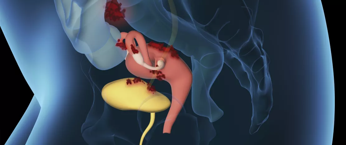
The theories about the origin of endometriosis have not yet been clarified. According to the hypothesis which is prevalent today, fragments of the endometrium migrate out of the uterus into the peritoneal cavity during retrograde menstruation and implant themselves into the surrounding tissues. However, although 90% of women have retrograde menstruation only 10% have endometriosis. In addition, the current treatments for the disease have side effects and do not prevent relapses.
In order to offer women new treatment solutions, other factors contributing to the alteration of the peritoneal environment and the development of lesions must be identified. In this context the intestinal microbiota has aroused the attention of researchers. In fact, the intestinal microbiota of women suffering from endometriosis presents a lower alpha diversity and a changed bacterial composition compared to women without endometriosis. In addition, the metabolites produced by the colonic flora of a mouse model for endometriosis are different to those of the control mice. This is important because it is through the metabolites from the transformation of dietary fibres that the intestinal microbiota provides its benefit to the human body. Amongst these, the short-chain fatty acids (SCFA), such as butyrate, acetate or propionate, in particular have anti-proliferation and anti-inflammatory effects. The authors of the study published in Life Science Alliance therefore considered the role of these SCFAs in endometriosis in vivo on a mouse model for endometriosis and in vitro on cells of endometriotic lesions.
Butyrate inhibits the growth of lesions by activating several mechanisms
The initial results show that endometriosis upsets the balance of the intestinal microbiota of mice by causing a reduction in the production of butyrate. The team also observed that butyrate (and not other SCFAs such as acetate or propionate) inhibits the growth of endometriotic lesions. Butyrate acts through at least three mechanisms: by activating membrane receptors coupled to G-proteins (GPCRs): GPR43 and GPR109A, by inhibiting the enzyme histone deacetylase (HDAC) and activating Rap1GAP (protein activating GTPase Ras-proximate-1). Rap1GAP blocks the Rap1 signal pathway involved in the proliferation, migration and adhesion of cells. It is already known to be a tumour suppressor, including in endometrial cancer.
New studies must now determine whether, in women suffering from endometriosis, the faecal butyrate level is lower than in women who do not suffer. If this is the case, different approaches intended to prevent the development of lesions could be tested: diet, butyrate analogues, butyrate-based supplements or probiotics inducing the production of butyrate.
Are you dreaming of having a (very) good reason to eat dark chocolate during the Christmas and New Year festivities? An unpublished study is handing it to you on a plate! Cocoa could increase intestinal microbial diversity and cause a virtuous feedback loop to our brain which is seen as a sustainable “feel good” effect. Food lovers, don’t feel guilty any more!
The characteristics of mood disorders are feelings of sadness, helplessness, despair and irritability. In order to prevent and treat these disorders better, amongst other things scientific research is looking at nutrition and the intestinal microbiota, our second brain. Certain foods, such as chocolate, may regulate our mood. However, the results are often controversial. For the first time a clinical trial seeks to verify and explain the positive effects of dark chocolate on our mood. So let’s open the box together and it will be explained to you.
Dark chocolate and good humour: the scientific proof
After three weeks the participants who consumed 85% cocoa dark chocolate showed a significant reduction in negative feelings, whereas the 70% group did not show any notable change. The effects of cocoa on our good humour therefore seems to depend on the dose consumed. Be careful, we are talking about cocoa here, the praline sweets we eat at Christmas contain less than 50%!
Intestinal microbiota and chocolate: a guilty pleasure that does some good?
The scientific study has also been able to show that 85% cocoa dark chocolate would increase the diversity of the microbial communities in the intestine. For the authors, it is the large quantities of polyphenols in the cocoa which have a positive action on the intestinal flora slowing the growth of pathogenic bacteria and encouraging the growth of those that are beneficial. If the intestine and chocolate seem to work to the benefit of our health rather than vice versa, one question remains: what is the link with our good humour? Surely the brain is the control tower for our emotions?
From intestine to brain: a communication network worth of Charlie and the Chocolate Factory!
The bioaccumulation of medicines by the intestinal bacteria changes their availability and the bacterial secretion of metabolites. With, into the bargain, possible dysbiosis and implications in terms of pharmacokinetics, adverse events and responses to medicines.
We know: medicines have an influence on the intestinal microbiota. But did you that there are also interactions in the other direction? With, into the bargain, a positive or negative effect on the efficacy of medicines. Take for example lovastatin and sulphasalazine, they are chemically transformed by intestinal bacteria into their active forms, whereas digoxin is inactivated by bacterial metabolism. More than 100 molecules have recently been notified as being affected by the intestinal microbiota in this way. And according to the results from a research team, the mechanisms in play are far from limiting themselves to a single biotransformation...
Biotransformation and above all bioaccumulation
The study in question has sifted through the interactions between 25 representative strains of human intestinal bacteria and (sidenote:
12 molecules administered orally and 3 controls: digoxin (highly specific interaction with Eggerthella lenta), metronidazole and sulphasalazine, medicines which are known to be metabolised by several intestinal bacteria
). The results? The in vitro cultures of 15*25= 375 bacteria-medicines pairs show 70 bacteria-medicines interactions, 29 of which (18 species, 7 medicines) were unknown until now. Above all, 12 of these 29 new interactions are explained by biotransformation phenomena. All the other cases, i.e. 17 interactions (14 species, 4 medicines), are based on bioaccumulation: the bacteria store the medicine in their cells without modifying it, and in most cases without any effect on the growth of the bacteria. Amongst the medicines that are exclusively bioaccumulated, (sidenote:
Duloxetine
Antidepressant, a selective serotonin and norepinephrine reuptake inhibitor
) and the antidiabetic, rosiglitazone, are noted. However, bioaccumulation is not routine: some molecules (montelukast, roflumilast) can be bioaccumulated by some bacterial species and biodegraded by others.
The case of duloxetine
As an example, the team studied the bioaccumulation of duloxetine more closely. Duloxetine binds to numerous bacterial enzymes and modifies the secretion of metabolites by the bacteria concerned. When it is tested in a microbial community of 5 bacterial species containing both accumulating and non-accumulating bacteria, duloxetine greatly changes the composition of the community. In fact, apart from the sequestration of this medicine which is harmful for some bacteria, the bioaccumulation of this medicine causes the secretion of metabolites by some species (Streptococcus salivarius) which will serve as a substrate to nourish others (Eubacterium rectale), thus greatly increasing their abundance. In this way medicines intended for humans seem capable of modulating the intestinal microbial communities, not only by direct inhibition but also by creating synergies of cross-feeding. The results were confirmed on the model, Caenorhabditis elegans: bioaccumulating bacteria reduce the effect of duloxetine on the movement of this worm.
The results of this study indicate that the bioaccumulation of medicines within the intestinal bacteria would modify their availability and bacterial metabolism. As this could cause individual repercussions within the composition of the intestinal microbiota, and also for the pharmacokinetics and the drug response. The authors suggest routinely establishing search for reciprocal interactions between bacteria and medicines so as to estimate the side effects in the best possible way.
Less diverse and low on certain bacteria: telltale signs in the gut microbiota of people with type 2 diabetes. Valuable new data open up new directions for preventing and curing this scourge of global health.
A leading cause of blindness, kidney failure, stroke, and amputations1, diabetes is a serious disease that happens when the pancreas does not produce enough insulin (type 1 diabetes2) or, in the majority of cases, when the body develops a resistance to the hormone (type 2 diabetes, T2D3). Faced with a fourfold rise in the number of cases over the past 40 years, with over 420 million people now affected around the world1, public authorities and international organizations are taking action to improve access to care. On November 14, every year since 1991, World Diabetes Day aims to raise awareness of the disease.
Microbiota specific to T2D...
Faced with these facts, researchers are doing everything they can to work out not only the causes of the disease, but also how to prevent it. An increasing amount of attention is being paid to the gut microbiota. And for good reason, since several studies have spotted a link between its composition and the risk of T2D. Most recently, researchers brought out the big guns and managed to assemble over 2,000 subjects (compared with just a few hundred for previous studies) to compare the microbiota of people with and without diabetes.
...with changes evident even at the early stages
The results are enlightening: T2D is less common in people whose microbiota is diverse and rich in certain bacteria able to produce a particular short-chain fatty acid (SCFA) known as (sidenote:
SCFAs
Short Chain Fatty Acids are a source of energy (fuel) for the cells of the individual. They interact with the immune system and are involved in the communication between the intestine and the brain.
Sources:
Silva YP, Bernardi A, Frozza RL. The Role of Short-Chain Fatty Acids From Gut Microbiota in Gut-Brain Communication. Front Endocrinol (Lausanne). 2020;11:25.
), which is beneficial for metabolism and cell activity, and has anti-inflammatory properties. But the researchers went further still and demonstrated, for the first time, that these people are also less affected by (sidenote:
Insulin-resistance
An altered response of cells to the action of insulin (a hormone that helps the body use sugar for energy), insulin resistance results in poor regulation of blood sugar levels.
Sources Inserm. La résistance à l’insuline, une histoire de communication. 2018. Centers for disease control and prevention. Diabetes - Resources and Publications -Glossary), a phenomenon that appears very early on in the development of T2D. The bottom line? There are telltale signs in the microbiota even at the very early stages of development of the disease.
These results are very promising and provide a new insight into our understanding of the factors involved in the pathogenesis of diabetes. Better still, they could pave the way for new targeted treatments for the rising number of patients.
A new study of over 2,000 subjects has found that a diverse intestinal microbiota and an abundance of 12 butyrate-producing taxa are linked to a lower risk of type 2 diabetes and insulin resistance.
Even though some studies had already made a link between the intestinal microbiota and type 2 diabetes (T2D), they were limited by a small sample size and there was ongoing debate as to the exact taxonomic units involved (results not always reproducible, insufficient power, etc.) This is why researchers decided to embark upon a large-scale study of 2,166 subjects taken from two Dutch cohorts, to investigate links between the composition of the intestinal microbiota (analyzed using bacterial 16S RNA gene sequencing and amplification on fecal samples) and T2D (verified by two physicians using WHO criteria (blood glucose, use of anti-diabetic drugs, etc.).
Even though it used a cross-sectional design (i.e., data about the microbiota and T2D collected at the same time), the analyses were adjusted for several confounders, such as energy intake, body mass index, education level, etc. Another strength and novel aspect of the study was the investigation of links between the microbiota and insulin resistance (IR), measured using fasting insulin and blood glucose, a significant subclinical parameter indicating an early stage in the pathogenesis of T2D.
Does a diverse microbiota strengthen protection?
Several indicators of microbiota (sidenote:
α diversity
A measure indicating the diversity of a single sample, i.e. the number of different species present in an individual.
Hamady M, Lozupone C, Knight R. Fast UniFrac: facilitating high-throughput phylogenetic analyses of microbial communities including analysis of pyrosequencing and PhyloChip data. ISME J. 2010;4:17-27. https://www.nature.com/articles/ismej200997) were linked to lower IR and a reduced prevalence of T2D. In turn, (sidenote:
β diversity
A measure indicating the species diversity between samples, it allows to assess the variability of microbiota diversity between subjects.
Hamady M, Lozupone C, Knight R. Fast UniFrac: facilitating high-throughput phylogenetic analyses of microbial communities including analysis of pyrosequencing and PhyloChip data. ISME J. 2010;4:17-27. https://www.nature.com/articles/ismej20099) was linked to IR. Finally, an abundance of seven taxonomic units - belonging to the Christensenellaceae and Ruminococcaceae families and the genus Marvinbryantia - were linked to a lower risk of IR, and an abundance of five other taxa - the Clostridiaceae and Peptostreptococcaceae families, and the genera Clostridiumsensu stricto, Intestinibacter and Romboutsia – to a lower risk of T2D.
Butyrate-producing taxa that reduce the risk of T2D
These 12 taxonomic units, 10 of which have been linked for the first time to a lower risk of T2D or IR, are known to produce butyrate, a short-chain fatty acid produced by the breakdown of dietary fibers by bacteria. Enhanced mitochondrial activity, improved energy metabolism, and reduced endotoxemia and inflammation are some of the mechanisms put forward to explain these effects. However, ad hoc studies are still needed to validate its role in glucose metabolism and the prevention of diabetes.
Nevertheless, the links that have been identified between, on the one hand, microbiota diversity and an abundance of butyrate-producing bacteria, and on the other hand, a lower risk of IR and T2D, shed further light on the etiology, pathogenesis, and treatment of type 2 diabetes.
Language is like fashion. What was popular twenty years ago isn’t popular now, but it might make a comeback down the line. Once frequently used, the term flora, implying the gut flora, has gradually given way to microbiota in the plural sense, since microbiota exist not only in the gut but also in the nose, mouth, vagina, skin, etc. At the same time, microbiota shouldn’t be confused with microbiome. See below for further detail on a debate not limited to semantics.
As soon as we look into the minuscule world that resides in our body, certain words tend to pop up. Such words are not always clearly defined or appropriately used. Some definitions are therefore necessary.
From the ancestral term “flora”...
The term “flora” has certainly been around the longest. It once generally referred to the digestive system (the “gut flora”) and denoted, according to the Larousse medical dictionary, the “collection of microorganisms that normally resides in the intestine”. At that time, it was thought that the gut flora contained mainly bacteria1. So when the term “flora” was used, it generally referred to the bacterial population living in the gut.
... to the contemporary term “microbiota”
As science progressed, this view of the flora proved to be far too simplistic. On the one hand, our digestive system hosts far more than bacteria. Viruses, fungi (including yeasts), and parasites also make it their home2.
On the other hand, the gut is far from being the only part of the body that hosts such microbial communities. For example :
(and no, urine isn’t sterile!) all have their own flora.…3
Little by little, another term, “microbiota”, has come into use. This term unambiguously refers to all communities of microorganisms (and not only bacteria). “Microbiota” is always accompanied by an adjective that specifies its location (skin microbiota, oral microbiota, etc.), since each microbiota has its own characteristic set of microorganisms.
And what about “microbiome”?
Sometimes the difference hangs on thread, or rather a couple of letters. Indeed, just two letters separate “microbiota” from “microbiome”. And yet these two terms are false friends. The first denotes the population of bacteria, viruses, etc., residing in a specific area of our body but the second refers to something completely different: the genetic material of this community taken as a whole, in other words everything that the microorganisms residing there know how to do (produce specific molecules, make specific types of membrane). Imagine putting all the microorganisms that make up a microbiota into a blender, thereby erasing their individual identity, and only being left with the genetic material of the resulting microbial soup. In a village, the “microbiote” would be the list of inhabitants and the “microbiome” the list of what these inhabitants collectively know how to do (make bread, build a house, etc.).
But be careful!
In English, the terms “microbiota” and “microbiome” are often used without distinction and some articles translated from English into other languages confuse the two terms. At least now you know how to tell the difference.
Stomatitis, cavities, gingivitis and other gum disease… Even with antiretroviral therapy, HIV-infected individuals are at an increased risk of oral diseases. The immune disturbances behind this vulnerability are not fully understood, but persistent inflammation and an imbalance of the oral microbiota appear to be involved.
December 1 is World AIDS Day. To mark the occasion it's worth bearing in mind that the human immunodeficiency virus (HIV) attacks specific white blood cells called CD4+ T cells. The weakening of the immune system allows opportunistic infections, cancer, and other diseases of the organs to develop. Today, antiretroviral therapy can restore CD4+ T cell counts and lower the viral load to undetectable levels. Unfortunately, this does not protect against all of HIV's adverse effects. Inflammation of the oral cavity is particularly common HIV-infected individuals and is often associated with an imbalance in the oral microbiota.
Gut imbalance plays role in immune response to infection
HIV and oral diseases: initial hypotheses and solutions
Why does HIV infection weaken the oral mucosa and increase the risk of oral diseases, even when controlled by treatment? One possible explanation is that HIV impairs the production of secretory immunoglobulin A antibodies in the saliva, which are considered the first line of defense against oral pathogens. It may also be because the loss of CD4+ T cells, as in the gut microbiota, results in inflammation and oral dysbiosis. However, few studies have analyzed the oral microbiotacomposition of HIV-infected individuals and the results of such studies are not always consistent. In addition, it is currently very difficult to assess the impact of the virus itself, versus that of the treatment, on oral bacterial communities.
(sidenote:
A scientific review published in September 2021 provides an update.
). Such research is essential to the discovery of therapies that relieve the distressing oral symptoms of HIV. "Probiotic" approaches have already been proposed for the prevention of oral diseases. One example is the displacement of one bacterial strain that causes cavities with another that is engineered to have low pathogenicity. Well-conducted clinical trials are now needed to assess the efficacy of using probiotic strains and antimicrobial agents to improve oral immune functions.
Sources
Coker, Modupe O et al. “HIV-Associated Interactions Between Oral Microbiota and Mucosal Immune Cells: Knowledge Gaps and Future Directions.” Frontiers in immunology vol. 12 676669. 20 Sep. 2021.
A less mature gut microbiota, affecting immune development, may result from intrauterine life.This finding may lead to the early identification of infants at risk of allergyand may even help us better prevent the development of allergic sensitization.
Gut microbiota maturation begins immediately after birth. This maturation continues through the first few years of life, in parallel with that of the immune system. Gut microbiota maturation and immune development are both implicated in allergic diseases, with prenatal factors suspected in each case. Meconium, the first stool of an infant after birth, contains metabolites produced in utero. It reflects perinatal influences, since it begins forming by gestational week 16, while it also contains the starting material for the initial microbiota. Hence this study attempted to link metabolic signatures within the meconium, microbiota maturation and immune system development.
Less maturation, more atopy
After analyzing (via 16S rRNA sequencing) stool collected at 3 months and 1 year from 950 children in the (sidenote:
Canadian CHILD Cohort Study
Canadian Healthy Infant Longitudinal Development study, a prospective study of children recruited before birth between 2008 and 2012
) cohort, the researchers made a first discovery: the gut microbiota of the future allergic children was less mature, even before the appearance of the atopy. Thus, the 212 infants who subsequently developed atopy at 1 year of age had a less mature microbiota at 3 months than those who did not become atopic. The relative abundance of 13 of the 15 taxa most involved in maturation of the microbiota was lower in atopic infants.
Influence of prenatal exposures
To understand the origin of this difference in maturation, the researchers went back in time and analyzed meconium samples in a subgroup of 100 children. They observed lower bacterial diversity in the future atopic children. Metabolic diversity was also reduced, with fewer molecules associated with the metabolism of amino acids, vitamins and hormones. This suggests that differences influencing the development of the microbiota and, ultimately, immune development, exist from birth. Thus, atopy at 1 year of age is associated both with a metabolically less rich meconium at birth and with a reduction in the diversity and maturation of the microbiota at the beginning of life. A potential mechanism of action is suggested: meconium metabolites, which reflect prenatal exposures, may be metabolized and fermented by bacteria. Thus, the gut microbiota at the beginning of life, and ultimately immune development, would be impacted by intrauterine life.
A better understanding of the prenatal determinants of meconium composition, and of the direct and indirect effects of its metabolites on immune development and bacterial colonization in newborns, may ultimately help prevent the development of allergies. It may even allow us to predict – via metabolic signatures – the risk of developing allergies, even if the researchers’ first studies (combining meconium data and clinical data from mother and child) gave results that are certainly encouraging but still far from precise.
The latest scientific research suggests that our microbiota form an interconnected network linking the gut, lungs, skin, mouth, urinary tract, and genital tract. Communication between these ecosystems may greatly influence our health and susceptibility to disease.
The gut microbiota is the most well-known of the body’s microbiota. However, the body hosts many other microbial ecosystems, each of which receives considerable attention in scientific publications and the media. Each new study shines further light on the decisive role they appear to play in our health. Let’s take a closer look at this vast “network”.
Human microbiota: a community of microorganisms...
The human microbiota is made up of the set of microorganisms – mainly bacteria, but also viruses and fungi – that live in our body. The vast majority (70%) are found in the digestive system, which is home to 1.5 kg of bacteria. But five other organs also (sidenote:
Host
The body provides food and shelter to the microorganisms that colonize it.
) a microbiota: the skin, mouth, lungs, urinary tract, and genitalia. The microorganisms in the microbiota are (sidenote:
симбиотические
Тесная и взаимовыгодная связь между двумя или более организмами
). We provide them with the necessary conditions for survival and in return they contribute to the proper functioning of our body (digestion of food, protection against infection, synthesis of vitamins). A win-win situation! A balanced microbiota helps ensure good health. On the other hand, an unbalanced microbiota – or dysbiosis – is conductive to numerous diseases.
... within a network of interconnected ecosystems
At first glance, the body’s various microbial ecosystems appear to influence only their respective host organ. However, numerous studies have shown that an imbalance in one organ can have an impact on the others. For example, a disturbed gut microbiota has been linked with certain diseases of the skin (dermatitis, psoriasis) or lungs (asthma, chronic bronchitis, cancer), while poor oral hygiene increases the risk of developing lung infections. The latest scientific research suggests that these different ecosystems form an interconnected network with the gut at the center. Gut-brain axis, gut-skin axis, gut-lung axis, gut-mouth axis, gut-liver axis... and so on. The gut channels information and acts as a relay with peripheral ecosystems.
Is the gut the only control tower?
At the same time, some studies suggest that exchanges between different regions of the body can take place without the gut’s involvement (e.g. lung-mouth axis or exchanges linked to anatomical proximity, such as between urinary and genital microbiota). Thus, the various microbiota distributed among the six organs form a network in constant communication which significantly influences our health.
Promising implications for research and medicine
Research on the microbiota is still in its infancy. To confirm the hypothesis of an interconnected network of microbiota centered around the digestive system, more research is needed on the microbiota as a whole. Despite this, the applications in medicine seem promising: by restoring the balance of one ecosystem, it may be possible to modify the balance of another, thus leading to innovative treatment strategies.
A literature review published in August 2021 in Nutrients hypothesizes that the human microbiota is organized as an interconnected network around the digestive tract and between different regions of the body. The study suggests that a dysbiosis in one organ can unbalance other microbiota and contribute to the development of multiple health problems.
The human microbiota is known to play a key role in host health. It is distributed between the digestive tract (70% of human microbiota), skin, lungs, mouth, urinary tract, and genital tract. These microbiota appear to be compartmentalized, hosting different microorganisms. However, a dysbiosis at one site appears to have remote consequences, leading to metabolic, inflammatory, immune, neoplastic, cognitive, degenerative, and genetic disorders. The authors of this review analyzed the communication axes between the various human microbiota with the aim of exploring the link between dysbiosis and illness.
Microbiota: communication axes centered around the gut?
Numerous studies report a link between gut dysbiosis and infectious (tuberculosis, pneumonia), genetic (cystic fibrosis), inflammatory (asthma, COPD) and neoplastic respiratory diseases. The composition of the gut microbiota varies according to the disease but a proliferation of Proteobacteria and Firmicutes is observed in each case. In addition, SARS-CoV-2 has been associated with a gut dysbiosis that may persist after recovery.
Gut-liver axis
Small intestinal bacterial overgrowth (SIBO) has been found in more than half of patients suffering from liver cirrhosis and is also associated with endotoxemia.
Gut-skin axis
Numerous studies have shown a link between a gut microbiota imbalance and skin diseases such as inflammatory dermatitis and psoriasis.
Gut-mouth axis
Some studies have reported an oral dysbiosis in patients with colorectal cancer and liver cirrhosis, which may be caused by a migration of periodontal bacteria to the gut. Oral and digestive dysbiosis may also be associated with systemic disorders such as rheumatoid arthritis and lupus.
Mouth-lung axis
The respiratory microbiota is mainly formed by the migration of microorganisms from the mouth during the first weeks of life. Several studies have associated oral dysbiosis with asthma and lung disease. Furthermore, SARS-CoV-2 accumulates in the oral cavity, resulting in dysbiosis. Covid-19 patients in ICU are at greater risk of pulmonary superinfection by bacteria migrating from the oral microbiota.
Vaginal-urinary axis
The urinary and vaginal microbiota are contiguous and communicate with each other, sharing many bacteria. One study has found changes in the urinary microbiota in cases of bacterial vaginosis.
Oral-genital-rectal axis
In women, the rectal and vaginal microbiota are connected, with certain rectal microbiota acting as risk factors for bacterial vaginosis. In men, disturbances of the seminal microbiota observed in infertile patients have been associated with changes in the rectal microbiota. The microbiota of the male and female genitalia communicate during intercourse. Several studies have linked the microbiota with the risk of contracting an STI. At the same time, the microbiota of the penis may also influence the occurrence of bacterial vaginosis. Moreover, other studies suggest that the oral microbiota may also be involved in the development of bacterial vaginosis, with a dysbiosis of the oral and vaginal microbiota frequently observed in such patients.
Mechanisms of communication between microbiota
Several non-exclusive mechanisms may explain the connections between the various microbiota:
Systemic diffusion of immunomodulatory metabolites from dietary fiber fermentation, particularly short-chain fatty acids (SCFAs). These metabolites may reach other microbiota through the bloodstream. For example, an accumulation of SCFAs in the respiratory tract may be responsible for lung inflammation and major susceptibility to allergens.
Systemic circulation of bacterial fragments, including extracellular bacterial vesicles.
Migration of entire bacteria:
Due to contiguity (e.g. between oral cavity and respiratory tract, or between urinary and genital tracts).
Through systemic passage where epithelial barriers lose integrity (particularly translocation to digestive tract).
New avenues for research
The authors conclude their study by highlighting the need for further research to clarify the interconnections between the microbiota and particularly whether dysbiosis is a cause or effect of disease. Multiomic research integrating global data (genomes, transcriptomes, metabolomes, proteomes, microbiomes, phenotypes) should allow a better understanding of the relationships between microbiota, host organs and human disease, leading to new therapeutic approaches.