The impact of western diet on the mucus layer

by Dr. Larissa Celiberto

Fiber ingestion helps ensure regular bowel movements. Moreover, since fiber is not digestible by human enzymes, it can also serve as a key nutrient for the gut microbiota as these microbes produce distinct enzymes that are able to ferment and degrade these fibers into important metabolites such as SCFAs.28

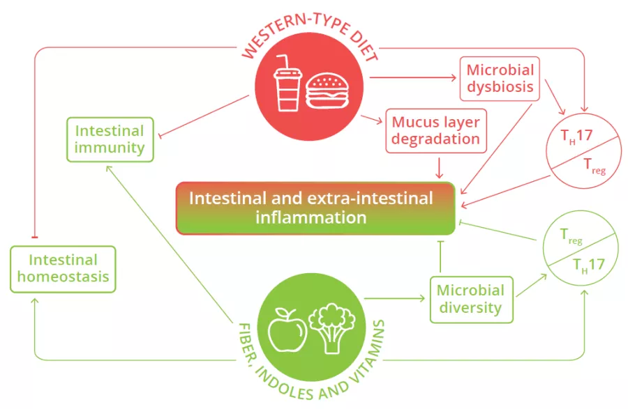
Microbial dysbiosis, mucus layer degradation and alteration of the balance of pro- and anti-inflammatory T cells in the intestine are observed in individuals consuming Western-type diets, leading to intestinal and extra-intestinal inflammation.26

FIGURE 4: Impact of Western diet versus diets rich in fiber and vitamins on local and systemic homeostasis and immunity.

Adapted from Siracusa F et al, 2019.26

The intestinal mucus layer can also serve as an alternative energy source for certain gut microbes (80% of its mass being composed of sugars) when the diet is lacking in fiber.29 This increase in mucus foraging by gut bacteria can prove detrimental as animal studies have shown that mice fed a diet with no fiber are more susceptible to intestinal infections and inflammation. This susceptibility was due to the resident microbiota eroding the mucus layer, such that it could no longer protect the underlying epithelium from invading pathogens.29 Western diets shift the microbiota composition away from fiber degrading bacteria in favor of bacterial species that thrive on mucus (Fig 4).30 Thus, our Western diets may be leading to the loss of protective microbes and the expansion of microbes that weaken key defenses and barriers in the intestine, thereby helping trigger chronic intestinal inflammation.

What is western diet?

Western style diets largely consist of specific dietary fats, sugars and processed foods, environmental pesticides and are lacking in fiber. Consumption of the Western style diet has been linked to obesity as well as inflammatory and metabolic conditions such as type 2 diabetes, insulin resistance and IBD.26 Aside from low quality food with high calories, it is also largely devoid of fiber due its lack of fruits, vegetables, legumes and whole grains, which makes achieving the recommended daily fiber intake of 28–35g27 for adults extremely difficult.

Summary
Off
Sidebar
On
Migrated content
Désactivé
Updated content
Désactivé
Hide image
Off
Dossier detail

Factors influencing microbiota development and maturation of the immune system early in life

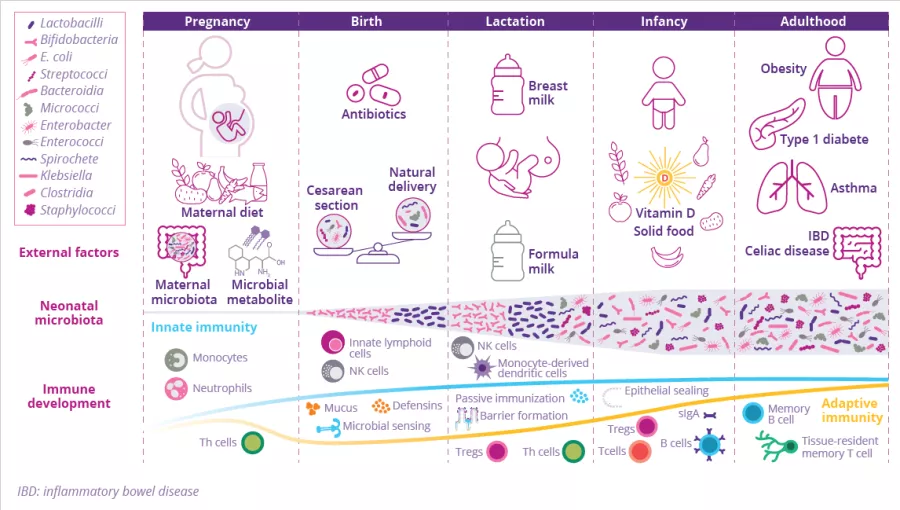
Birth represents the biggest substantial environmental change in life as the newborn is exposed for the first time to a countless variety of microbes which colonize all body surfaces, leading to the establishment of the commensal microbiota in parallel with the immune system. Many factors shape the composition of the gut microbiota and the maturation of the newborn immune system (Fig 3). Discrepancies in the microbiota and immunity crosstalk during each developmental stage can have long-term effects on disease susceptibility.13

Birth impacts gut microbiota composition...

by Dr. Travis J. De Wolfe

The mode of delivery impacts what type of bacteria from the mother are transmitted to the neonatal intestine.14 Babies delivered via the birth canal often carry many gut bacteria that synthesize lipopolysaccharide (LPS), a major membrane component of Gram-negative bacteria that can properly train the human immune system to properly respond to microbial threats.15 In contrast, children delivered by caesarean section are predisposed to being colonized by opportunistic pathogens that circulate in hospitals.14


…As well as maturation of immune structure

These differences in initial microbial colonization can affect the subsequent maturation of the local innate lymphoid structures and alter the population of protective regulatory T cells (Treg), resulting in long-term effects on human intestinal physiology. Maturation of T cells and induction of immune factors can protect against, or in some cases, contribute to autoimmune-mediated diseases (diabetes, multiple sclerosis…) that develop later in life.15,16

Antibiotics impact on immune responses

By Dr. Pascal Lavoie

Antibiotics are essential to treat serious bacterial infections, however unnecessary antibiotic exposure can have serious adverse health consequences and should be avoided (ie. when the infection is due to a virus). In older adults, prolonged antibiotic use can lead to the overgrowth of a gut bacterial pathogen called Clostridioides difficile, with potentially life-threatening health consequences, particularly in the elderly.17 Overusing antibiotics can also promote antimicrobial resistance, which can limit treatment options for future infections.18 In animal models, antibiotic based perturbation of the gut microbiome alters immune functions and immune response thresholds.19 Data in humans suggest that unnecessary antibiotic use may increase the risk of developing chronic health problems, like type I diabetes, asthma, allergies or even obesity.20 Prolonged antibiotic use (> 1 week) is known to reduce the diversity of the gut microbiome, with babies born prematurely being the most vulnerable to perturbations in their gut microbiome. Prolonged, broad spectrum antibiotic use in the mother or premature infant reduces gut bacterial diversity, increasing the risk of sepsis and necrotizing enterocolitis.21 Overall, the data in humans support the concept that the gut microbiome plays a major role helping babies develop into healthy adults. While the risks of excessive antibiotic exposure in adults are less severe, they may still impact the development of their immune responses, therefore antibiotic usage at any age should be restricted to those cases where they are necessary.

Dysbiosis is not universal and is defined for each individual, according to their state of health. A common definition describes it as a compositional and functional alteration in the microbiota that is driven by a set of environmental and host-related factors that perturb the microbial ecosystem.9

FIGURE 3: Environmental factors influencing the development of the newborn microbiota and mucosal immune system.

Adapted from Kalbermatter C et al, 202113

Throughout pregnancy, microbial metabolites (originating from the maternal microbiota and diet) influence fetal immune development. At birth, microbiota colonization starts in parallel with development of the immune system. At this stage, the newborn is still dependent on maternal protection, which is ensured through breastfeeding: maternal milk contains mother-derived bacterial antigens that stimulate the maturation of the innate mucosal immune system. Regarding gut microbiota colonization, Enterococcacae, Clostridiaceae, Lactobacillaceae, Bifidobacteriaceae, Streptococcaceae dominate in the first weeks of life. The introduction of solid food in an infant’s diet leads to an increase in gut microbiota diversity, evolving to a more adult-like microbiota: the abundance of Bifidobacteriaceae decreases, while Bacteroides, Ruminococcus, and Clostridium become more prevalent. Birth mode, breast milk, solid food, and the intake of antibiotics are factors that shape the early life microbiota and the neonatal immune system.

PROOF OF CONCEPT: gut microbiota colonization is essential for immune system development

by Dr. Travis J. De Wolfe

Studies with germ-free mice have demonstrated the important role that the microbiota plays in preventing a faulty immune system.22 Germfree mice are impaired in the production of CD4-positive T helper immune cells, whereas selectively colonizing these mice with Clostridia, a commensal bacterial group, can induce the production of these cells that subsequently promote antimicrobial defenses in the gut and protect against pathogen infection.23 IgA antibodies are another critical component of the immune system, that are deficient in germ-free mice. These antibodies bind to commensal bacteria and prevent them from escaping the GI tract. Selective colonization of germ-free mice with an Escherichia coli strain, or distinct Bacteroides strains trigger a rapid restoration/normalization of IgA.24,25

Sources

13 Kalbermatter C, Fernandez Trigo N, Christensen S, et al. Maternal Microbiota, Early Life Colonization and Breast Milk Drive Immune Development in the Newborn. Front Immunol. 2021 May 13;12:683022. 

14 Shao Y, Forster SC, Tsaliki E, et al. Stunted microbiota and opportunistic pathogen colonization in caesarean-section birth. Nature. 2019 Oct;574(7776):117-121.

15 Wampach L, Heintz-Buschart A, Fritz JV, et al. Birth mode is associated with earliest strain-conferred gut microbiome functions and immunostimulatory potential. Nat Commun. 2018 Nov 30;9(1):5091. 

16 Vatanen T, Kostic AD, d'Hennezel E, et al. Variation in Microbiome LPS Immunogenicity Contributes to Autoimmunity in Humans. Cell. 2016 May 5;165(4):842-53. 

17 Guh AY, Kutty PK. Clostridioides difficile Infection. Ann Intern Med. 2018 Oct 2;169(7):ITC49-ITC64.

18 Costelloe C, Metcalfe C, Lovering A, et al. Effect of antibiotic prescribing in primary care on antimicrobial resistance in individual patients: systematic review and meta-analysis. BMJ. 2010 May 18;340:c2096.

19 Konstantinidis T, Tsigalou C, Karvelas A, et al. Effects of Antibiotics upon the Gut Microbiome: A Review of the Literature. Biomedicines. 2020 Nov 16;8(11):502.

20 Sarkar A, Yoo JY, Valeria Ozorio Dutra S, et al. The Association between Early-Life Gut Microbiota and Long-Term Health and Diseases. J Clin Med. 2021 Jan 25;10(3):459.

21 Walker WA. The importance of appropriate initial bacterial colonization of the intestine in newborn, child, and adult health. Pediatr Res. 2017 Sep;82(3):387-395. 

22 Sommer F, Bäckhed F. The gut microbiota--masters of host development and physiology. Nat Rev Microbiol. 2013 Apr;11(4):227-38. 

23 Ivanov II, Atarashi K, Manel N, et al. Induction of intestinal Th17 cells by segmented filamentous bacteria. Cell. 2009 Oct 30;139(3):485-98.

24 Hapfelmeier S, Lawson MA, Slack E, et al. Reversible microbial colonization of germ-free mice reveals the dynamics of IgA immune responses. Science. 2010 Jun 25;328(5986):1705-9. 

25 Yang C, Mogno I, Contijoch EJ, et al. Fecal IgA Levels Are Determined by Strain-Level Differences in Bacteroides ovatus and Are Modifiable by Gut Microbiota Manipulation. Cell Host Microbe. 2020 Mar 11;27(3):467-475.e6

Summary
Off
Sidebar
On
Migrated content
Désactivé
Updated content
Désactivé
Hide image
Off
Dossier detail

The infant's gut at the heart of immunity

By Dr Travis J. De Wolfe

Development of innate immune barriers

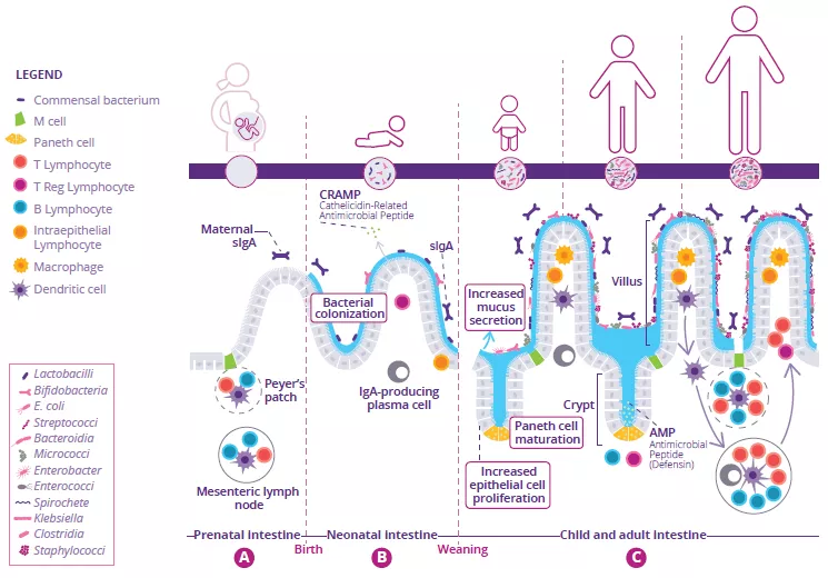
Development of the intestinal immune system begins before birth and continues throughout neonatal weaning. In utero, immature lymphoid structures including Peyer’s patches and mesenteric lymph nodes are generated (Fig 1A). Since these structures are not fully functional until later in development, to compensate, antimicrobial peptides (AMP) are produced by the gut epithelium and function as a defense barrier in response to the first bacterial colonizers (Fig 1B).1 Mucus is another important barrier structure which is produced by goblet cells and secreted at the apical surface of the GI tract. Together, these innate immune barriers play a key role in limiting direct contact of the gut microbiota with host epithelial cells, especially as the microbiota establishes itself within the infant intestine.

80% At least 80% of the body Ig-producing cells are located in the gut.

Neonatal adaptive immune system is also critical during development 

Immunoglobulin A (IgA) are produced with varying affinity toward members of the microbiota as well as specific food antigens ingested by the neonate. Secreted IgA act to cross-link these targets in the intestinal lumen and limit their ability to adhere to and/or penetrate the gut epithelium (Fig 1B).2 Correspondingly, during weaning, the neonatal gut microbiota becomes increasingly diverse and concentrated in response to a changing diet and development of crypt-villous architecture. This requires further protection of the epithelial barrier through maturation of local lymphoid structures. Activated Paneth cells begin to produce host defense proteins (defensins) at the base of small intestinal crypts, allowing other epithelial cells to transition away from producing AMP at baseline. Lastly, proliferation of epithelial cells increases alongside the increased secretion of mucus (Fig 1C).

FIGURE 1: Development of gut microbiota and intestinal immune system before birth (A), before (B) and after weaning (C).

Adapted from Brandtzaeg P, 20173 and Ximenez C et al, 20176

Importance of intestinal homeostasis

At least 80% of the body’s Ig-producing cells are located in the gut:3 this is the largest effector organ of humoral immunity. Specialized antigen-sampling epithelial cells (M cells) have a gatekeeping function by facilitating the transport of antigens - arising from commensal bacteria, diet or pathogens - from the gut lumen to the underlying lymphoid cells. These antigens will then be digested by dendritic cells (DC) and presented to the adaptive immune system.

Together, the different components of intestinal immunity promote homeostasis through two anti-inflammatory strategies (Fig 1C):

1) Immune exclusion of foreign antigens limits/prevents the gut microbiota from colonizing or penetrating the intestinal mucosa. This is performed by sIgA.3

2) Oral tolerance acts to limit local and peripheral immune responses to innocuous antigens that come in contact with the epithelial barrier.4 This depends on Treg cells with regulatory functions (Fig 2).3

When these strategies are operating properly, immune system regulation along with the actions of commensal microbiota in the development and training of this system will lead to the establishment of a durable and homeostatic host-commensal relationship that has long-term implications for human health.5

Beyond immune cells: the importance of intestinal mucus barrier

by Dr. Larissa Celiberto

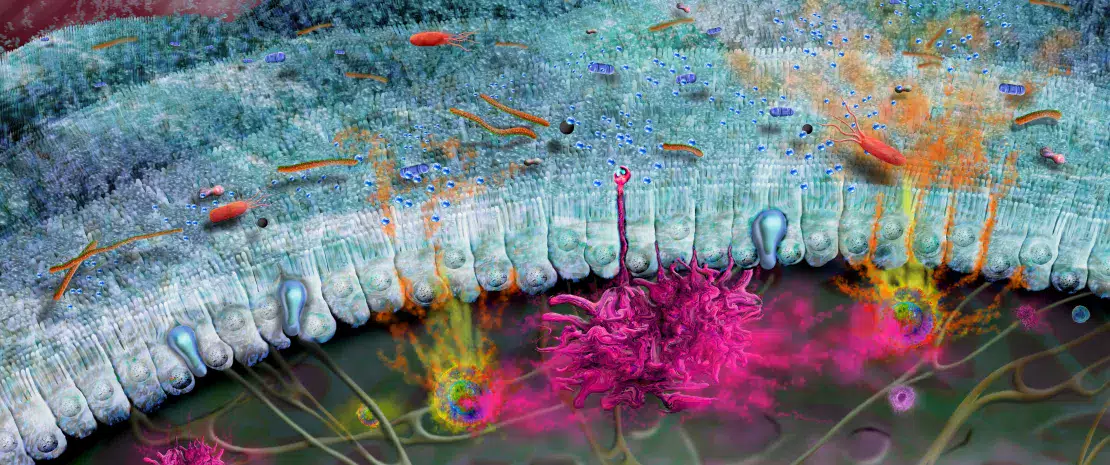
The gut is lined by a single layer of cells, called the intestinal epithelium, over top of which lies a dense mucus layer (Fig 1). Together these barriers confine microbes within the gut lumen, as well as protect the underlying immune system from unnecessary activation by the microbiota.3 Intestinal mucus generate and release the mucin 2 (MUC2), a sugar-coated glycoprotein that provides structure to the mucus. Recent studies have shown that the maturation and function of the mucus layer are strongly influenced by the gut microbiota, while the types of sugars found on MUC2 can also influence which bacteria are able to bind to it or use it, and its sugar chains as a nutrient source.7 Notably, a mucus barrier that is disrupted or dysfunctional can lead to increased penetration or passage of potentially harmful bacteria out of the lumen (e.g. leaky gut), resulting in systemic infection and inflammation.8 Moreover, a defective mucus layer, and a corresponding gut microbiota dysbiosis,9 has been observed in several diseases (such as inflammatory bowel disease (IBD),10,11 diabetes…12) thus highlighting the importance of this protective barrier to human health.

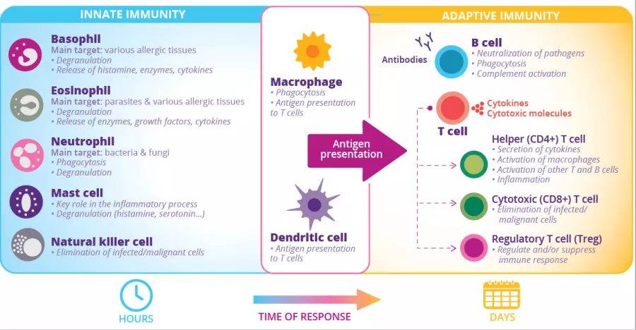
FIGURE 2: Innate and adaptive immune cells and functions.

Sources

Kai-Larsen Y, Bergsson G, Gudmundsson GH, et al. Antimicrobial components of the neonatal gut affected upon colonization. Pediatr Res. 2007 May;61(5 Pt 1):530-6.

Corthésy B. Multi-faceted functions of secretory IgA at mucosal surfaces. Front Immunol. 2013 Jul 12;4:185.

3 Brandtzaeg P. (2017) Role of the Intestinal Immune System in Health. In: Baumgart D. (eds) Crohn's Disease and Ulcerative Colitis. Springer, Cham. 

Commins SP. Mechanisms of Oral Tolerance. Pediatr Clin North Am. 2015 Dec;62(6):1523-9.

Belkaid Y, Hand TW. Role of the microbiota in immunity and inflammation. Cell. 2014 Mar 27;157(1):121-41. 

Ximenez C, Torres J. Development of Microbiota in Infants and its Role in Maturation of Gut Mucosa and Immune System. Arch Med Res. 2017 Nov;48(8):666-680.

Schroeder BO. Fight them or feed them: how the intestinal mucus layer manages the gut microbiota. Gastroenterol Rep (Oxf). 2019 Feb;7(1):3-12.

Miner-Williams WM, Moughan PJ. Intestinal barrier dysfunction: implications for chronic inflammatory conditions of the bowel. Nutr Res Rev. 2016 Jun;29(1):40-59.

Levy M, Kolodziejczyk AA, Thaiss CA, et al. Dysbiosis and the immune system. Nat Rev Immunol. 2017;17(4):219-232.

10 Swidsinski A, Loening-Baucke V, Theissig F, et al. Comparative study of the intestinal mucus barrier in normal and inflamed colon. Gut. 2007 Mar;56(3):343-50.

11 Johansson ME, Gustafsson JK, Holmén-Larsson J, et al. Bacteria penetrate the normally impenetrable inner colon mucus layer in both murine colitis models and patients with ulcerative colitis. Gut. 2014 Feb;63(2):281-91.

12 Chassaing B, Raja SM, Lewis JD, et al. Colonic Microbiota Encroachment Correlates With Dysglycemia in Humans. Cell Mol Gastroenterol Hepatol. 2017 Apr 13;4(2):205-221.


Summary
On
Sidebar
On
Migrated content
Désactivé
Updated content
Désactivé
Hide image
Off
Dossier detail

Aspirin: antibiotic effect in colorectal cancer?

Known for its anti-inflammatory effects, aspirin also acts as an antibiotic on certain bacteria involved in colorectal cancer. It may even prevent tumorigenesis, both in vitro and in vivo.

There is increasing understanding of the clinical correlations and potential mechanistic roles of specific members of the gut and tumoral microbiota in colorectal cancer (CRC) initiation, progression, and survival. Despite this, we are still a long way from defining microbially-informed diagnostic, preventive, or therapeutic approaches. However, a step forward has just been made, with a team recently showing that aspirin, a chemopreventive agent recommended by the (sidenote: United States Preventive Services Task Force An independent, volunteer panel of US experts in disease prevention and evidence-based medicine. 
 
)
 for the prevention of CRC, has specific effects on (sidenote: Fusobacterium nucleatum Increased presence in human colonic adenomas and CRCs, responsible for tissue proliferation in vitro and in animal models. ) , a bacterium associated with this disease.

In vitro and in vivo effect

US researchers have recently shown that aspirin disrupts the growth of the Fn7-1 strain of F. nucleatum, and even kills it, in vitro, in human colonic adenoma tissue cultures. At levels that do not inhibit bacterial growth, aspirin influenced the gene expression of Fn7-1: 55 genes were upregulated and 155 genes downregulated.

To assess aspirin’s role as a modulator of F. nucleatum growth, the researchers also conducted in vivo experiments. In a murine model, Fn7-1 was orally inoculated daily to induce an intestinal tumor, but mice receiving an aspirin-enriched diet saw inhibited tumorigenesis when compared to those on a normal diet. The protective effect of aspirin was also found with other strains of F. nucleatum, including some isolates from human CRC tissues, the latter proving more sensitive than the Fn7-1 strain. Conversely, this protective effect was much milder with other CRC-associated microbes, such as enterotoxigenic Bacteroides fragilis and colibactin-producing Escherichia coli.

Lastly, a (sidenote: Quantitative PCR  Specific PCR (polymerase chain reaction) method used to measure the initial quantity of DNA. 
 
)
 performed on adenoma DNA samples of patients taking aspirin daily showed a 2 to 3-fold lower fusobacterial abundance than in samples taken from control patients. This result suggests that the modulatory effect observed in vitro also occurs in humans.

Antibiotic effect in addition to anti-inflammatory effect

These data confirm the direct antibiotic effect of aspirin on strains of F. nucleatum. Its protective effect against colorectal cancer and adenomas thus exceeds its anti-inflammatory role. Consideration of the potential effects of aspirin on the microbiome holds promise in optimizing risk-benefit assessments for the drug’s use in CRC prevention and management. However, it is unlikely that the anti-inflammatory effects of aspirin alone are enough to stop tumorigenesis in its entirety. Further research is required before its use to improve the prognosis of CRC – the second leading cause of cancer death worldwide – can be considered.

Summary
Off
Sidebar
On
Migrated content
Désactivé
Updated content
Désactivé
Hide image
Off
News Gastroenterology Oncology

Allergies: the role of intrauterine life

Intrauterine life influences the risk that a child will subsequently develop an allergy. How? A less rich meconium affects the development of the gut microbiota and, ultimately, the immune system.

Allergic rhinitis Asthma and microbiota Allergic eczema Food allergies

While some people can read the future in coffee grounds, researchers are beginning to use (sidenote: Meconium Earliest “stool” of the newborn, containing the amniotic liquid absorbed in utero. The meconium helps identify microorganisms lining the gastrointestinal tract of the fetus.
 
)
 –  the “tarry” first stool of infants – to predict the risk of developing allergies. Eczema, food allergies, asthma, allergic rhinitis: today, nearly one in three children suffers from an allergy. However, many things may be at play even before birth. Hence researchers had the idea of studying meconium, which begins forming in utero by gestational week 16. 

Signs of allergy even during pregnancy?

Their results support the idea that the onset of an allergy begins well before the first symptoms appear: at three months of age, future allergic infants have a less diverse and less mature gut microbiota than peers. The researchers therefore looked back in time, at their very first stool, the much-vaunted meconium: the same observation was made, a lower diversity of bacteria and a low diversity of molecules produced by these microorganisms. The appearance of an allergy may therefore be explained by the following mechanism: during pregnancy, environmental factors favoring an allergy modify the composition of the meconium, which is less rich in metabolites at birth. Since the first bacteria to colonize the infant’s digestive tract feed on these metabolites, a less rich meconium means the microbiota is less diverse and matures more slowly early in life. 

Prevent... and predict?

These discoveries have multiple implications. On the one hand, the researchers hope one day to be able to prevent these allergies. This will require a better understanding not only of what affects the composition of meconium in utero, but also of how the different metabolites of meconium influence bacterial colonization in newborns. At the same time, they hope to be able to predict the risk of developing an allergy based on the composition of a newborn’s meconium. In the meantime, the only recommendation to be made is for women to adopt a healthy lifestyle during pregnancy.

Summary
Off
Sidebar
Off
Migrated content
Désactivé
Updated content
Désactivé
Hide image
Off
News

A new, unanticipated marker for predicting the clinical course of prostate cancer: gut microbiota

According to a new study published in Cancer Science, the composition of the intestinal microbiota could be used as a marker of increased risk of prostate cancer.

Having demonstrated that intestinal bacteria and their metabolites (short chain fatty acids, SCFA) promote the growth of cancer cells in mouse models of prostate cancer, the researchers in this new study wished to further explore the link between intestinal microbiota (IM) and the prognosis for prostate cancer in men. Their findings are surprising, to say the least.

A “discovery” cohort and a “test” cohort 

The study included 152 Japanese men who had undergone a prostate biopsy (96 positive and 56 negative). The men were randomised to two cohorts: “discovery” (114 patients) and “test” (38 patients). Two comparison groups were defined in each cohort: a high grade group (men with grade 2 prostate cancer or above) and a negative/grade 1 group (men with a negative biopsy or grade 1 prostate cancer). Samples were taken during rectal examination prior to administration of prophylactic antibiotics and prostate biopsy. ARNr 16S gene sequencing was used to determine the composition of the gut microbiota.
 

A greater abundance of specific bacteria indicates a high grade

While there no significant difference was observed in bacterial diversity between the patient groups, three bacterial taxa, Rikenellaceae, Alistipes and Lachnospira, were present in greater numbers in patients with a high grade prostate cancer. There was no link between the presence of such bacteria and patients’ metastatic status. Microbial data were also used to predict the functional profiles of patients’ microbiota: (sidenote: Starch and sucrose metabolism, phenylpropanoid biosynthesis, phenylalanine, tyrosine and tryptophan biosynthesis, cyanoamino acid metabolism and histidine metabolism )  were more prevalent in patients with high grade prostate cancer.

A faecal indicator for the prostate 

The researchers then investigated whether microbial profiles could be used to identify high-risk PCa patients in the test cohort. It was not possible to use the three bacteria identified above on their own to discern the men with a high grade prostate cancer. The LASSO regression model was employed to identify a further 18 operational taxonomic units. These bacterial groups were highly correlated (positively or negatively) with high-risk PCa in the discovery cohort and were used to create a Fecal Microbiome Prostate Index (FMPI). In the test cohort, not only was the FMPI significantly higher in patients with high-grade PCa (P < 0.001), but it also detected these patients with greater accuracy than conventional serum prostate-specific antigen (PSA) assay. 

While these results are highly encouraging, the study cohort only included Japanese men living in an urban area and with similar lifestyles. The scope of the research will need to be widened to additional populations to corroborate these initial findings.

Summary
Off
Sidebar
On
Migrated content
Désactivé
Updated content
Désactivé
Hide image
Off
News Gastroenterology

Understanding the complex interplay between the microbiota and immunity is only just beginning

By Dr Bruce Vallance

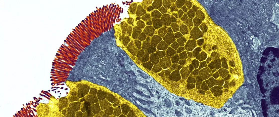
Goblet cells. Coloured transmission electron micrograph (TEM) of a section through goblet cells in the lining of the small intestine,part of the digestive tract.

Your body is home to trillions of bacteria that together with viruses, fungi and other organisms, collectively make up the human microbiota.

These microbes play an important role in promoting our health, as well as controlling our susceptibility to disease, by influencing different aspects of our daily lives. For example, the metabolic activity of our gut microbiota determines whether certain medications like acetaminophen are toxic to our livers.1 Specific members of the microbiota can also change and evolve in response to new dietary sources of carbohydrates, allowing us to digest foods like sushi2 or produce important and protective chemicals such as short chain fatty acids (SCFA).3 Other microbes selectively shape our immune systems to become reactive, or tolerant to invading organisms, thereby controlling our risk of severe gastrointestinal (GI) infections.4


During the 1000 first days of life, the critical window of early childhood growth and development (period from conception to 2 years of age), any interference with microbiota establishment in the neonatal gut may potentially lead to negative health outcomes.5

1000 first days of life critical window of early childhood growth and development

Although scientists have established the importance of the microbiota in maintaining human health, our understanding of the complex interplay between the microbiota and immunity is only just beginning.

Summary
Off
Sidebar
On
Migrated content
Désactivé
Updated content
Désactivé
Hide image
Off
Dossier detail Gastroenterology Pediatrics

Faecal microbiota transplantation: a new treatment for irritable bowel syndrome?

A new study reveals that faecal microbiota transplantation improves the symptoms of irritable bowel syndrome, as well as patient quality of life, even one year after treatment. However, it is essential that the donor is in good health.

The gut microbiota Functional gastrointestinal disorders Fecal transplant
IBS

Pain, cramps, bloating, diarrhoea, constipation: irritable bowel syndrome is a disease characterised by a range of abdominal symptoms that come and go throughout the patient's life. These symptoms can be exacerbated by stress, changes in emotional state and certain foods, and have a considerable impact on patients' quality of life. While there are no anatomical or structural problems with the bowels of people who suffer from this syndrome, the finger is often pointed at the gut microbiota.

A single donor with a super microbiota?

The researchers involved in this study performed a clinical trial to test the efficacy of faecal microbiota transplantation by using stool samples from a single Caucasian man aged 36 years1 who ticked all the "super donor" boxes: in good health, normal BMI, undertaking regular exercise, born vaginally and breastfed. Even better, he was not taking any medication, had only received three antibiotic treatments during the course of his life and was regularly taking dietary supplements. In this clinical trial, faecal microbiota transplantation was found to be effective in patients with irritable bowel syndrome. However, these results were only observed three months after the transplantation, and several questions still need to be answered, particularly whether the clinical effects of the transplantation continue into the long term. In the current study, the researchers continued to monitor these patients for one year. 

Benefits still present after one year 

Most of the patients who responded to faecal microbiota transplantation after three months maintained their response after one year. Another encouraging result was also observed: their abdominal symptoms, fatigue and quality of life were clearly improved compared to three months after the transplantation. Even better, between 32 and 45% of the patients, depending on the group, experienced complete remission over the course of the one-year follow-up. Complete analysis of the patients' gut microbiota revealed changes in the gut bacteria profile and a significant reduction in the dysbiosis index.

In conclusion, faecal microbiota transplantation from a "super donor" can restore the gut microbiota and reduce the symptoms of patients suffering from irritable bowel disease.


1. El- Salhy M, Hatlebakk JG, Gilja OH, et al. Efficacy of faecal microbiota transplantation for pa-tients with irritable bowel syndrome in a randomised, double- blind, placebo- controlled study. Gut. 2020;69(5):856- 867.

Summary
Off
Sidebar
On
Migrated content
Désactivé
Updated content
Désactivé
Hide image
Off
News

Antibiotic resistance: an odd “souvenir” from exotic destinations

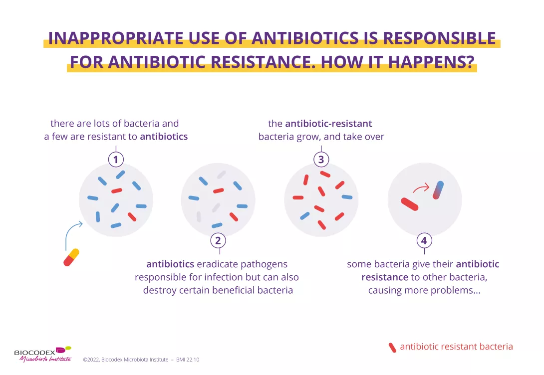
An incredible medical breakthrough becomes the victim of its own success. Since they were first discovered in the early 20th century, antibiotics have saved millions of lives. However, the wide scale and sometimes inappropriate use of these drugs is making them less and less effective at treating infections. The result is that many bacteria have now become resistant! But their excessive and incorrect use cannot take the whole blame for antibiotic resistance.

One study1 has revealed that international travel is increasing the acquisition of antimicrobial resistance genes and could be helping the spread of antibiotic resistance. Head straight to the departure lounge for more information.

Every year since 2015, the World AMR Awareness Week (WAAW) has been raising awareness of the rise in (sidenote: Antimicrobial Resistance Antimicrobial Resistance occurs when bacteria, viruses, fungi, and parasites change over time and no longer respond to medicines. Antibiotics and other antimicrobial medicines stop working, and infections become harder and even impossible to treat. This resistance to microbes increases the risk of disease spread, severe illness, and death. Antibiotic resistance means the resistance of bacteria to antibiotics. Source: Antimicrobial resistance. October 26, 2020. ) . Linked to the incorrect and overuse of antibiotics, this is when a bacterium is able to resist the action of an antibiotic. In 2020, the WAAW broadened the scope of its message to include (sidenote: Antimicrobials Medicines – including antibiotics, antivirals, antifungals and antiparasitics – used to prevent and treat infections in humans, animals and plants. WHO Antimicrobial Resistance; Nov 2023 )  - antivirals, antifungals and antiparasitics, etc., that is medicines that are essential for fighting (sidenote: Pathogens A pathogen is a microorganism that causes, or may cause, disease. Pirofski LA, Casadevall A. Q and A: What is a pathogen? A question that begs the point. BMC Biol. 2012 Jan 31;10:6. )   (sidenote: Microorganisms Living organisms too small to see with the naked eye. This includes bacteria, viruses, fungi, archaea, protozoa, etc., collectively known as ’microbes’. Source: What is microbiology? Microbiology Society. ) . By reducing our options for treating infections, antibiotic resistance has become a threat to everyone’s health2. Several studies are therefore underway to understand how this phenomenon is spreading so we can better control it, or even stop it altogether.

Antibiotics save life! Did you know that they also have an impact on your microbiota? Did you know that the misuse and overuse of antibiotics can lead to antibiotic resistance? Have you heard about the World AMR Awareness Week
(WAAW)? All the answers in this dedicated page:

Antibiotics: what impact on the microbiota and on our health?

Learn more

Antibiotic resistance flying beneath the radar

We now know that antibiotic resistance is mainly due to the overuse of antibiotics in human medicine, as well as in livestock and agriculture2. However, a recent study has revealed an unexpected cause of this spread: holidays and business trips to far-flung destinations! In fact, international travel encourages the spread of (sidenote: Gene A gene is the basic physical and functional unit of heredity. Genes are made up of DNA. Source: What is a gene? MedlinePlus.gov )  in the gut. Researchers studied 190 Dutch travelers, divided into four subgroups based on destination. They were all destined for areas badly affected by antibiotic resistance, namely South East Asia, South Asia, North Africa, and East Africa The aim was to determine whether international travel to these regions could encourage the spread of resistance to areas less badly affected. In order to measure the abundance of these genes in the gut, a stool sample was taken from each participant before and after travel.

Image

A diplomatic bag for antibiotic resistance in the gut?

Using cutting edge technology ( (sidenote: Metagenomics A method of studying the genetic material in samples taken directly from complex natural environments (intestines, oceans, soil, air, etc.), as opposed to samples grown in a laboratory. It produces a description of the genes contained in the sample, as well as an insight into the functional potential of the microbial community.
Source: Riesenfeld CS, Schloss PD, Handelsman J. Metagenomics: genomic analysis of microbial communities. Annu Rev Genet. 2004;38:525-52.
)
), the team observed an increase in the number of antibiotic resistance genes between departure and return, especially in travelers coming back from South East Asia. A total of around fifty antibiotic resistance genes was detected following travel. These included genes with well-known and long-standing resistance to antibiotics (such as β-lactams, tetracyclines and fluoroquinolones) as well as new, never-before seen genes.

Could travel pose a danger for public health?

The results of this study are clear: international travelers, colonized by resistance genes whilst on their travels, could unwittingly be bringing back antibiotic-resistant bacteria along with the rest of their luggage. Faced with the risk of this resistance spreading, the authors sound the alarm and highlight the importance of taking rapid action in countries particularly affected by antibiotic resistance. This resonates with the WHO's campaign.

What is the World AMR Awareness Week?

Each year, since 2015, the WHO organizes the World AMR Awareness Week (WAAW), which aims to increase awareness of global antimicrobial resistance.

Antimicrobial resistance occurs when bacteria, viruses, parasites and fungi change over time and no longer respond to medicines. As a result of drug resistance, antibiotics and other antimicrobial medicines become ineffective and infections become increasingly difficult or impossible to treat, increasing the risk of disease spread, severe illness and death.
Held on 18-24 November, this campaign encourages the general public, healthcare professionals and decision-makers to use antibiotics, antivirals, antifungals and antiparasitics carefully, to prevent the further emergence of antimicrobial resistance.

Summary
Off
Sidebar
On
Migrated content
Désactivé
Updated content
Désactivé
Hide image
Off
News