Contrary to popular belief, obesity is not necessarily synonymous with excess. Vitamin B7 (biotin) deficiencies can trigger a vicious circle between gut bacteria and obesity. Explanation.
Although we need only very small quantities, vitamins are no less essential to our health—if we do not get enough vitamin C, for example, we can develop scurvy, a disease from which many sailors died when a lemon would have been enough to save them. To a lesser extent, cognitive disorders, numbness or a persistent state of fatigue can also be signs of a vitamin B deficiency. Moreover, the etymology of the word reveals something of its “vital function”: “vitamin” comes from the Latin “vita,” meaning life.
The B7 engine
Among the many vitamins essential for our body to function properly are the B vitamins, including the famous vitamin B7, also called (sidenote:
Biotin
Called vitamin B7, or B8 in certain countries. This vitamin plays a key role in the metabolism of carbohydrates, lipids and amino acids. It is also involved in the biosynthesis of other vitamins (B9 and B12). Many foods are good sources of biotin (whole grains, eggs, milk, nuts, etc.). This vitamin is also synthesized by the bacteria in gut flora.
Biotin_NIH National Cancer Institut). One of our main sources of biotin is food, but that’s not all! The bacteria in gut microbiota also produce it... or not, as the case may be. And this has consequences for our health, especially in the case of obesity.
x11
Severe obesity increased 11-fold in men
x3
and 3-fold in women worldwide between 1975 and 2014.
The vicious circle of obesity
A team of researchers has just shown that the gut bacteria that produce and transport biotin are absent in severely obese patients ( (sidenote:
Body Mass Index (BMI)
Body Mass Index (BMI) assesses the corpulence of an individual by estimating the body fat mass calculated by a ratio between weight ((kg) and height squared (m2).
https://www.nhlbi.nih.gov/health/educational/lose_wt/BMI/bmicalc.htmhttps://www.euro.who.int/en/health-topics/disease-prevention/nutrition/a-healthy-lifestyle/body-mass-index-bmi)> 35). Their obese host is thus deprived of this additional non-dietary source, despite actually needing more than the average person (vitamin B being necessary for well-balanced adipose tissue). Experiments in animals show that the (sidenote:
Western diet
Diet rich in processed foods, refined sugar, salt, saturated fats (red meats) and trans fats (pastries)
Zinöcker MK, Lindseth IA. The Western Diet-Microbiome-Host Interaction and Its Role in Metabolic Disease. Nutrients. 2018 Mar 17;10(3):365. ), well known for promoting obesity, reduces both the quantity of biotin-producing gut bacteria and the circulating levels of this vitamin in serum. Also, paradoxically, the gut inflammation observed in obese patients limits the absorption of biotin supplied by the diet.
In cases of severe obesity, a vicious circle is thus set in motion, since gut dysbiosis is believed to aggravate inflammation (and obesity) as well as tissue biotin deficiency.
How can we break this vicious circle? Weight-loss surgery (bariatric surgery), which improves metabolism and inflammation, stimulates growth of biotin-producing bacteria, resulting in an increase—at least during the first year—in the level of biotin circulating in the body. Another approach involves supplementing with prebiotics (fructo-oligosaccharide type fibers) and biotin. This has been tested in mice fed a high-fat diet, and was found to improve the diversity of gut microbiota and boost bacterial production of biotin and other B vitamins, while limiting weight gain and deterioration of blood sugar levels.
Multiple sclerosis (MS) is an autoimmune neurodegenerative disease that affects 2.5 million people worldwide. A recent study published in eBioMedicine shows a network of associations between meat consumption, an increase in certain blood metabolites and pro-inflammatory markers, and fewer polysaccharide-digesting bacteria in patients with this disease. Explanations.
Recently, research has revealed the involvement of gut microbiota (GM) in the pathogenesis of MS, yet little is still known about its mechanisms.
A novel multiomic approach
For the first time, a research team has investigated interactions between diet, immune system, metabolism and gut microbiota in the development and progression of MS. For this, the researchers adopted a multiomic methodology.
This novel approach was the subject of a 6-month study. Blood and stools from the 49 participants (24 untreated MS patients and 25 non-diseased individuals) were collected at baseline and 6 months later. The metabolic profiles and GM of all participants remained stable for more than 6 months. Participants were also asked to record what they ate at each meal.
An altered relationship between gut microbiota and the immune system in MS patients
The results were similar to those of previous studies: on the whole, both groups share a similar bacterial community structure. However, MS patients had a lower abundance of bacteria with immunomodulatory properties. Moreover, the microbiota of patients differed depending on the extent of their disability, suggesting a possible link between certain species and disease severity.
The results of the multiomic analyses also suggest dysfunctional homeostasis of microbiome–immune system interactions, because a disrupted immune cell–microbiome relationship and modification of the blood metabolic profile were observed in the group of MS patients.
Meat consumption: a possible explanation?
Study of the participants’ food diaries played a key role in this research.
The researchers found that MS patients consumed more meat. This consumption was correlated with an increase in the quantity of Th17 cells, involved in the autoimmune process, and of S-adenosyl-L-methionine (SAM), a metabolite produced from methionine (an amino acid enriched in meat) and involved in the activation of Th17 cells. Conversely, MS patients had reduced proportions of B. thetaiotaomicron bacteria, known for being highly capable of digesting polysaccharides.
A new therapeutic approach?
This data points to pathways of interaction between diet, gut microbiota, immune response and blood metabolome through B. thetaiotaomicron, Th17 and SAM. For the authors, these results suggest that decreasing dietary consumption of meat or methionine could reduce the number of circulating pro-inflammatory Th17 cells in MS patients.
This study, and its promising results, could pave the way for a better understanding of the regulatory pathways between diet, metabolites, immune response and microbiota, to prevent multiple sclerosis from worsening and possibly open up avenues for new therapeutic targets. This study has been widely reported in the medical press and is of particular clinical interest. It will be discussed on World MS Day on May 30.
Microplastics appear to be linked to Inflammatory Bowel Disease (IBD). More precisely, their fecal concentration, size and chemical origin vary according to whether patients suffer from Inflammatory Bowel Disease (IBD) or not, and the severity of the condition.
Microplastics (MPs) are now everywhere, including in what we eat and the air we breathe. Does this increased human exposure present a health risk? Although the answer to this question is still largely unknown, researchers suspect a link, in the digestive system, with an increased risk of inflammation, oxidative stress, increased intestinal permeability and microbiota dysbiosis. This is an already long list, to which we may have to add IBDs, such as Crohn’s disease and ulcerative colitis, if we are to believe the results of a study comparing the characteristics of fecal MPs in 52 IBD patients and 50 healthy subjects from 10 Chinese provinces.
Microplastics in stools: Significant differences
First observation: the researchers detected MPs in all stool samples. Most were much smaller than 300 µm and shaped in the form of sheets (1 MP in 2) or fibers (1 MP in 3).
In contrast, MP concentrations differed according to the person’s state of health. The stools of IBD patients contained:
50% more MPs (41.8 pieces/gram of dry fecal matter, vs 28.0 in healthy subjects);
more very small MPs (<50 μm), whereas those of healthy participants contained MPs measuring 50 to 300 μm;
a different relative abundance of each type of MP in terms of its chemical nature, among the fifteen or so types found. In the stools of healthy participants, the researchers found mainly PET, a plastic typical of water bottles (22.3%), and to a lesser extent polyamides from textiles (8.9%) and polypropylene typical of food packaging (8.7%).
In IBD participants, PET (34.0%) and polyamides (12.4%) were more abundant, followed closely by the type of PVC found in pipes, PVC floors, etc. (10.3%), and ahead of polypropylene (9.5%).
Finally, the researchers demonstrated a positive correlation between fecal MPs and IBD severity. Two hypotheses are therefore possible: either exposure to MPs contributes to the disease; or the disease influences MP retention.
Bottled water and fast food are suspected
So where do these MPs come from? Based on the answers to their questionnaire, the team showed that the concentration of MPs in stools increased twofold when drinking bottled water (vs boiled tap water). This is because bottled water contains 22 times more MPs, in particular PET, than tap water.
22
Bottled water contains 22 times more MPs, in particular PET, than tap water.
Other factors that went hand in hand with a near twofold increase in fecal MP concentration were consumption of fast food (vs homemade food) and exposure to dust at work or in daily life.
Although we have a long way to go before discovering all its secrets, we do know that allergic rhinitis is associated with a respiratory microbiota imbalance. Better characterizing this dysbiosis may help us to develop targeted and individualized treatments.
40%
Allergic rhinitis is thought to affect up to 40% of the world’s population, with a high prevalence.
1 person in 4
in industrialized countries.
An unbalanced respiratory microbiota
To find out, a Chinese team compared respiratory microbiota in nasal samples taken from 28 people suffering from acute episodes of seasonal allergic rhinitis with those of 15 non-allergic subjects. They found no difference between the two groups in terms of microorganism diversity and abundance, but important disparities in their composition. The bacterial genera Moraxella, Haemophilus, Streptococcus and Flavobacterium, predominant in the respiratory microbiota of healthy individuals, had been replaced in allergic individuals by the genera Klebsiella, Prevotella and Staphylococcus. In total, the researchers identified 10 bacterial genera that were over-represented in the latter.
Hay fever
Hay fever (or allergic rhinitis) is a very common chronic condition that affects both children and adults.
It is an inflammatory disease of the nasal mucosa that is accompanied by one or more nasal symptoms, including nasal pruritus (itching, tingling), sneezing, rhinorrhea (runny nose) and nasal congestion (runny nose).
The combined results of these two approaches confirm the hypothesis that inflammatory reactions of allergic origin influence the balance of respiratory microbiota. More importantly, they provide important candidate biomarkers of potential use in the diagnosis of allergic rhinitis. The authors therefore suggest continuing this work to refine the identification of different subtypes of allergic rhinitis (seasonal/perennial, intermittent/persistent, mild/moderate/severe), which may pave the way for the development of individualized treatments... and an end to the ordeal of thousands of people.
The downside of the good resolution to stop smoking: additional kilos. Good news a recent study shows that it is not inevitable but a simple consequence of the imbalance of the microbiota caused by tobacco. So all that is left is to rebalance the microbiota!
Unfortunately impedes some attempts to stop smoking: former smokers tend to put on weight. On average an extra 4.5 kg on the scales 6–12 months after the last cigarette. Something that discourages the best intentions. Unless our gut microbiota offers us welcome assistance? In any event, this is what is suggested by a recent study in mice.
Leading avoidable cause
Smoking is the most avoidable cause of disease and death in the world.
7.2 million
Each year, smoking is responsible for over 7.2 million deaths worldwide, killing more people than AIDS, malaria and tuberculosis combined.
1 in 4 Europeans
According to the WHO, Europe has the highest prevalence of smoking among adults (28%), that is 1 in every 4 Europeans.
Smoking cessation: a microbiota that carries weight
As in humans, mice exposed regularly to cigarette smoke tend to gain weight after stopping smoking. After a long series of experiments, the researchers seem to have identified the mechanism potentially in play. Some compounds in tobacco (nicotine?) are thought to be able to reach the digestive system of “smoker” mice after traveling in the blood. They are then thought to modify the composition of the gut microbiota. And in fact, it is enough to transplant the microbiota of smoker mice into non-smoker mice to make them gain weight. Something that designates this microbiota as being partially responsible for the kilos gained.
But in practice, how is this possible? It would appear that smoking disrupts the delicate balance between the molecules that promote weight gain and others that restrict it. In smokers, the molecule that promotes weight gain is thought to be produced in greater quantities, whilst the molecule that blocks it becomes increasingly scarce. So why don't they get fat? Because the mechanism is gradual, allowing the body time to adjust by associating every cigarette lit to the necessity to eat less. Except that on stopping smoking, this appetite-suppressant effect of the tobacco disappears immediately, while the imbalance in the microbiota that encourages weight gain lasts much longer. Direct consequence: the scales go into panic mode!
Supporting ex-smokers
“The compounds that we identified could lead to new treatments that will help people to avoid gaining weight when they stop smoking”
said Prof. Evan Elinav who led the research team. While waiting to find out how to repair the microbiota of ex-smokers (diet? microbial therapy, postbiotics?) in order to limit weight gain after quitting smoking, this study makes a “weighty” argument for never smoking your first cigarette, or exposing those around you to passive smoking: protecting the equilibrium of the microbiota.
Do you know what a plastic bottle, a fast food container and a polyamide sweater have in common? They are all thought to be sources of microplastics that end up in our intestines. With nevertheless differences depending on whether or not you suffer from Inflammatory Bowel Disease (IBD). Bon Appétit…
Fish, coral reefs, shellfish and marine bacteria are not the only casualties of microplastics derived from the degradation of plastic bags. Microplastics are now everywhere: in the air we breathe, in the water we drink, and in the food we eat. No-one can escape them, as shown by a research team who found them in 100% of the stools of patients with IBD and also in those of healthy individuals.
5g plastic/week
Humans are thought to ingest 5g of plastic each week, the equivalent of a credit card.
CIBD: stools full of microplastics
From homo erectus... to homo plasticus! Although we have all unwittingly become consumers of microplastics, it seems that we are not all in the same boat. So, depending on the health of our intestines, our stools do not contain the same number, size or type of plastic particles. This study showed that in people with Inflammatory Bowel Diseases (or IBD) such as Crohn's disease or ulcerative colitis, these microplastics were:
more numerous (around 42 pieces/gram of dry fecal matter vs. 28 in healthy subjects),
generally smaller (<50 μm),
and of different origin, with PET (a plastic typically used in bottles of water), polyamide (derived particularly from synthetic textiles) or PVC (pipes, plastic flooring) being more abundant.
Crohn's disease
Crohn's disease is an Inflammatory Bowel Disease (IBD) whose cause is not yet known. This chronic inflammatory condition can affect every part of the digestive tract. It is characterized by damage to the intestinal wall, in which often deep lesions alternate with healthy areas. It progresses in flares interspersed with periods of remission. The intestinal microbiota seems to be implicated: a deterioration in the diversity and composition of the flora is observed in patients.
The team also noticed that the greater the quantities of microplastics present in the stools of IBD patients, the more severe was the disease. For all that, this does not necessarily mean that microplastics are responsible for IBD. Other explanations are possible. For example, the disease could cause greater retention of microplastics in the intestines, to such an extent that they are found in greater quantities in the stools. The researchers are still working to determine which is the consequence of the other, microplastics or IBD.
Ulcerative colitis
Ulcerative colitis is an Inflammatory Bowel Disease (IBD) characterized by ulceration of the surface of the mucous membrane of the colon. Its cause is not yet known. The gut microbiota is thought to be involved in the pathological process of the disease
As for knowing where these tiny pieces of plastic come from, the team points to three sources:
the consumption of bottled water, which goes hand in hand with a doubling of the quantity of plastic in the stools. This is not surprising, if you consider that bottled water contains 22 times more microplastics (especially PET) than tap water.
the consumption of fast food, doubtless due to the plastic packaging;
and exposure to dust, whether at work or elsewhere in life.
Another reason, if it was needed, to prefer home cooking and inert containers (glass jars): not only is it good for the planet, but good for our bodies too.
A recurring complaint among ex-smokers, weight gain discourages many of these. Hence the importance of recent studies that highlight the role of the microbiota, damaged by years of smoking. With into the bargain possible solutions for avoiding weight gain.
An additional 4.5 kg in the 6–12 months following smoking cessation, indeed more than 10kg in one year in 13% of ex-smokers: weight gain represents a major obstacle to giving up cigarettes. A team of researchers used a mouse model to assess the potential role of the gut microbiota in this weight gain.
Tobacco, dysbiosis and weight gain
First observation: rodents regularly exposed to cigarette smoke stayed in shape, even with a high-fat, high-sugar diet. On the other hand, as in humans, smoking cessation led to weight gain, unless the mice were given broad-spectrum antibiotics that depleted their microbiota. In question? Compounds linked to tobacco, such as nicotine, appear to penetrate the digestive system of “smoker” mice, causing long-term changes (after cessation) in the bacterial composition of the gut microbiota. With into the bargain, a metabolism better able to extract energy from foods (fewer calories in their feces).
4,5kg
An additional 4.5 kg in the 6–12 months following smoking cessation
10kg
more than 10kg in one year in 13% of ex-smokers
Transfer of the microbiota of “smoker” or “ex-smoker” mice confirms the role of the gut microbiota: the recipient mice ( (sidenote:
Germ-free mice
mice that have no microbes at all, raised in sterile conditions.
) and never exposed to smoke) gradually gained weight, unless they had previously been given antibiotics (markedly lower weight gain).
Two metabolites at issue
There remained the task of determining which metabolites were involved. Two molecules with opposite effects were isolated from the thousands of bioactive compounds whose levels varied at the time of cessation:
dimethylglycine (DMG), manufactured by the intestine and liver from dietary choline, which increases weight gain;
acetylglycine (ACG) which has the opposite effect.
Whereas the two antagonistic molecules allowed the “non-smoker” mice to stay in shape, smoking gradually disrupted this equilibrium (increased DMG production and less ACG production). According to the authors, an “anorexic reaction”, leading to reduced food intake is thought to be set up, to avoid calorie overload. The problem: with smoking cessation, this appetite-suppressant effect disappears whilst the obesogenic dysbiosis and the accumulated metabolites are thought to be slow to reverse. Hence the weight gain.
And in humans?
In humans, a preliminary study showed dysbiosis in smokers and modifications of microbial metabolites similar to those observed in mice. The fact also remains that smoking is a voluntary behavior, doubtless involving additional mechanisms. Nevertheless, this study provides proof of concept for the role of the microbiota in post-smoking weight gain. And opens up the possibility of rebalancing the intestinal flora (dietary, biotic measures) in order to limit kilos gained after giving up tobacco and to avoid jeopardizing smoking cessation.
Symptoms, diagnosis, treatment, potential links to the microbiota... To mark Endometriosis Awareness Month, the Microbiota Institute is handing the floor to three experts. This article deciphers a long-neglected chronic inflammatory disease that is still poorly diagnosed.
“The diagnosis of endometriosis starts by talking to the patient”
Dr. Erick Petit
(sidenote:
Dr. Erick Petit, radiologist, founding head of the Endometriosis Center at the Paris St. Joseph Hospital, President of RESENDO (community-hospital endometriosis network), member of the steering committee of the specialist endometriosis group EndoSud-IDF and of the endometriosis national strategy steering committee, co-author of: Tout sur l’endométriose, soulager la douleur, soigner la maladie (Editions Odile Jacob, 2019) [Everything there is to know about endometriosis, easing the pain, treating the illness].
)
Do we know when endometriosis dates back to?
Erick Petit : Endometriosis has a long and sinuous—if not tumultuous—history. Even though the symptoms have been described for 4,000 years, it was not until the end of the 19th century that it was recognized as an organic disease. Unbelievably, that means it was left undiagnosed for almost 4,000 years. The first clinical description of endometriosis dates back to 1855 BC, concerning an Egyptian woman. The disease was next listed in the Greek body of clinical literature around the year 500 BC.
That was when the symptoms were clearly catalogued and the association was made with menstruation. The disease was subsequently considered to be a condition of the feminine psyche and was consigned to limbo until the Renaissance. Hysterikos being Greek for “uterus,” the physicians of the time had a field day with this so-called illness that they believed had been entirely made up by what they referred to as “hysterical” women. The pain, however, was very real...
For centuries, women were imprisoned in the belief that pain was inevitable. They were committed to specially created institutions and marginalized. It was only in the 19th century, thanks to the work of Carl von Rokitansky, a Bohemian pathologist working in Vienna, that endometriosis was histologically confirmed for the first time in 1860.1
Why is the diagnosis of endometriosis so long and complicated?
E. P. : The gold standard test is still the endovaginal ultrasound (or an MRI for young girls who have never had sexual intercourse, although this technique is less sensitive and less specific), but I remain convinced that medical imaging does not tell us the whole story. The scans must be compared with the clinical data and the patient should be listened to carefully. That is why, in the (sidenote: https://www.resendo.fr/) network, we use a clinical questionnaire that contains specific questions and gives a better picture of the pain experienced. In nine out of ten cases, the diagnosis of endometriosis is confirmed. We believe that, first and foremost, listening to the patient and having a real conversation is the basis of the diagnosis. Time constraints mean that nobody can spend even 15 minutes talking with a patient anymore.
Yet asking patients the right questions is exactly how we can establish a reliable diagnosis and treat women who have in some cases been left undiagnosed for a whole decade!2 Not enough large-scale epidemiological studies have been conducted to date, but there are tangible signs suggesting that prevalence has been on the rise over recent years. It is commonly said that one in ten women suffers from endometriosis. It is in fact more likely to be one in seven, or even one in five, women of reproductive age.2
1 out of 10
It is commonly said that one in ten women suffers from endometriosis
10 years
Some women are in diagnostic wandering for many years, sometimes more than 10 years.
#1
Endometriosis is the leading cause of hyperfertility
Does a typical profile exist for women affected by endometriosis? What are the consequences?
E. P. : It is a complex pathology involving many factors. So there is no typical profile. I would say that there are as many forms as there are patients. There is no correlation between the anatomical and clinical findings for this disease. Certain women can have very severe endometriosis anatomically speaking, without experiencing too much pain. Conversely, others with only mild endometriosis may suffer from debilitating symptoms. This illness is the number one cause of hypofertility,2 which is the second biggest consequence of endometriosis after pain. We have in fact observed a correlation between the extent of the lesions and fertility. But this is not necessarily linked to pain.
What are the early signs?
E. P. : The disease appears at menarche. This is why a young girl should be carefully observed at this time. Is the level of pain intense? Does she remain bedridden during menses? Non-attendance at school is also a good indication. Early menarche (before the age of 11) and having a mother or sister who also suffers from endometriosis are risk factors. To avoid leaving the condition undiagnosed and starting treatment too late, I have been campaigning for years to raise awareness about endometriosis among girls between the ages of 11 and 13 during their medical appointments.
In addition, almost 100% of patients suffering from endometriosis are also affected by some form of irritable bowel syndrome. These symptoms in the digestive system can also be a warning sign of the disease, and they are sometimes the only sign. It is therefore crucial to make gastroenterologists more aware of the condition.
How is it treated?
E. P. : The available care is still very inadequate and is mainly based on hormone treatments. The condition requires multidisciplinary care:
Hormonal treatment
Hormonal treatment will stop menses, thereby preventing pain and halting the progression of the disease.
Surgery
For more severe forms, surgery may be beneficial in order to remove the endometriosis lesions (this concerns around 1/3 of patients).
Pain relief
Prescription drugs, and also recourse to alternative medicine, which has proved very effective: hypnosis, osteopathy, acupuncture, electrical nerve stimulation, etc.
Nutritional therapy
Nutritional therapy also helps to reduce pain and to significantly improve the continuous functional bowel disorders: this is an essential component.
“Certain clinical signs support the hypothesis of a link between the microbiota and endometriosis”
Vanessa Gouyot
(sidenote:
Vanessa Gouyot: dietician with 20 years’ experience, micronutrition specialist for nutritional therapy treating endometriosis within the RESENDO community-hospital network. I have extensive experience working in hospitals since 2003 and I have participated in different research projects. I now run a private practice in Levallois-Perret and at the Landy clinic in the town of Saint-Ouen-sur-Seine. I qualified as a biochemist at Université Paris XII and a specialist in micronutrition at the Faculty of Medicine in Dijon. I am also a media expert in nutrition and have made contributions to two books about endometriosis with RESENDO.
)
What links between endometriosis and the microbiota do we know of?
Vanessa Gouyot : Although they are increasingly tangible, these links have not yet been confirmed. To date, no scientific study has been able to formally identify the links between endometriosis and the dysbiosis observed within the various microbiota in the human body.3 However, medical practice has highlighted clinical signs4 that support the hypothesis. We therefore now know, from a dietary perspective, that 90% of women affected by endometriosis also suffer from associated digestive disorders (irritable bowel syndrome or poor digestion in particular). At my practice, I see a great many patients who state that they have an imbalanced microbiota, whether it concerns their oral, gastric and/or gut microbiota. One hypothesis seems to be emerging: endometriosis is an inflammatory disease that could be feeding on the “fertile” inflammatory environment in the digestive tract (i.e., the low-grade inflammation in the gut) in order to develop.
90%
of women affected by endometriosis also experience associated digestive disorders”
43%
Of women know that the intestinal microbiota influences the vaginal microbiota.
Could the microbiota nevertheless accelerate the diagnosis of the illness?
V. G. : Endometriosis is a chronic inflammatory disease that is complex and often diagnosed late. The pathophysiology of endometriosis has inspired numerous hypotheses but it has not yet been possible to determine which is the most robust. The gut microbiota is a promising line of investigation that is offering new perspectives for research to enhance our understanding of what causes this pathology.5 Eventually, what we might consider is not necessarily an assessment of endometriosis via the microbiota, but instead improved diagnosis of digestive inflammation6,7 through the assessment of the microbiota.
Endometriosis can only be diagnosed by taking a holistic approach. Today when treating a new patient suffering from endometriosis, we review her whole lifestyle, i.e., her diet, including liquid intake, the quality of the air in her living environment, etc. We even go right back to birth, because we know that the first months of life are pivotal to the formation of the microbiota. When I take their history, I also question my patients about any digestive system disorders existing prior to menarche. Nearly 90% of my patients who suffer from endometriosis were also affected by disorders of the digestive system before menarche, although the statistic should be analyzed objectively.
My mission is to enable my patients to understand that the digestive tract is a passage which is permanently under assault. This aggression may alter the digestive system and lead to inflammation. This holistic approach should be accompanied by a multidisciplinary component including family doctors, gynecologists, pain specialists and osteopaths, etc. Every effort should be made to avoid leaving patients undiagnosed. I am convinced that a coordinated and multidisciplinary care pathway is the key to early diagnosis and improved treatment for patients with endometriosis.
Vanessa Gouyot :
“The pathophysiology of endometriosis has inspired numerous hypotheses but it has not yet been possible to determine which is the most robust. The role of the microbiota is one hypothesis among many.”
Could the microbiota eventually be used in the search for future treatments?
V. G. : Research into the microbiota8,9is making rapid advances. It is a source of much hope and should, in the medium term, reduce the time to diagnosis for endometriosis patients suffering from disorders of the digestive system. Probiotics currently represent one of the solutions that can be offered to restore the gut flora and reduce inflammation. The problem is the knowledge deficit regarding their use.
It should be pointed out that taking probiotics alone is not a cure for intestinal hyperpermeability. It can help, but it does not restore the condition of the gut. Certain patients judge that they do not need them, others take them but not regularly, and others have abandoned their course of priobiotics because they felt that the treatment had no effect. Time must be set aside to explain, reassure, and also adjust treatment according to needs. The objective of probiotic therapy is to give our patients the tools to manage their condition, so that they become more attentive to signals from their body. Helping our patients live a normal life with less symptomatic pain is the ultimate victory for us.
Image
41%
Only 41% of women surveyed say they have taken probiotics and/or prebiotics (either orally or vaginally)
“Diet plays a major role in relieving a painful digestive system associated with endometriosis”
Dr Laetitia Viaud Poubeau
(sidenote:
Dr. Laetitia Viaud Poubeau : Doctor of medicine, specialized in functional medicine and nutrition.
She graduated with a specialist doctorate degree in general medicine and added to her qualifications through various training courses in micronutrition. Finding the link between the impact that the condition of the microbiota can have on what are referred to as diseases of civilization has become her passion. All her acquired expertise has enabled her to address the needs of her patients more effectively.
)
In the case of endometriosis, can nutrition play a role in restoring balance to the microbiota?
Laetitia Viaud Poubeau : An anti-inflammatory diet, such as a Mediterranean diet for example, can only be beneficial to the gut microbiota for those suffering from endometriosis.
This type of diet is rich in vegetables, fruits, pulses and whole grains, but also in Omega-3 fatty acids, which are both prebiotics and anti-inflammatory, and it encourages the development of a flora that is eubiotic, rich in bifidobacteria and lactobacilli.10-12
Such nutrition optimizes the synthesis of short-chain fatty acids, such as butyrate, which fuels the microbiota and the cells in the gut.13,14
There are three benefits: it promotes a balanced gut microbiota, combats intestinal permeability and thereby reduces the underlying low-grade inflammation.
What foods should be excluded from the diets of endometriosis patients?
L. V.-P. : The “Western diet,”11,15 which is rich in processed foodstuffs, refined sugar, salt, saturated fats (red meat for example) and trans fat (such as pastries),16 is detrimental to the balance of microbes in the gut. This type of diet causes gut dysbiosis and low-grade inflammation. Drinks like sodas, fruit syrups, fruit juice and strong alcoholic beverages should also be avoided.
On the other hand, the consumption of dairy products does not seem to put individuals at a higher risk of developing endometriosis.19,20 The levels of growth hormones that they contain may, however, be conducive to the relative hyperestrogenism of patients suffering from endometriosis.21 Additionally, hypersensitivity to milk proteins maintains low-grade inflammation.11
Attention should also be paid to the impact of additives, endocrine disruptors, antibiotics used in the agri-food industry, pesticides and other chemical pollutants. Many of these are found in our foods and affect the balance of our microbiota.
Foods to be avoided by endometriosis patients:
processed foodstuffs
refined sugar
salt
saturated fat (red meat, etc.)
trans fat (pastries, etc.)
sodas, syrups, fruit juice
strong alcoholic beverages
reduce gluten intake
What are the consequences of a Western-style diet on the gut microbiota? Is this diet responsible for the gastrointestinal disorders observed in endometriosis?
L. V.-P. : Gut dysbiosis caused by a Western diet is conducive to the development of gram-negative bacilli. These bacteria have a lipopolysaccharide (LPS) molecular pattern that causes metabolic endotoxemia and triggers low-grade inflammation via the activation of the TLR4 receptor.11,22,23
This induced dysbiosis results in discomfort in the gut, which can range from constipation to diarrhea. We have also observed bloating, intestinal spasms and flatulence that can be odorous to varying degrees, which all add to the discomfort of patients suffering from endometriosis.
In this case, a diet excluding FODMAPs (Fermentable Oligo- Di- Monosaccharides and Polyols) or a digestive-sparing diet can be introduced during the initial phase of balancing patients’ dietary habits in order to provide rapid relief.24 The digestive-sparing diet is a model that aims to reduce inflammation and help the gut mucosa to heal. It is based on a set of simple dietary and lifestyle rules: excluding raw vegetables and raw fruits; limiting citrus fruits and cruciferous vegetables; cutting out lactose, gluten, and irritant beverages like coffee, strong alcohol and sodas. It can be followed for 4 to 6 weeks, is less restrictive than the FODMAP-free diet and above all does not alter the gut flora balance like the FODMAP-free diet does, which reduces the endoluminal concentration of bifidobacteria.25
In France, throughout the month of March, the Microbiota Institute and the Foundation for Endometriosis Research are mobilizing to raise awareness among the general public and health professionals about the possible links between the microbiota and endometriosis. The Foundation for Endometriosis Research under the aegis of the FRM supports research projects on endometriosis. By making a donation to the Foundation for Research on Endometriosis, you contribute to the opening of new research projects necessary to better understand the disease and potentially the links with the microbiota.
2. Kvaskoff M. Epidémiologie de l’endométriose. In : Petit E, Lhuillery D, Loriau J, Sauvanet E. Endométriose : Diagnostic et prise en charge. Issy-les-Moulineaux : Elsevier Masson ; 2020. P.9-14.
Reinforced concrete leads to a weakened microbiota. This is the paradox of urban spaces: the nature deficit has caused a surge of autoimmune diseases among our children. Researchers have proposed greening schoolyards to rebalance the microbiota. Not only does it work, but the benefits are long-lasting.
Our increasingly urban lifestyles mean that our children have less and less contact with nature in their day-to-day surroundings. This has consequences for the microbiota, with the microbiota of children living in rural areas differing from that of children in urban areas. This could partly explain the higher incidence of autoimmune diseases in city children.
In 2021, the same team unveiled new results. They studied 61 children in six different centers and looked at the effects on their microbiota over two years.
Microbiota: nature always wins
This study of the microbial composition of floor surfaces in daycare centers and of children’s gut, salivary, and skin flora showed that greening these spaces had a positive impact on microbial composition. The children’s gut, oral, and skin microbiota saw long-term shifts towards a new equilibrium involving increases in the relative abundance of beneficial bacteria.
Their microbiota also contained fewer potentially pathogenic microorganisms.
This is important, since we know that a balanced microbiota contributes to the proper functioning of the immune system.
These effects observed after two years are very promising and potentially form the basis of a strategy for optimizing urban spaces. Reintroducing biodiversity into urban environments could help reduce the quantity of pathogenic microorganisms in cities, which may in turn lower the incidence of diseases linked to microbiota imbalances. However, further studies are needed to confirm any real impact on the incidence of immune-related diseases. In the meantime, feel free to let your children roll around in the grass!
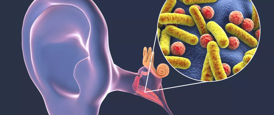
A study combining 16S rRNA sequencing and large-scale bacterial culture (“culturomics”) has documented the nasal microbiota characteristics associated with the ear and nose health of indigenous Australian children (2-7 years), a population at high risk of otitis.
By analyzing the nasal microbiota of 101 indigenous Australian children using 16S rRNA gene sequencing and a more extended bacterial culture, the researchers studied the associations between nasal microbiota composition and the children’s ear and nasal health.
Moraxella, a marker of previous otitis?
They found a greater relative abundance of Moraxella in children who had previously had an ear infection. This was so even in children who were free of otitis at the time of the analysis and may be due to a lasting remodeling of the nasal microbiota following a previous case of otitis. Moreover, the abundance of Moraxella in the nasal microbiota was negatively correlated with that of Staphylococcus, a bacterial genus found in greater abundance in children with no infectious nasal discharge. In vitro data suggest that certain species of Staphylococcus may inhibit Moraxella, which could explain the negative correlation observed.
A protective duo of microorganisms?
Furthermore, in the children not suffering from an ear condition at the time of the study, a positive correlation was observed between Dolosigranulum and Corynobacterium. This correlation was also found in children with no infectious nasal discharge, leading the authors to consider this co-colonization as potentially protective against pathogens such as S. pneumoniae and guaranteeing the health of the upper respiratory tract and ear.
Towards the identification of new otopathogens
In contrast, Ornithobacterium was found in greater abundance in children with serous otitis than in the children who had never had otitis. It may thus be a new otopathogen. Its presence was correlated with that of two other bacterial genera, Dichelobacter and Helcococcus, whose effects on nasal and ear health have yet to be defined.
This study combining 16S rRNA sequencing and culturomics was the largest ever conducted on indigenous populations. It has described associations between the nasal microbiota and ear and nasal health, identifying potential synergies (and antagonisms) between microorganisms, and new otopathogenic candidates, which will now have to be studied in greater detail.