Antenatal depressive symptoms impair offspring neurodevelopment by inducing maternal gut microbiota dysbiosis during pregnancy

COMMENTED ARTICLE - Children’ section

By Prof. Emmanuel Mas

Gastroenterology and Nutrition Department,
Children’s Hospital, Toulouse, France

Comments on the original article by Zhou et al.¹

Commented adult article

In a cohort of more than 2,000 pregnant women, the authors of this article found that depressive symptoms occurring during any trimester of pregnancy were associated with delayed neurocognitive development in infants. Analysis of the maternal gut microbiota revealed a reduction in butyrate-producing bacteria and disruptions in amino acid metabolism among women experiencing antenatal depression. These alterations were correlated with poorer cognitive outcomes in their children. To investigate causality, the microbiota from women with depressive symptoms was transplanted into germ-free pregnant mice. This transfer led to impaired intestinal barrier function, increased foetal brain inflammation, and metabolic disturbances in the developing brain, ultimately resulting in cognitive deficits in the offspring. Maternal supplementation with butyrate partially reversed these abnormalities. Overall, the findings suggest that antenatal depression may influence foetal brain development through changes in the maternal gut microbiota and its metabolites, highlighting potential preventive and therapeutic targets.

What do we already know about this subject?

Antenatal depression (AND) prevalence is around 20% worldwide and is associated with increased risk of miscarriage, preterm birth and low birth weight. However, the link between AND and neurodevelopmental issues has not been clearly established. Studies show that depression can alter the composition of the gut microbiota (GM). According to some studies, maternal mood disorders during pregnancy can also disrupt both their own GM and that of their offspring. Disruption to maternal GM during pregnancy could therefore alter brain development.
Image
Figure 1 MAS

What are the main insights from this study?

This was a prospective study of 2,053 women who gave birth between July 2019 and July 2022. Participants were recruited during the preconception period or the first trimester of pregnancy. Depression was assessed using a questionnaire (Edinburgh Postnatal Depression Scale – EPDS). Three hundred and twenty-six women (15.9%) had EPDS ≥10 (AND group). A nested case-control study was conducted to compare 135 women at risk of AND with 135 women matching on age, pre-pregnancy BMI, education level, parity, delivery mode, gestational age, Group B Streptococcus (GBS) infection, and use of corticosteroids, antibiotics, and probiotics.

Neurodevelopment was assessed up to the age of one year. Infants in the AND group scored lower for problem-solving at the age of 3 and 6 months (p = 0.039 and 0.040), and communication (p = 0.039) and motor skills (p = 0.013) at 12 months.

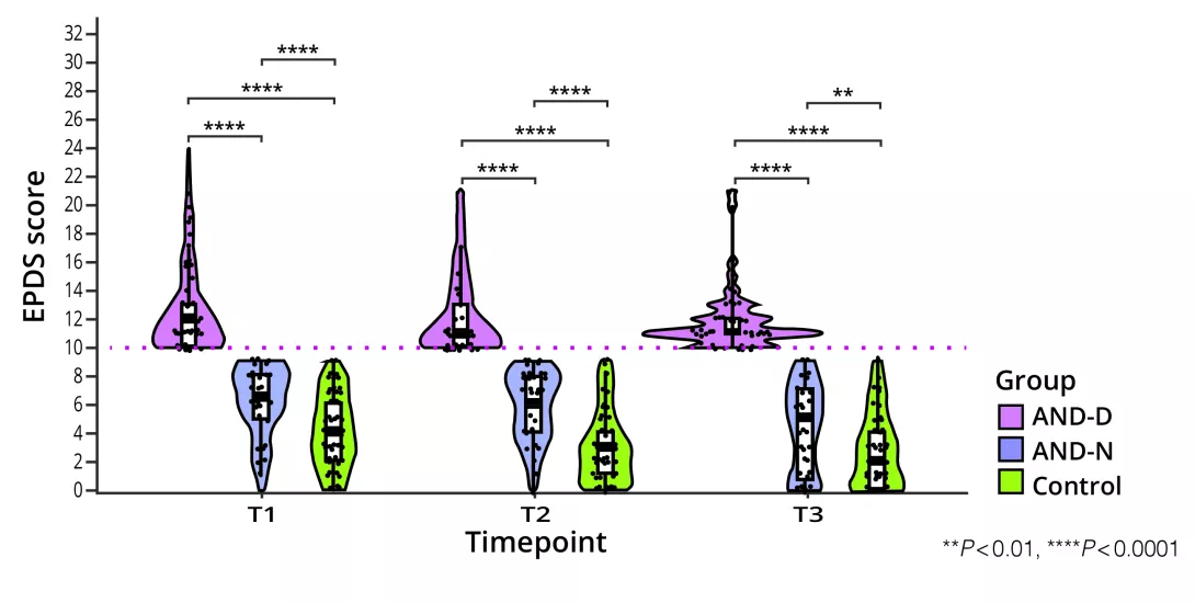
A GM analysis was conducted on 504 maternal faecal samples, divided into three groups based on the EPDS score at the time of the sample collection: AND-D (EPDS ≥10), AND-N (EPDS <10 but considered as at-risk of AND due to a high score at another stage of pregnancy) and controls (EPDS <10 in each trimester of pregnancy) (figure 1). The factors influencing the GM were, firstly, the trimester of pregnancy and, secondly, AND, with significant differences between the AND-D group and the control group, and between the AND-N group and the control group.

Compared with the control groups, the abundance of Eubacterium, Ruminococcaceae, Faecalibacterium, and Oscillospiraceae, which produce butyrate, was reduced, while Streptococcus abundance was increased in the AND-D and AND-N groups. Two hundred and ninety faecal samples were collected. A 16S RNA analysis of GM showed that AND had no effect. However, a correlation existed between maternal GM and infant neurodevelopment.

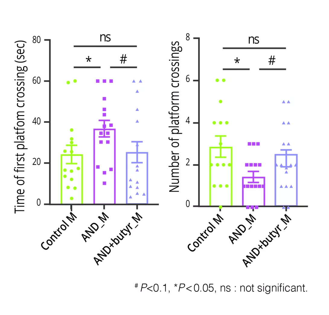
To assess the effect of maternal GM, prior to mating the authors transferred GM taken from AND at-risk mothers (AND-M group) and controls (Control-M group) to germ-free mice. The GM composition of the mice was similar prior to mating while at E18.5 (late foetal stage) it was similar to that of the respective donors. AND-M mice exhibited impaired performance in neurodevelopmental tests (escape time, platform crossing) (figure 2).

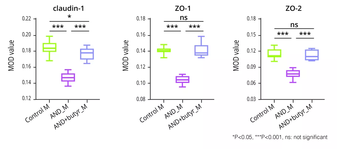
A foetal brain metabolomic analysis showed reduced activity of polyunsaturated fatty-acid (PUFAs) pathways and amino-acid pathways in AND-M mice. An increase in inflammatory cytokines (TNF-α and IL-1β) was also observed in the AND-M group. Finally, mothers in the AND-M group had increased intestinal permeability, as assessed by immunohistochemistry, which revealed lower expression of tight junction proteins (claudin-1, ZO-1, and ZO-2) (figure 3), as well as higher inflammatory cytokine levels in serum (TNF-α and IL-1β).

Butyrate supplementation given to mice during pregnancy not only improved the neurodevelopment of their offspring but also increased PUFA and amino-acid levels, improved maternal gut barrier function and reduced the levels of inflammatory cytokines in foetal brains and maternal serum.

What are the consequences in practice?

The effects of antenatal depression on the developing foetal brain may be mitigated through butyrate supplementation.

Key points ​​​​​​​

  • Antenatal depression is associated with altered infant neurodevelopment 
  • Maternal gut microbiota could play a role in this association 
  • In mice, butyrate mitigated the anomalies observed and improved neurodevelopment

Assessment of neurodevelopment in the offspring of germ-free mice having received faecal matter from control mothers (Control_M), AND mothers (AND_M) and AND mothers treated with butyrate during pregnancy (AND+butyr_M). Tests assessed mobility on a platform and showed that mobility was impaired in the AND_M group and improved after receiving butyrate supplementation during pregnancy.
Image
Figure 2 MAS

Measurement of tight junction protein expression (claudin-1, ZO-1, ZO-2) between colonocytes, reflecting gut permeability in germ-free mice having received faecal matter from control mothers (Control_M), AND mothers (AND_M) and AND mothers treated with butyrate during pregnancy (AND+butyr_M). Permeability was impaired in the AND_M group and improved with butyrate supplementation during pregnancy.
Image
Figure 3 MAS

Conclusion

Antenatal depression impairs neurodevelopment in children, linked to changes in the maternal GM. Experiments conducted in mice showed that maternal gut dysbiosis increases intestinal permeability, which in turn is associated with foetal brain inflammation and lower activity of metabolic pathways (PUFAs and amino acids). Butyrate supplementation during pregnancy mitigated these effects.

Summary
Off
Sidebar
On
Migrated content
Désactivé
Updated content
Désactivé
Hide image
Off
Commented article Off

Functional dyspepsia and the gut microbiota

By Prof. Lucas Wauters
Gastroenterology and Hepatology, University Hospitals Leuven, Leuven, Belgium

Functional dyspepsia (FD) refers to chronic epigastric symptoms, with postprandial distress syndrome and epigastric pain syndrome as the major subgroups. It is part of the disorders of gut-brain interaction, with visceral signaling (gut-brain) via duodenal hyperpermeability and immune activation, potentially triggered by an altered microbiome composition or dysbiosis. Interestingly, first-line therapy with proton pump inhibitors (PPI) may improve both symptoms and duodenal alterations in the short term but adversely influence the duodenal environment in the long-term. In addition, other treatments, including prokinetics and neuromodulators, have limited efficacy or raise safety issues. Therefore, alternative treatments have been studied, including the spore-forming Bacillus coagulans MY01 and Bacillus subtilis MY02. Besides the clear efficacy and safety in a randomized, double-blind, placebo-controlled trial, beneficial immune and microbial changes were found and more pronounced in FD patients on PPIs, including a reduction in small intestinal bacterial overgrowth. These and other studies have provided insights into potential underlying mechanisms of FD, including the role of gut microbiota.

Definition and diagnosis

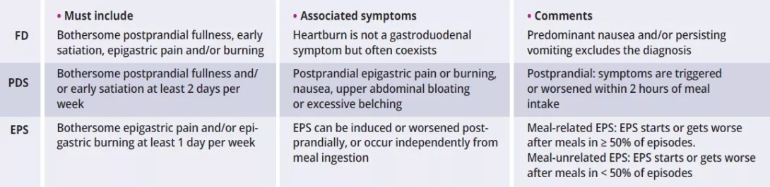
Dyspepsia refers to chronic upper abdominal symptoms originating from the gastroduodenal region with a significant impact on patients’ quality of life. According to the Rome criteria, functional dyspepsia (FD) is characterized by bothersome epigastric symptoms that remain unexplained after routine investigations¹⁻³. Disorders of gut-brain interaction (DGBI), including FD, are related to any combination of dysmotility, visceral hypersensitivity, altered luminal or central factors. In contrast, organic dyspepsia may result from erosive esophagitis, peptic ulcer disease or cancer in a minority of patients¹⁻³.

Functional dyspepsia is characterized by bothersome epigastric symptoms that remain unexplained after routine investigations.

Image
table 1 rome V

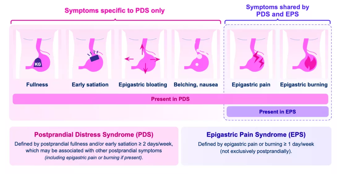
Two subgroups of FD were proposed by the Rome III consensus and reiterated in the Rome IV version:

  • postprandial distress syndrome (PDS) with postprandial fullness or early satiation,
  • epigastric pain syndrome (EPS) with epigastric pain or burning¹. 

The recent Rome V version specifies that symptoms must be severe enough to affect usual activities with a minimal frequency of 1 (EPS) or 2 (PDS) days per week², which is lower than the previous Rome IV criteria for PDS (3 days per week) (table 1). As PDS is characterized by meal-related symptoms (occurring < 2 hours after meal ingestion), postprandial epigastric pain in the presence of PDS symptoms should also be characterized as PDS and not EPS according to Rome V². In the absence of PDS, epigastric pain symptoms could be further subdivided into meal-related or meal-unrelated EPS, which requires further studies (figure 1). Nausea (especially postprandially) and vomiting can coexist in PDS and EPS, but predominant nausea or recurrent vomiting is more common in chronic nausea and vomiting syndrome or gastroparesis².

Using the Rome IV criteria, PDS was consistently more prevalent (61% vs. 18% for EPS and 21% for overlap) with a similar distribution across the USA, UK and Canada⁴. In addition, FD often coexisted with gastroesophageal reflux (GER) and/or irritable bowel syndrome (IBS). Of note, bowel disorders should be considered when epigastric symptoms are consistently relieved by bowel movements¹⁻³. 

While the exclusion of structural, systemic, or metabolic diseases is necessary for confirmation of FD, the yield of routine endoscopy is low in (young) patients with dyspeptic symptoms and no alarm or risk factors (see below). The prevalence of dyspepsia was higher in women, smokers, users of nonsteroidal anti-inflammatory drugs (NSAIDs) and after Helicobacter pylori (H. pylori) infection¹⁻³. Thus, a non-invasive “test & treat” strategy (i.e., urea breath test or H. pylori fecal antigen detection) is common, with endoscopy for alarm factors (e.g., age > 55 years old, or lower in certain regions of the world, evidence of gastrointestinal bleeding or iron-deficiency anemia, dysphagia, unintentional weight loss and family history of gastric or esophageal cancer)¹⁻³. Nevertheless, the Rome criteria for FD do not exclude the presence of microscopic pathology in the gastroduodenal region or the stomach and proximal small intestine.

Image
Synthese figure 1

Duodenum and dysbiosis

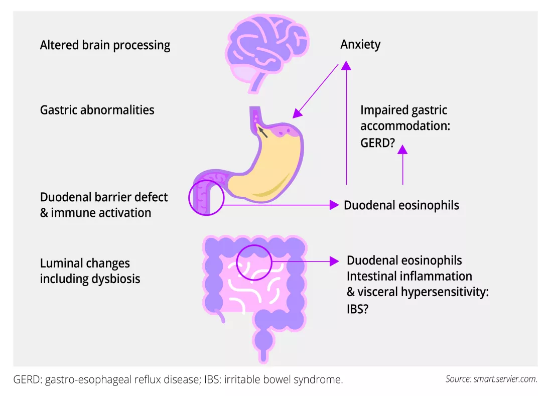
Abnormalities of gastric function in FD include impaired accommodation or expansion of the stomach, hypersensitivity to distention and delayed emptying, but these changes correlate poorly or not at all with symptoms¹⁻². Both visceral sensory signaling (gut to brain) and central modulation (brain to gut) can be abnormal in FD, as up to 50% of subjects identify stress as a trigger for their symptoms, and anxiety and depression are common. Recent research focused on mucosal and luminal alterations in the proximal small intestine or duodenum⁵. Interestingly, duodenal mucosal eosinophilia was linked to impaired gastric accommodation, reflux symptoms and anxiety, which may influence gastric function⁵. Overlapping IBS symptoms in FD may also be explained by more extensive intestinal inflammation with visceral hypersensitivity (figure 2).

Image
Figure 2 synthese

Current and novel therapies

Despite the recognition of meal-related symptoms, no specific dietary factors have been convincingly linked with the pathophysiology of FD. In the absence of controlled dietary intervention studies, standard dietetic advice includes frequent small size meals with avoidance of high‐fat food items, alcohol or coffee and smoking cessation¹⁻³. Acid-suppressive therapy with PPI is recommended as first-line therapy in H. pylori-negative patients or after eradication if H. pylori-positive³. Indeed, PPIs are more effective than placebo in the reduction of global symptoms of FD with a number needed to treat (NNT) of 11 in a Cochrane meta-analysis⁹. This includes a standard once-daily dose of PPI during 4 to 8 weeks, with discontinuation of therapy in case of insufficient improvement. While PPIs are beneficial for overlapping reflux symptoms, we have also shown beneficial effects of short-term PPI-therapy on duodenal permeability and immune activation in FD patients⁷.

Recent research focused on mucosal and luminal alterations in the proximal small intestine or duodenum.

Duodenal mucosal changes in FD patients include a barrier defect and immune activation, with increased numbers and degranulation of eosinophils and mast cells. Even if a mucosal T-helper (Th) 2-signal was not commonly found, increased interleukin (IL)5/13-production after stimulation of peripheral blood mononuclear cells (PBMC) from FD patients suggested a shift from Th1- to Th2-type inflammation⁵. In addition, increased IL1β-production of cultured PBMC pointed to a Th17-response, and production of GM-CSF from Th17 cells may also drive mucosal eosinophil recruitment in FD (see below). Moreover, CD4+ T cells co-expressing integrin α4β7 and CCR9, indicative of small intestinal mucosal migration, were upregulated in FD and correlated with gastric emptying. This also suggests a primary role for duodenal mucosal changes, but the luminal or other triggers are unknown⁵.

Duodenal luminal changes include an altered microbial composition or “dysbiosis”.

We found a lower abundance of Neisseria and Porphyromonas and microbial diversity in duodenal brush samples of FD patients compared to healthy controls, which inversely correlated with symptoms and duodenal eosinophils⁶. In contrast, microbial changes were associated with the effects of proton pump inhibitors (PPI) on duodenal bile salt concentrations in healthy controls, which persisted after long-term PPI-therapy in FD patients⁶⁻⁷. The presence of intestinal-like bacteria in the gastric fluid of FD patients also suggested small intestinal bacterial overgrowth (SIBO) in a subset, even if a standardized diagnosis of SIBO is not available yet and the majority of studies have used non-invasive breath tests in FD with different substrates (glucose or lactulose), doses or type of exhaled gases (hydrogen and/or methane)⁸. 

We and others have hypothesized that the duodenal barrier defect may lead to mucosal immune activation through antigen presentation with a predominant Th2- or Th17-response and recruitment and degranulation of eosinophils, triggering visceral hypersensitivity and dysmotility. Possible causes include the duodenal microbiome, which may also be a therapeutic target⁵’⁸. 

A significant reduction in dyspeptic symptoms was also found with prokinetics, with a NNT of 12 when cisapride (withdrawn from the market due to cardiac adverse events) was excluded in a systematic review and meta-analysis¹⁰. However, QT-prolongation (domperidone) or extrapyramidal side effects (metoclopramide) limit their chronic use and other prokinetics such as itopride or acotiamide are not widely available. In addition, amitriptyline (tricyclic antidepressant) but not escitalopram (selective serotonin-reuptake inhibitor) was effective for ulcer-like FD, with threefold higher odds of reporting adequate relief of symptoms compared to placebo¹¹. Another systematic review and network meta-analysis confirmed the superior efficacy of antidepressants or neuromodulators for symptom improvement, and that symptom resolution was most common with standard-dose PPIs¹². 

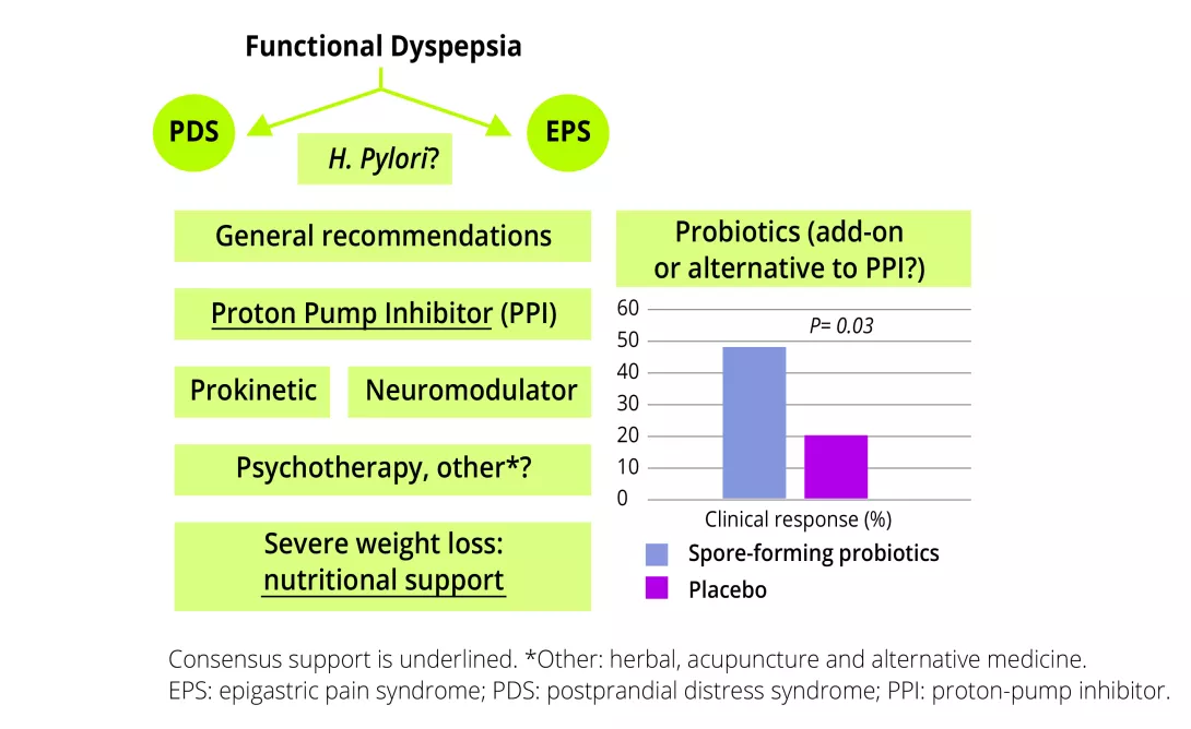
However, inappropriate and prolonged use of PPIs is frequently reported, and may increase the risk of dysbiosis. Probiotics are live microorganisms that exert a health benefit on the host, and spore-forming probiotic strains may outperform traditional supplements because of gastric-acid resistant endospores with improved survival in the small intestine. We showed that Bacillus coagulans MY01 and subtilis MY02 spore-forming probiotics (daily dose of 5·10⁹ colony-forming units or CFU) were effective and safe in FD patients as mono- or add-on therapy to long-term intake of PPI¹³. Importantly, PDS (primary outcome), EPS and individual symptoms significantly decreased with spore-forming probiotics compared to placebo. We extended the clinical efficacy during an open-label phase (total 16 weeks), with symptom-improvement in the original placebo-group.

Inappropriate and prolonged use of PPIs is frequently reported and may increase the risk of dysbiosis.

Interestingly, longer-term probiotic treatment decreased systemic Th17-signaling, including Th17 cells and IL-17A, which were associated with clinical efficacy. Additional decreases were found in Th2, gut-homing and GM-CSF+ T cells in FD patients on-PPI, suggesting that the probiotic effects may be more pronounced in case of PPI-related dysbiosis. Moreover, a reduction in SIBO was found in FD patients on-PPI after 8 weeks. Finally, despite the absence of major shifts in relative or quantitative fecal microbiota community composition, a proportional increase in the beneficial Faecalibacterium was also associated with probiotic efficacy. Another RCT found a higher clinical efficacy for the probiotic Bifidobacterium animalis subsp. lactis BL-99 in a high (5·10¹⁰ CFU/day) compared to low dose (1·10¹⁰ CFU/day) or positive-drug control (rabeprazole) and placebo after 8 weeks¹⁴. Interestingly, this effect was sustained 2 weeks after stopping therapy, including the persistence of the probiotic strains in stool samples. Moreover, an increase in short-chain fatty acid (SCFA)-producing microbiota such as Faecalibacterium and Roseburia was also found with the probiotic, with increased levels of SCFA in stool and blood samples, pointing to underlying microbial mechanisms. 

While the use of probiotics was not yet included in the European consensus on FD³, agreement or consensus support was only reached for PPI and nutritional support in case of severe weight loss (figure 3). Based on the novel evidence, probiotics (particularly spore-forming) could be considered as add-on or alternative to (long-term) PPI-therapy.

Image
Figure 3 synthese

Conclusion

Emerging data increasingly point towards the duodenum as a key integrator in FD, including the microbiome of the small intestine¹⁵. While first-line or PPI-therapy may be effective in the short term, long-term therapy is frequently reported with an increased risk of dysbiosis. Alternative therapies include probiotics, with not only beneficial clinical but also immunological and microbial effects of spore-forming probiotics. Although these could be considered as monotherapy, changes in immune activation were more pronounced in FD patients on-PPI, suggesting additional effects on the observed chronic alterations with PPI-therapy, which is especially useful in case PPI-withdrawal is not feasible.

Sources:

1. Stanghellini V, Chan FK, Hasler WL, et al. Gastroduodenal Disorders. Gastroenterology 2016; 150: 1380–92.

2. Törnblom H, Carbone F, Hasler WL, et al. Gastroduodenal Disorders. Gastroenterology 2026; 170: 1240-60.

3. Wauters L, Dickman R, Drug V, et al. United European Gastroenterology (UEG) and European Society for Neurogastroenterology and Motility (ESNM) consensus on functional dyspepsia. United Eur Gastroenterol J 2021; 9: 307-31.

4. Aziz I, Palsson OS, Törnblom H, et al. Epidemiology, clinical characteristics, and associations for symptom-based Rome IV functional dyspepsia in adults in the USA, Canada, and the UK: a cross-sectional population-based study. Lancet Gastroenterol Hepatol 2018; 3: 252-62.

5. Wauters L, Talley NJ, Walker MM, Tack J, Vanuytsel T. Novel concepts in the pathophysiology and treatment of functional dyspepsia. Gut 2020; 69: 591-600.

6. Wauters L, Tito RY, Ceulemans M, et al. Duodenal dysbiosis and relation to the efficacy of proton pump inhibitors in functional dyspepsia. Int J Mol Sci 2021; 22: 13609.

7. Wauters L, Ceulemans M, Frings D, et al. Proton pump inhibitors reduce duodenal eosinophilia, mast cells and permeability in patients with functional dyspepsia. Gastroenterology 2021; 160: 1521-31.e9.

8. Marasco G, Fiocca M, Cremon C, et al. Therapeutic role of probiotics for the treatment of dyspepsia: a review of the literature. Neurogastroenterol Motil 2025; 37: e70057.

9. Pinto-Sanchez MI, Yuan Y, Hassan A, et al. Proton pump inhibitors for functional dyspepsia. Cochrane Database Syst Rev 2017; 11: CD011194.

10. Pittayanon R, Yuan Y, Bollegala NP, et al. Prokinetics for functional dyspepsia: a systematic review and meta-analysis of randomized control trials. Am J Gastroenterol 2019; 114: 233-43.

11. Talley NJ, Locke GR, Saito YA, et al. Effect of amitriptyline and escitalopram on functional dyspepsia: a multicenter, randomized controlled study. Gastroenterology 2015; 149: 340-9.e2.

12. Ford AC, Moayyedi P, Black CJ, et al. Systematic review and network meta-analysis: efficacy of drugs for functional dyspepsia. Aliment Pharmacol Ther 2021; 53: 8-21.

13. Wauters L, Slaets H, De Paepe K, et al. Efficacy and safety of spore-forming probiotics in the treatment of functional dyspepsia: a pilot randomised, double-blind, placebo-controlled trial. Lancet Gastroenterol Hepatol 2021; 6: 784-92.

14. Zhang Q, Li G, Zhao W, et al. Efficacy of Bifidobacterium animalis subsp. lactis BL-99 in the treatment of functional dyspepsia: a randomized placebo-controlled clinical trial. Nat Commun 2024; 15: 227.

15. Wauters L, Burns G, Ceulemans M, et al. Duodenal inflammation: an emerging target for functional dyspepsia? Expert Opin Ther Targets 2020; 24: 511-23.

Summary
Off
Sidebar
On
Migrated content
Désactivé
Updated content
Désactivé
Hide image
Off
Article Off

Can prenatal antibiotics leave a mark on a child's skin?

We often treat antibiotics in pregnancy as a short trade: infection cleared, problem solved. A meta-analysis of 30 studies¹ suggests the story may reach the child's skin, with a modest but consistent rise in atopic dermatitis after prenatal exposure.

Antibiotics are among the most common drugs in pregnancy: an earlier meta-analysis of about 34 million pregnancies put exposure at 23.6%. Their effect on the mother is well documented: antibiotics induce a gut microbiome disruption. Their effect on the child's early immune development is far less clear. A new systematic review in Scientific Reports, from researchers at the University of Szeged, tackles that gap for atopic dermatitis, a condition your patients' children may already know well.

Peripartum prophylactic antibiotic therapy decreases bifidobacterium levels in breast milk

https://www.biocodexmicrobiotainstitute.com/fr/pro/une-antibiotherapie-prophyla…

Thirty studies, one modest signal

Searching four databases up to August 2025 under a registered PROSPERO protocol, the team pooled 30 observational studies covering more than 4.1 million mothers and 4.3 million children, 26 studies assessing maternal antibiotic use at any point in pregnancy. The available data did not allow reliable comparisons between exposure in the first trimester, second trimester and third trimester. Prenatal exposure was associated with higher odds of atopic dermatitis in childhood: an adjusted odds ratio (aOR) of 1.32 (95% CI 1.12 to 1.56) across 14 studies, and a pooled adjusted hazard ratio of 1.10 (1.05 to 1.14) across four. The age of children at diagnosis also varied between studies. Evidence of a potential publication bias was detected, so the authors applied a trim-and-fill correction. The aOR fell to 1.22 (1.03 to 1.44) but stayed significant: the raw figure may have overstated the effect without erasing it. The association remained statistically significant in leave-one-out analyses, even when individual studies at serious risk of bias were excluded. The authors themselves call the effect modest. These relative effect estimates do not quantify the absolute increase in risk or imply that every exposed child will develop atopic dermatitis.

Prenatal vs intrapartum exposure

Prenatal exposure means antibiotics taken at any point in pregnancy. Intrapartum exposure means prophylaxis given during labour and delivery, as in the group B Streptococcus prophylaxis studies included here. In this meta-analysis, the first showed a modest association with atopic dermatitis and the second did not.

Atopic dermatitis vs eczema

Atopic dermatitis is a chronic inflammatory skin disease with red, dry, itchy skin, driven by a defective skin barrier. Eczema is the broader, non-specific term, but the two are often used as synonyms, which is why the authors counted both as outcomes.

Adjusted odds ratio (aOR)

An odds ratio recalculated to account for confounders such as infant sex, maternal age, maternal allergy or atopy, and smoking in pregnancy. An aOR of 1.32 means the odds of atopic dermatitis were 32% higher in exposed children, after those adjustments.

Where the signal holds, and where it fades

Risk rose with the number of antibiotics courses. Pooled adjusted hazard ratios were 1.07 for one course, 1.10 for two and 1.16 for more than two, although only three studies contributed. Geography mattered: European studies showed a significant association (aOR 1.40, 95% CI 1.08 to 1.83), while Asian and US studies did not reach significance. Of course, an absence of significance does not imply an absolute absence of association between prenatal antibiotic use and eczema in those regions. Results were also stronger with parent-reported eczema diagnoses than with medical records, and for the largest studies (over 10,000 children), the pooled adjusted association was not statistically significant. Overall, heterogeneity was very high (I² up to 99.7%) and GRADE certainty was low. Intrapartum exposure behaved differently: a pooled analysis of four intrapartum studies found no significant association (OR 1.64, 95% CI 0.84 to 3.17). However, individual findings were inconsistent, heterogeneity was substantial, and the certainty of evidence was very low. Findings also differed by birth mode, with some studies reporting different results for vaginal and caesarean deliveries. The window of intrapartum exposure is much narrower, which may mean only a transient effect on the infant's microbiota, though one study did find a signal when exposure exceeded 24 hours.

First 1,000 days

The period from conception to about age 2, when the microbiome and immune system are taking shape. It is the window in which the exposures discussed here are thought to matter most.

Trim-and-fill

A statistical method that estimates which studies may be missing because of publication bias and recalculates the pooled effect. Here it lowered the aOR from 1.32 to 1.22, which stayed significant.

GRADE

A framework that rates how much confidence we can place in a body of evidence. The prenatal findings were rated low and the intrapartum findings very low, because of bias, inconsistency and imprecision.

A plausible mechanism, not a proven one

The meta-analysis tested exposure and outcome, not the microbiota itself. It runs like this: antibiotics alter the mother's gut and vaginal communities, those communities act as the "starter culture" for the newborn's gut at birth, and a different start could shape early immune programming during the first 1,000 days. Birth mode and feeding practices may also influence how that microbiota develops. Reduced intestinal diversity has already been tied to atopic dermatitis in some other studies. The caveats are real. Postnatal antibiotic use, a plausible driver in its own right, was not consistently adjusted for, and observational data cannot establish causality. The authors urge antibiotics only when clearly indicated, for the shortest effective course, favouring narrow-spectrum antibiotics when appropriate. The takeaway for us is not to withhold antibiotics from pregnant patients with an infection, which is still essential to treat, but to treat each prescription as a risk-benefit decision, with a possible effect on the child's skin now part of the equation.

Source

1. Petrás S, Vargané Szabó B, Kiss T, Bahar MA, Csupor D, Tóth B. Prenatal maternal antibiotic use increases the risk of childhood eczema: a systematic review and meta-analysis. Sci Rep. 2026;16:21156. doi:10.1038/s41598-026-51441-x.

Summary
Off
Sidebar
Off
Migrated content
Désactivé
Updated content
Désactivé
Hide image
Off
News Obstetrics Gynecology Pediatrics Dermatology General Medicine On

Antibiotics in pregnancy: what's the link to baby's eczema?

An infection during pregnancy needs treating. But could the antibiotic that clears it leave a faint trace on your baby's skin? A new analysis of over 4 million children found a small but consistent link with eczema.

Antibiotics in pregnancy: what's the link to baby's eczema?

Atopic dermatitis, the most common form of eczema, affects about one child in five. Skin turns red, dry and itchy because a weakened barrier lets water escape and irritants in. It usually appears before the age of two and often fades by itself, but it can be the first step toward other allergies, such as food allergy, asthma and hay fever. Strictly speaking, "eczema" is the wider term, though most people use the two interchangeably. In children, this can be an early stage of what doctors call the "atopic march".

Atopic dermatitis vs eczema

Atopic dermatitis is a chronic, itchy skin condition linked to a weakened skin barrier. Eczema is the broader word, and the two are often used for the same thing.

The first 1,000 days

From conception to a child's second birthday, a window when the microbiota and immune system are being built. What happens here can echo for years. 

A small link, found again and again

Nearly one pregnancy in four involves antibiotics, according to an earlier review of about 34 million pregnancies. Researchers1 from Hungary and Indonesia combined 30 studies on prenatal antibiotics exposure and more than 4.1 million mothers and 4.3 million children. After adjusting for factors such as the mother's age, allergies and smoking, children exposed before birth had 22% to 32% higher odds of eczema, depending on how the data were corrected for publication bias. This association concerns the odds of eczema, not a precise prediction of an individual child's risk. The link grew slightly with each additional course of antibiotics, and even a single course was associated with a small increase.

In three studies, the association grew with repeated antibiotic exposure, although the authors caution that the evidence is limited. This is an association, not proof of cause, and the authors rate the evidence as low certainty. The studies also lacked comparable data to distinguish exposure in the first trimester, second trimester or third trimester, so no firm conclusion can be drawn about the timing of prenatal treatment. It was clearest in European studies, and not seen in the US ones. The association was also not statistically significant in analyses restricted to larger studies or to eczema diagnosed from medical records. For one child the increase is small, the authors say, but with so many pregnancies and so many children affected, it may still matter across a population.

Prenatal vs intrapartum antibiotics

Prenatal means taken during pregnancy. Intrapartum means given during labour and delivery. This study found a small link with the first, and none with the second. 

Why would a medicine reach the skin?

The leading hypothesis involves the microbiota. Antibiotics can disturb the microbes of a mother's gut and vagina, and a baby inherits its first microbes from her at birth. This early microbial exposure may help shape the child's developing immunity, making maternal microbiota a possible link between pregnancy and later skin health. Think of them as the first teachers of a young immune system: if the early lessons change, so might what it learns. The studies did not measure this directly. Nor could the researchers separate the effects of different antibiotic types, doses or treatment durations. Antibiotics given during labour showed no significant link, though only four studies looked. That finding does not rule out a birth-related association: the evidence for antibiotics around delivery was rated very low certainty.

6 essential things you should know about antibiotics

Learn more

None of this is a reason to refuse a treatment your doctor prescribes, since antibiotics remain essential for maternal infections. The authors recommend using antibiotic treatment in pregnancy when clearly needed, with an appropriate duration and choice of medicine. Eczema also has other roots, such as family history and variants of the filaggrin gene, which help the skin build its protective layer.

Maternal microbiota

The community of microbes living in a mother's gut and vagina. The vaginal microbiota in particular helps shape the newborn's gut microbiota, which is why antibiotics taken in pregnancy are being studied.

Source

1. Petrás S, Vargané Szabó B, Kiss T, Bahar MA, Csupor D, Tóth B. Prenatal maternal antibiotic use increases the risk of childhood eczema: a systematic review and meta-analysis. Sci Rep. 2026;16:21156. doi:10.1038/s41598-026-51441-x

Summary
Off
Sidebar
On
Migrated content
Désactivé
Updated content
Désactivé
Hide image
Off
News On

Chlorinated water and antibiotic resistance in infants: is there a link?

Chlorination helps control harmful microorganisms in drinking water. But could residual chlorine also affect infant gut microbiome and contribute to antibiotic resistance? A study involving 197 infants explored this question.1

The gut microbiota

Antibiotic resistance is increasing worldwide and remains a major public health challenge. Between 2018 and 2023, more than 40% of the pathogen–antibiotic combinations monitored by the WHO showed increasing resistance.2 Efforts to address this issue have primarily focused on reducing antibiotic use and promoting vaccination to prevent infections. However, environmental factors may also play a role in the development of antibiotic resistance. Australian researchers investigated one such factor that has received relatively little attention: the consumption of chlorinated drinking water.

Nearly 2 out of 5 people have been informed by a healthcare professional that taking antibiotics could negatively affect the balance of their microbiome.

Only 1 out of 4 people reported receiving all the essential information about the microbiome when antibiotics were prescribed.

Chlorinated vs. filtered water: a 12-month study involving 197 infants

Chlorination disinfects drinking water, helps ensure its microbiological safety, and reduces the risk of gastrointestinal infections. Chlorine levels in drinking water are regulated, and the concentrations used in this study (0.3–1.1 mg/L) remained below the 5 mg/L limit. However, residual chlorine, which remains in tap water after disinfection, may affect infants’ gut environment by favoring bacteria carrying antibiotic resistance functions. To test this hypothesis, 197 six-month-old infants were randomly assigned to one of two groups for 12 months: one group received filtered tap water, from which chlorine and certain other chlorine-based disinfectants had been removed, while the other received conventional chlorinated tap water. Stool samples were collected at the beginning and end of the study to analyze the DNA of the microorganismses present in the gut.

Tap water and baby bottles: what French health authorities recommend.⁴

Tap water can be used to prepare baby bottles, with a few precautions: 

- Let the water run for a few seconds, use only cold water, and clean the tap aerator regularly.

- ANSES* advises against using home-filtered or softened water to prepare baby bottles, as these systems can promote the growth of microorganisms.


*ANSES: French Agency For Food, Environmental And Occupational Health & Safety.

An overall unchanged microbiome, but greater antibiotic resistance potential

Overall, the gut microbiota profiles of the two groups were similar, with no significant differences in bacterial content, diversity, or overall community structure. However, the group that drank chlorinated water had a more extensive resistome—that is, a greater abundance of functions associated with antibiotic resistance, including enzymes capable of breaking down antibiotics.3

An additional analysis suggested that this increase in antibiotic resistance functions may be more pronounced among children who both consumed chlorinated water and received antibiotics during the study. The authors therefore hypothesize that these two exposures may act together, potentially increasing the risk of selecting more resistant bacteria.

Resistome

The set of antibiotic resistance genes carried by microorganisms within a given environment, in this case, the gut microbiota.3

Residual chlorine

Chlorine that remains in tap water after disinfection.

Should we stop drinking tap water?

These findings do not challenge the practice of drinking water chlorination, which has well-established health benefits. However, the authors concluded that chlorinated tap water could strengthen antibiotic resistance functions in the infant gut microbiota, although no associated clinical risk to infant health has yet been demonstrated. Further research is needed to determine whether adjusting the timing or method of introducing tap water could mitigate this effect and to assess its potential impact on children’ health, particularly for those exposed to multiple courses of antibiotics.

The study1 was partly funded by Water Corporation WA and received in-kind support from Pure Water Systems. Neither partner was involved in the study design, data analysis, or decision to publish.

Sources

1. Parkin K, Christophersen CT, Verhasselt V, et al. Chlorinated drinking water exposure enriches antimicrobial resistance pathways in the infant gut microbiome: a randomized trial. Commun Med. 2026;6:405. doi:10.1038/s43856-026-01626-2.

2. World Health Organization. WHO warns of widespread resistance to common antibiotics worldwide. Geneva: WHO; 13 October 2025. https://www.who.int/news/item/13-10-2025-who-warns-of-widespread-resistance-to-common-antibiotics-worldwide. 

3. D'Costa VM, McGrann KM, Hughes DW, Wright GD. Sampling the antibiotic resistome. Science. 2006;311(5759):374-377. doi:10.1126/science.1120800.

4. ANSES. Biberon : comment le préparer et le conserver ? Maisons-Alfort : Anses ; article du 17/01/2013. https://www.anses.fr/fr/article/biberon-comment-preparer-conserver  (consulté le 05/10/2026).

Summary
Off
Sidebar
Off
Migrated content
Désactivé
Updated content
Désactivé
Hide image
Off
News Off

Chlorinated water and the infant gut resistome

Even at concentrations compliant with health standards, chlorinated drinking water may enhance antibiotic resistance functions in the infant gut microbiome. A randomized, double-blind trial involving 197 Australian infants revealed a potential risk of selectively favoring resistant bacterial populations.1

Antibiotic resistance –a major component of antimicrobial resistance– is a global public health priority and a growing health concern. Between 2018 and 2023, more than 40% of the pathogen–antibiotic combinations monitored by the WHO showed increasing resistance.2 While efforts to combat antibiotic resistance have primarily focused on reducing antibiotic use and promoting vaccination, an Australian study explores a less investigated environmental factor that may impact the gut microbiota: the consumption of chlorinated drinking water. Although chlorination is essential for water disinfection and is subject to regulatory limits (residual chlorine levels ranged from 0.3 to 1.1 mg/L in the study, below the 5 mg/L limit), residual chlorine and other chlorine-related disinfection by-products may exert selective pressure on the infant gut microbiome, potentially favoring resistant bacteria. According to the authors, this is the first randomized, double-blind controlled trial evaluating this hypothesis in a high-income country, where drinking water is subject to treatment and disinfection standards.

Resistome :

The set of antibiotic resistance genes carried by microorganisms within a given environment, in this case, the gut microbiota.4 

197 infants followed for 12 months

A total of 197 six-month-old infants from the ORIGINS Project cohort were randomly assigned to two groups for a 12-month intervention: one received conventional chlorinated tap water, while the other received tap water dechlorinated through filtration, which significantly reduced levels of chlorine and certain disinfection by-products. Stool samples were collected at the beginning and end of the study period and analyzed using metagenomic sequencing. This enabled researchers to assess changes in the composition and functional characteristics of the gut microbiota under different levels of exposure to chlorine.

Summary table of key results

Evaluated Criterion: overview

Overview: number and variety of bacteria (abundance, diversity).

Observed Result (chlorinated water vs. dechlorinated water): 
No significant difference between the two groups.

Evaluated Criterion : detailed view

Detailed view: quantity of certain bacteria (genera and species).

Observed Result (chlorinated water vs. dechlorinated water):
Despite the overall similarity, subtle differences were observed, with considerable variability between infants. 

Less abundant in the chlorinated-water group: Bacteroides, Clostridium, and Blautia (taxa associated with a healthy gut microbiome). 
More abundant: Streptococcus and Klebsiella pneumoniae (opportunistic pathogens under certain circumstances), as well as Faecalibacterium prausnitzii and Bifidobacterium (beneficial taxa).

Evaluated Criterion : antibiotic resistance functions (functional potential)

Observed Result (chlorinated water vs. dechlorinated water):
Significantly more abundant in the chlorinated-water group; 
The specific resistance genes involved were not individually identified.

Evaluated Criterion : clinical exposure to antibiotics (stratified analysis)

Observed Result (chlorinated water vs. dechlorinated water):
Greater enrichment of resistance functions was observed among children who had received antibiotics, but the difference was not statistically significant after adjustment. Further investigation is needed.

Gut microbial diversity remained unchanged, but antibiotic resistance functions were enhanced

No significant differences in bacterial richness or overall diversity were observed between the two groups. At the taxonomic level, differences between the two groups were identified in a small subset of bacterial taxa. However, these were rarely detected and showed considerable variability between infants. The main finding concerned the functional potential of the gut microbiota: functions associated with antibiotic resistance, including efflux pumps and antibiotic-degrading enzymes, were significantly enriched in infants exposed to chlorinated drinking water. The authors nevertheless acknowledge an important limitation: the specific antibiotic resistance genes involved were not individually identified. An additional analysis stratified by antibiotic exposure suggested a more pronounced enrichment among infants who had received antibiotics (n=40). However, this trend was not statistically significant after adjustment and requires further investigation. Secondary health outcomes, including allergies, wheezing, and eczema, were available for approximately half of the children. No significant differences were observed between the groups, although a clinical effect cannot be ruled out.

Stratified analysis:

A statistical approach in which analyses are performed separately within subgroups defined by a specific characteristic (in this case, antibiotic exposure) to assess whether an observed effect varies according to that factor.

These findings do not challenge the practice of drinking water chlorination, which has well-established health benefits. However, they highlight the need to investigate, within the broader context of the infant exposome, whether the timing or methods of water introduction could be optimized to minimize exposure to chlorinated disinfectants, alongside the appropriate use of antibiotics in pediatric care. The results should nevertheless be interpreted as exploratory. The COVID-19 pandemic reduced the number of children who completed the study, and the sample size was insufficient to adequately assess the role of antibiotic exposure. Consequently, the study cannot yet establish whether chlorinated drinking water selects for antibiotic-resistant bacterial populations or whether such changes have health consequences.

Exposome:

The totality of environmental exposures an individual experiences from conception onward, including diet, lifestyle, and endogenous factors.5

The study1 was partly funded by Water Corporation WA and received in-kind support from Pure Water Systems. Neither partner was involved in the study design, data analysis, or decision to publish.

What type of water should be used to prepare infant formula? Recommendations from the French Agency for Food, Environmental and Occupational Health & Safety (ANSES)3

Tap water (public water supply)

ANSES recommendation : 
Suitable for use, provided certain precautions are taken: let the water run for a few seconds, use only cold water, and clean the tap aerator regularly.

Home-filtered or softened water

ANSES recommendation : 
Not recommended, as these systems may promote microbial growth.

Water from lead pipes (homes built before 1948)

ANSES recommendation : 
Consult your local town hall or competent health authority before use.

Sources

1. Parkin K, Christophersen CT, Verhasselt V, et al. Chlorinated drinking water exposure enriches antimicrobial resistance pathways in the infant gut microbiome: a randomized trial. Commun Med. 2026;6:405. doi:10.1038/s43856-026-01626-2.

2. World Health Organization. WHO warns of widespread resistance to common antibiotics worldwide. Geneva: WHO; 13 October 2025. 

3. ANSES. Biberon : comment le préparer et le conserver ? Maisons-Alfort : Anses ; article du 17/01/2013.

4. D'Costa VM, McGrann KM, Hughes DW, Wright GD. Sampling the antibiotic resistome. Science. 2006;311(5759):374-377. doi:10.1126/science.1120800

5. Wild CP. Complementing the genome with an "exposome": the outstanding challenge of environmental exposure measurement in molecular epidemiology. Cancer Epidemiol Biomarkers Prev. 2005;14(8):1847-1850. doi:10.1158/1055-9965.EPI-05-0456

Summary
Off
Sidebar
On
Migrated content
Désactivé
Updated content
Désactivé
Hide image
Off
News Off

IVF: could your gut play a part in implantation?

The embryo looked good, the transfer went as planned, and yet the test came back negative. Again. For women facing repeated implantation failure, Italian researchers suggest looking somewhere unexpected: the gut.

The gut microbiota
IVF: could your gut play a part in implantation?

Why do some embryos never settle?

Infertility affects about one adult in six worldwide (17.5%). If you are going through IVF, you know how much hope rides on each embryo transfer. Yet fewer than half of in vitro fertilization cycles lead to a birth, and even an embryo with a normal set of chromosomes fails to implant in 40 to 50% of cases. Doctors may speak of recurrent implantation failure when repeated embryo transfers do not lead to positive pregnancy test. Investigations usually look at the embryo, the uterine lining and, more recently, the vaginal microbiota. The gut microbiome was rarely on the list.

In vitro fertilization (IVF)

A form of assisted reproduction in which eggs are fertilized in the laboratory before one embryo is placed in the uterus. That moment, the embryo transfer, is where implantation either begins or, too often, does not.

Recurrent implantation failure

In this study, recurrent implantation failure referred to women who had experienced at least two unsuccessful embryo transfers, warranting further investigation. All 20 women in the study's first group had been through this, with an average of 2.5 transfers that did not take.

Practices that put your vaginal microbiota at risk

Learn more

What 41 women's gut bacteria revealed

A team1 at Tor Vergata University in Rome compared stool samples from 20 women with recurrent implantation failure and 21 women who had already had a child, all around 38 years old. In women whose transfers had failed, the gut ecosystem was less diverse. Firmicutes, a large group that includes bacteria involved in maintaining gut health, made up 52.7% of their gut bacteria, against 65% in the other women. Among those less abundant were the bacteria that turn the fiber on your plate into short-chain fatty acids such as butyrate. These molecules feed the cells of the gut lining and keep it sealed, a little like grout between tiles.
Proteobacteria, a group often linked to inflammation, were 2.5 times more common (9.1% versus 3.6%). So was
Bilophila, a bacterium capable of producing hydrogen sulfide that may affect the protective intestinal mucus.

Microbial diversity

A measure of how many different kinds of bacteria live in your gut and how evenly they share the space. A varied community tends to be more stable and better at keeping inflammation in check, which is why its drop in women with implantation failure stood out. 

Short-chain fatty acids

Small molecules, butyrate being the best known, that some gut bacteria make when they ferment the fiber you eat. They nourish the cells lining your gut and help calm inflammation, and several of the bacteria that produce them were scarcer in women whose transfers had failed. 

Can the gut microbiota influence fertility?

The researchers see possible links. A weaker intestinal barrier could let bacterial fragments reach the bloodstream and contribute to chronic low-grade inflammation, hardly the calm an embryo needs. Some gut bacteria, also carry genes involved in estrogen metabolism, known as the estrobolome, help recycle estrogens. These hormones play an important role in preparing the uterus for pregnancy. For now these are hypotheses. The study is small and shows an association, not a cause, and because the comparison group had conceived naturally, the differences may reflect infertility in general rather than implantation itself. 

Intestinal barrier

The single layer of cells and the mucus covering it that separate the trillions of bacteria in your gut from the rest of your body. When it weakens, bacterial fragments can slip through and fuel low-grade inflammation. 

Estrobolome

The set of gut bacteria able to reactivate estrogens that the body was about to eliminate, sending them back into circulation. The study did not measure it, but it is one of the ways researchers think the gut could talk to the reproductive system. 

The hopeful part is that the gut microbiota, unlike the uterus, can be analyzed from a simple stool sample and can shift in a relatively short time. Larger studies now need to show whether caring for it can help an embryo settle. Until then, any change is best discussed with your fertility team.

Source

1. La Placa G, Fabozzi G, Pala B, Peluso D, Cimadomo D, Vaiarelli A, et al. Exploring the association between gut microbiota and infertility in women with multiple implantation failures: an exploratory study. Microorganisms. 2026;14(6):1334. doi:10.3390/microorganisms14061334

Summary
Off
Sidebar
On
Migrated content
Désactivé
Updated content
Désactivé
Hide image
Off
News Off

Implantation failure: is the gut microbiome the missing suspect?

Recurrent implantation failure workups focus on the embryo, the endometrium and, lately, the vaginal microbiota. A pilot study now looks at the gut and finds a less diverse ecosystem, poorer in protective, SCFA-producing bacteria.

Infertility affects about 17.5% of adults worldwide, and assisted reproduction still fails more often than it succeeds: fewer than half of IVF cycles end in a live birth. Even euploid blastocysts fail to implant in 40 to 50% of cases. Implantation remains the “black box” of IVF, and when failures repeat, the search usually turns to the embryo, the endometrium or the vaginal microbiota. A pilot study¹ from Tor Vergata University of Rome, published in Microorganisms, looked further from the uterus: at the gut.

Short-chain fatty acids in brief

When dietary fiber reaches the colon, bacteria such as Faecalibacterium, Roseburia and Anaerostipes ferment it into butyrate, propionate and acetate. Butyrate is the main fuel of colonocytes, it reinforces tight junctions and it promotes regulatory T cells. Several of these producers were depleted in the women with recurrent implantation failure in this study.

A narrower, differently organized ecosystem

The team1 compared 20 women with recurrent implantation failure (RIF), defined according to the 2023 ESHRE recommendations as repeated unsuccessful embryo transfers warranting further investigation, with 21 fertile controls of similar age (mean 38.5 years). Stool was collected within a year of the last failed transfer and profiled by 16S rRNA sequencing. Of 141 operational taxonomic units detected, 50 appeared only in controls and 32 only in women with RIF. Women with RIF also had lower alpha diversity (Shannon p = 0.003; inverse Simpson p = 0.002), and their communities clustered apart from those of controls (PERMANOVA R² = 0.16; p = 0.001). The separation held after adjustment for clinical covariates such as waist-to-hip ratio, though group then explained about 5% of the variance. 

Recurrent implantation failure (RIF)

Under the 2023 ESHRE good practice recommendations, RIF describes a history of at least two embryo transfers that did not lead to a positive pregnancy test, at a frequency high enough to warrant investigation. The women in this study met that definition, with an average of 2.5 blastocysts that had failed to implant.

Alpha and beta diversity

Alpha diversity tells us how many taxa live in one person's gut and how evenly they share the space; beta diversity compares whole communities between people. Here both pointed the same way: women with RIF had less diverse gut microbial communities, with a different overall composition compared with fertile controls.

Short-chain fatty acids (SCFAs)

Butyrate, propionate and acetate are produced when gut bacteria ferment dietary fiber. They feed the colonic epithelium, help keep the intestinal barrier tight and support immune tolerance, which is why the loss of SCFA-producing bacteria in women with RIF caught the authors' attention.

Fewer fiber fermenters, more inflammatory signals

Firmicutes fell from 65.0% to 52.7% of the community. Within that phylum, barrier-supporting genera such as Faecalibacterium and Roseburia made up a smaller share, the fiber-fermenting Lachnospiraceae and Peptostreptococcaceae families were depleted (the latter including Romboutsia, a genus involved in bile acid metabolism), and butyrate producers such as Anaerobutyricum, Anaerostipes and Fusicatenibacter were significantly reduced. Not every shift fits a simple good-versus-bad reading: Bacteroides, usually seen as a healthy commensal that produces acetate and propionate, was more abundant in the RIF group. In excess, it can degrade the mucus layer and add lipopolysaccharide to the gut. Proteobacteria, often associated with gut dysbiosis, rose from 3.6% to 9.1% (p < 0.001), with Enterobacteriaceae, Desulfovibrionaceae and the hydrogen sulfide producer Bilophila enriched. The authors suggest this microbial signature could sustain chronic low-grade inflammation through lipopolysaccharide signaling via TLR4, weaken the intestinal barrier and perhaps disturb estrogen recycling by the estrobolome. These pathways remain hypotheses: the study measured bacteria, not metabolites or immune markers.

Proteobacteria

A phylum of Gram-negative bacteria, including Escherichia and Klebsiella, whose outer membrane carries lipopolysaccharide, a potent trigger of inflammation. An expansion of this group is a common sign of dysbiosis; in this study it was about 2.5 times more abundant in women with RIF.

Intestinal barrier

The epithelium, its tight junctions and the mucus layer that keep gut microbes away from the bloodstream. Hydrogen sulfide released by bacteria such as Bilophila can erode the mucus layer, one of the routes through which the authors think gut dysbiosis might feed chronic inflammation.

Estrobolome

The collection of gut bacterial genes, notably those encoding β-glucuronidase, that reactivate estrogens excreted in bile and send them back into circulation. The study did not measure it, but it offers a plausible bridge between the gut microbiota and hormonal balance in women trying to conceive.

Marker, target or bystander?

The design limits what the data can say. Controls were naturally fertile women, not IVF patients with a successful transfer, so the signature may reflect infertility, ART exposure or reproductive history rather than failed embryo implantation itself. Pharmacological data such as metformin use were missing, and 16S profiling says nothing about function. Still, the gut has one practical advantage over the uterus: it can be sampled non-invasively and potentially modulated through diet, probiotics or lifestyle interventions, making it an interesting target for future research. For now, the gut microbiota belongs on the RIF research agenda, not yet on the prescription pad.

Source

1. La Placa G, Fabozzi G, Pala B, Peluso D, Cimadomo D, Vaiarelli A, et al. Exploring the association between gut microbiota and infertility in women with multiple implantation failures: an exploratory study. Microorganisms. 2026;14(6):1334. doi:10.3390/microorganisms14061334. 

Summary
Off
Sidebar
Off
Migrated content
Désactivé
Updated content
Désactivé
Hide image
Off
News Reproductive system IVF Off

Prioritizing the One Health approach in hospitals and veterinary clinics

Hospitals and veterinary clinics harbor distinct microbial communities, so monitoring both is essential to better understand reservoirs of antibiotic resistance and how pathogenic bacteria may spread among humans, animals and their shared environment.

Privilégier l’approche One Health en hôpital et clinique vétérinaire

Antibiotic resistance  (or antibiotic resistance) is a major challenge for global public health and human medicine. The “One Health” approach addresses it by bringing together human, animal and environmental health.To better understand the processes involved, researchers1 compared the microbial species, antibiotic resistance genes and plasmids detected in environmental samples—surfaces and wastewater—from a veterinary hospital for companion animals (48 samples) and a human hospital department (36 samples) in London. 

One Health :

An integrated, unifying approach that aims to sustainably maintain and optimize the health of human populations, animals and ecosystems. It recognizes that the health of humans, domestic and wild animals, plants and the wider environment (including ecosystems) is closely linked and interdependent.

This approach applies to the design and implementation of programs, policies, legislation and research that bring sectors of human and veterinary medicine together to communicate and collaborate on improving public health outcomes. It is particularly useful for tackling antimicrobial resistance (the emergence of microorganisms and bacteria resistant to antibiotic treatments). It also helps ensure food and water safety, improve nutrition, control zoonoses (diseases that can be transmitted between animals and humans) and manage pollution.
2,3

Greater richness and lower evenness in hospitals

The results? The two microbial communities prove to be very different. More species, particularly bacteria, were identified in the hospital samples (greater richness). Certain bacterial species dominated the hospital communities, whereas the veterinary samples showed a more uniform distribution (greater evenness). The veterinary microbiome was more diverse in terms of biological kingdoms (89.1% bacteria, 5.7% eukaryotes, 5.15% viruses, 0.1% archaea), whereas the classified sequences in the hospital samples were almost exclusively bacterial.
According to the authors, these differences are not so surprising: hospitals admit more people and use a wider variety of antimicrobials, including different classes of antibiotics, which sustain the dynamism and constant evolution of the bacterial pool and resistant bacteria, while veterinary facilities receive different species (animals and humans), maintaining greater evenness and a broader range of kingdoms. 

Human microbiomes: reservoirs of antimicrobial peptides

Learn more

Resistance genes…

Other differences: at the hospital, the total number of antibiotic resistance genes was higher; in the veterinary setting, genes conferring resistance to common surface disinfectants were ubiquitous. The resistomes of samples from both types of facilities frequently included genes conferring resistance to antibiotics in the β-lactam, aminoglycoside and sulfonamide classes. Hence the importance of strengthening infection prevention in both human and veterinary medicine (to reduce antibiotic use) and infection control when a dangerous resistant strain is detected (disinfection, wastewater management).

8,7 % According to a World Health Organization (WHO) report covering 55 hospitals in 14 countries, 8.7% of hospitalized patients had a hospital-acquired infection.⁴

2,9 % - 40 % The prevalence of these infections is estimated at between 2.9% and 5% in Europe and at 5% in North America, while it reached around 40% in some countries in Asia, Latin America and Africa.⁵

…and plasmids to spread them

Finally, hospital samples contained more plasmids associated with Enterobacterales, often linked to nosocomial infections and resistant bacteria. These plasmids associated with Enterobacterales are particularly concerning because they are highly mobile (transfer more easily from one bacterium to another) and more likely to carry high-risk antibiotic resistance genes (ARG), including extended-spectrum β-lactamases and carbapenemases.
Veterinary samples, in contrast, contained more plasmids associated with Gram-positive bacteria present in this animal environment. However, both types of plasmids promote the spread of antibiotic resistance in humans, animals or the environment.


The results highlight the importance of monitoring using metagenomic sequencing across different types of human and veterinary healthcare facilities. This integrated One Health approach would help identify reservoirs of antibiotic resistance and associated genes, track their evolution and better characterize potential pathways of spread between human and animal healthcare settings and their surrounding environment.

Summary
Off
Sidebar
Off
Migrated content
Désactivé
Updated content
Désactivé
Hide image
Off
News Off

Antibiotic resistance: hospitals and veterinary clinics under surveillance

To combat antibiotic resistance, we must address human, animal and environmental health together, as well as the challenges facing human and veterinary medicine. Coordinated monitoring of this resistance may be needed.

Antibiotic resistance is a major global public health challenge. It occurs when certain bacteria can withstand the effects of one or more antibiotics. To better understand how this resistance can spread between bacteria, researchers1 have adopted a “One Health” approach, which recognizes the close links between environmental health and human and animal health. Microorganisms, including certain bacteria, are part of our everyday lives: some studies have shown that, in some cases, tap water or shower water can carry pathogens, that clouds can carry antibiotic resistance genes, and that our gut microbiota can help combat resistance.

One Health

An integrated, unifying approach that aims to sustainably maintain and optimize the health of human populations, animals and ecosystems associated with both humans and animals. It recognizes that the health of humans, domestic and wild animals, plants and the wider environment (including ecosystems) is closely linked and interdependent.

This approach applies to the design and implementation of programs, policies, legislation and research that bring sectors of human and veterinary medicine together to communicate and collaborate on improving public health outcomes. It is particularly useful for tackling antimicrobial resistance (the emergence of microorganisms resistant to antibiotic treatments). It also helps ensure food and water safety, improve nutrition, control zoonoses (diseases that can be transmitted between animals and humans) and manage pollution.2,3

Different microbial communities

Researchers have therefore examined the microorganisms (particularly bacteria) and antibiotic resistance genes present in the environment (wastewater, surfaces) of a hospital and a veterinary clinic in London. How? Using metagenomic sequencing, a method that directly analyzes all the genetic material in a sample and, therefore, every gene it contains. It is a little like putting the entire contents of a sample into a blender and deciphering all the DNA and the genes associated with resistance.

The results show that the microbial communities in the two facilities are very different. A greater variety of species was found in the hospital, along with a few dominant bacteria (each bacterium contributing to the composition of the microbial community). By contrast, microorganisms were more evenly distributed in the veterinary (animal-related) environment, which harbored not only bacteria but also fungi and viruses. … Of all the dangerous bacteria monitored that could potentially cause an infection, only the bacteria responsible for salmonellosis (Salmonella spp.) and campylobacteriosis (Campylobacter spp.)—two foodborne illnesses—were found in both facilities. Researchers detected these bacteria through their DNA, though they could not establish whether the bacteria were alive.

8,7 % According to a World Health Organization (WHO) report covering 55 hospitals in 14 countries, 8.7% of hospitalized patients had a hospital-acquired infection (nosocomial infection).⁴

2,9 % - 40 % The prevalence of nosocomial infections is estimated at between 2.9% and 5% in Europe and at 5% in North America, while it reached around 40% in some countries in Asia, Latin America and Africa.⁵

Resistance genes… and their “carriers”

 Another finding: antibiotic resistance genes were found in both facilities, but with distinct signatures. At the hospital, antibiotic resistance genes considered to pose a high risk to human health were prevalent; in the veterinary setting, genes conferring resistance to common surface disinfectants were ubiquitous.  

The researchers also examined plasmids, small pieces of DNA capable of moving from one bacterium to another and carrying one or more genes, particularly antibiotic resistance genes. Once again, the profiles differed: hospital samples contained more types of plasmids associated with bacteria involved in nosocomial infections and capable of carrying antibiotic resistance genes (responsible for antibiotic resistance). Veterinary samples contained more types of plasmids associated with Gram-positive bacteria.

Nosocomial infections or hospital-acquired infections

A significant category of infections in human medicine that are not present in patients before their admission to hospital.

The need for environmental monitoring

The researchers believe that systematic metagenomic sequencing could help identify reservoirs of antibiotic resistance and improve understanding of the risks of its spread between humans, animals and the environment. This is at the very heart of the One Health approach.

Summary
Off
Sidebar
Off
Migrated content
Désactivé
Updated content
Désactivé
Hide image
Off
News Off