The first thousand days : a crucial time for Microbiota

By Dr. Marta Ardila Jimenez 
Coordinator, Pediatrics and Neonatalogy Department, Clínica de Marly Jorge Cavelier Gaviria, Bogota, Columbia
At the 56th congress of the ESPGHAN, in MIlan, Microbiota was one of the congress’ main protagonists, thanks to the increasing number of ascending studies and the imperative necessity to learn more about this topic every day. We are living in a new era in which microbiota plays a fundamental role in how we will fulfill our duty to reduce disease rates for future generations. But as fellow doctors, how can we teach this information to our patients in order to prevent constant disease?

When reflecting on how certain strategies could impact the evolution of the weight of disease carried by the general population, numerous research efforts have proven that the first 1,000 days of life are a critical time period that represents the opportunity to cause a positive intervention and prevent a lifetime of non-communicable diseases. Why is it so important? Well, this period is fundamental for intestinal colonization, and, subsequently, the establishment of microbiota. Hence, it has significant control over intestinal maturation and both metabolic and immunological programming. Microbial colonization of the gastrointestinal tract is fundamentally linked to metabolic programming, immunologic maturation any perturbations in colonization during infancy have been associated with increased risk for multiple conditions, including asthma, atopic dermatitis, food allergy, diabetes, inflammatory bowel disease, and obesity. 1 

Since the beginning of the embryonic phase, both development and growth rates are determined not only by genetic load but also by the environmental factors to which a child is going to be exposed. Epigenetic mechanisms such as the modification of histones, non-codifying RNA, and DNA methylation are heavily impacted by these critical factors, such as the consumption of specific substances, certain diets, and large amounts of stress. Every stage of growth and development is crucial for determining the positive effects microbiota can have on a patient, just like a puzzle requiring all of its pieces to be complete.

Image

Studies carried out on microbiota during the gestation period show changes in a mother’s macrobiotic composition in varying body sites, including the endometrium, vagina, and gut, in some cases even causing pregnancy complications. As we go further along during the first trimester of pregnancy, the microbiota’s intestinal constitution is similar to the one of healthy, non-pregnant women 2. Throughout the third trimester microbiota’s diversity exponentially increases, causing some weight gain, insensibility to insulin, higher concentration of fecal cytokines , which is reflected by inflammation. There will also be higher levels of Bacteroides and Staphylococcus found in feces of overweight pregnant women as compared to those who sustain a healthy weight. The vertical transition of bacteria from a mother to a newborn contributes to the development of microbiota on the intestinal tract and can affect the function and growth of that specific immune system. Notable differences between the mother’s and newborn’s microbiota born by a natural birth or a c-section babies born through a natural process. Surprisingly, several studies show that children born through a c-section have a stronger chance to experience immune disorders such as allergies and asthma. It is at the very moment of the first skin to skin contact that breastfeeding plays a crucial role in enriching the newborn with several nutrients that will help preserve its immune system 3, 4.

The timing of introducing solid foods also influences gut microbiota composition. In preterm infants the gut microbiota development is influenced by multiple factors, including the time since birth and the intake of breast milk, and interventions such as probiotics and prebiotics supplementation show promising results in reducing morbidity and mortality in this population.

Having a clear understanding on how these first thousand days impact the newborn’s life as an adult, our mission will be to conduct public policies to strengthen microbiota. We can achieve this goal by taking action, from caring and nurturing a lactating mother, to incentivizing the practice of breastfeeding and most importantly reporting and communicating information about healthy nutritional habits to all of our patients.

Summary
Off
Sidebar
On
Migrated content
Désactivé
Updated content
Désactivé
Hide image
Off
Congress review

Highlights from the DDW congress

By Dr. Purna C. kashyap
Division of Gastroenterology and Hepatology,
Bernard and Edith Waterman Microbiomics Program, Center for
Individualized Medicine, Mayo Clinic College of Medicine, Rochester, USA

In May 2024, more than 13,000 professionals interested in digestive diseases from over 100 countries attended the annual Digestive Disease Week. The meeting sponsored by the American Gastroenterology Association (AGA), American Association for the Study of Liver Diseases (AASLD), American Society of Gastrointestinal Endoscopy (ASGE) and the Society for Surgery of the Alimentary Tract (SSAT) included over 400 original lectures and 4,300 oral abstract and poster sessions encompassing innovation and cutting-edge research in areas of gastrointestinal and liver diseases. The dedicated Microbiome and Microbial Therapy section and the Center for Gut Microbiome education and Research within AGA hosted 11 scientific sessions with 33 invited talks by leading international experts and 52 oral abstract presentations highlighting cutting edge research and innovation in microbiome science. Below is a summary from some of the key presentations.

Microbiome and cancer

The inaugural Gail Hecht and David Hecht Distinguished Microbiome Lecture was delivered by Dr. Jennifer Wargo. She presented insights on the tumor microenvironment, the gut microbiome in cancer, microbial biomarkers of treatment response, and novel strategies targeting tissue, tumor, and gut microbiome to intercept and prevent cancer. Dr. Wargo summarized findings showing that gut and tumor microbiome diversity and composition are crucial prognostic markers for cancer outcomes, particularly following stem cell transplants and in patients receiving immunotherapy.

Image

Probiotics 

Dr. Wargo presented data suggesting that some probiotics may lead to worse cancer outcomes in a subset of patients, a finding also observed in animal models 1 (Spencer et al., Science, 2021). However, a live bacterial product, CBM588, combined with CTLA4 and PD1 blockade, showed benefits in treating metastatic renal cell carcinoma 2. Additionally, commensal bacteria like Bifidobacterium have been shown to promote antitumor immunity and enhance the efficacy of treatments like PD-L1 and CTLA4 blockade, highlighting the need for personalized and targeted probiotic approaches.

Antibiotics 

Similar to probiotics, patients receiving antibiotics before immune checkpoint inhibitor treatment had worse outcomes 3. Conversely, targeted antibiotic approaches, such as using ciprofloxacin or metronidazole to target intratumoral bacteria that mediate resistance to cancer treatment 4, 5 may improve antitumor immunity and treatment responses.

Image

Fecal Microbiota Transplantation (FMT)

FMT is emerging as a promising approach in cancer treatment. Small open-label clinical trials have shown that FMT can overcome resistance to immunotherapy in patients with metastatic melanoma 6, 7. FMT from healthy donors, combined with anti-PD1 treatment in treatment-naïve patients with metastatic melanoma, has been associated with high response rates.

Diet and the microbiome 

Diet plays a crucial role in modulating the gut microbiome and influencing cancer treatment outcomes. Patients consuming more than 20 grams of fiber per day had better outcomes with immune checkpoint blockade 1. Ongoing studies using high-fiber and other individualized dietary management strategies show promise in improving cancer treatment responses.

Image

Prebiotics 

Dr. Wargo shared encouraging findings from interventions such as the prebiotic food-enriched diet (PreFED) and prebiotic food sources like beans in the BEGONE trial, which show how prebiotics can modulate gut microbes and reduce systemic inflammation. Additionally, Dr. Tessa Anderman discussed lessons learned from a prebiotic trial in patients undergoing allogeneic hematopoietic cell transplantation (allo-HCT). The effectiveness of prebiotics and the production of short-chain fatty acids vary based on the prebiotic used and the individual’s microbiota composition, suggesting that a combination of different prebiotics may be more effective in promoting health.

Microbiome therapeutics 

Dr. Colleen Kelly outlined the recommendations made by the AGA clinical practice guideline on fecal microbiota–based therapies for gastrointestinal diseases. In immunocompetent adults, AGA suggests the use of fecal microbiota–based therapies upon completion of standard of care antibiotics but in mildly or moderately immunocompromised adults with recurrent C. difficile infection, or adults hospitalized with severe or fulminant C. difficile infection not responding to standard treatment. In addition, AGA suggests the use of conventional fecal microbiota transplant upon completion of standard of care antibiotics. In adults with ulcerative colitis, Crohn’s disease, pouchitis or irritable bowel syndrome, the AGA suggests against the use of conventional fecal microbiota transplant, except in the context of clinical trials. Dr. Jessica Allegretti outlined the current state of fecal microbiota-based therapies, noting recent updates from FDA wherein an investigation new drug application is required when using banked stool products and more comprehensive donor screening including screening for Sars-Cov-2 screening and extended spectrum betalactamase producing (ESBL) bacteria given reported systemic infection with ESBL bacteria after FMT in two immunocompromised patients. The treatment landscape for C. difficile infection is rapidly evolving with the FDA approval of two new fecal microbiota products for the prevention of recurrent CDI, REBYOTA (fecal microbiota, live –jslm) as a single-dose rectal installation or VOWST (fecal microbiota spores, live-brpk) 4 capsules taken orally once daily for 3 consecutive days, 3-4 days after standard of care antibiotics as well as an ongoing phase III trial of a rationally defined live bacterial consortia, not derived from donor stool.

A moderated debate on the role of probiotics in GI diseases included a comprehensive review of probiotic use in adults and children. The discussion focused on the path forward and while probiotics appear to lack efficacy in adult gastrointestinal disorders when considering the overall literature, their effects vary by species and strain and some patients may derive benefit from them. Additionally homemade fermented foods like yogurt, kimchi, and kefir were discussed as cost-effective alternatives. The differences in recommendation from different scientific societies regarding probiotic use appear to stem from different methodologies and the types of clinical studies that are considered when developing recommendations. Direct to consumer microbiome tests are gaining popularity among patients especially to guide probiotic therapy, but the panel concluded that currently there is no proven clinical benefit, and they should not be recommended. However, there is a potential for future uses such as to track microbiota changes in an individual after interventions. A central theme which emerged from all these discussions was centered around the need for personalized approaches to microbiome therapeutics.

Summary
Off
Sidebar
On
Migrated content
Désactivé
Updated content
Désactivé
Hide image
Off
Congress review

Cultivated Enterococcus faecium B6 from children with obesity promotes non-alcoholic fatty liver disease by the bioactive metabolite tyramine

COMMENTED ARTICLE Children’s section

By Pr. Emmanuel Mas
Gastroenterology and Nutrition Department, Children’s Hospital, Toulouse, France

Comments on the article by Wei et al. (Gut Microbes 2024) [1]

This article explores the relationship between the gut microbiota and non-alcoholic fatty liver disease (NAFLD) in children with obesity, in the context of the global increase in childhood obesity and this disease. The methodology is based on multi-omics analyses and studies of cohorts of children, combined with in vitro and in vivo experiments. The researchers discovered that Enterococcus faecium B6 isolated from these children promoted NAFLD via tyramine production, leading to lipid accumulation, inflammation and liver fibrosis. These findings validate the causal role of this bacterium in NAFLD progression and pave the way for therapeutic approaches based on microorganisms and/or their metabolites.

What do we already know about this subject?

Due to the obesity pandemic, metabolic overload diseases (e.g. NAFLD) have become the main type of liver injury ranging from steatosis, to NASH (non-alcoholic steatohepatitis) and even cirrhosis. NAFLD is now affecting an increasing number of children.

The pathophysiology of NAFLD is complex, but the gut microbiota is thought to play an important role. The effects of the gut microbiota could be mediated by various metabolites, including tyramine.

What are the main insights from this study?

In the first part of the study 156 children with obesity aged between 6 and 18 were enrolled, including 78 with NAFLD and 78 with isolated obesity. The two groups differed in terms of liver and metabolic parameters. Microbial alpha diversity was lower in the NAFLD group. The abundance of Enterococcus, Escherichia, Klebsiella, Dialister and Enterobacter was higher in the NAFLD group, while Faecalibacterium, Eubacterium_eligens_group, Roseburia, Fusicatenibacter, Clostridium, Coprococcus and Parasutterella abundance was lower. Enterococcus was correlated with serum levels of ALT, ASAT, triglycerides and total cholesterol.

Image

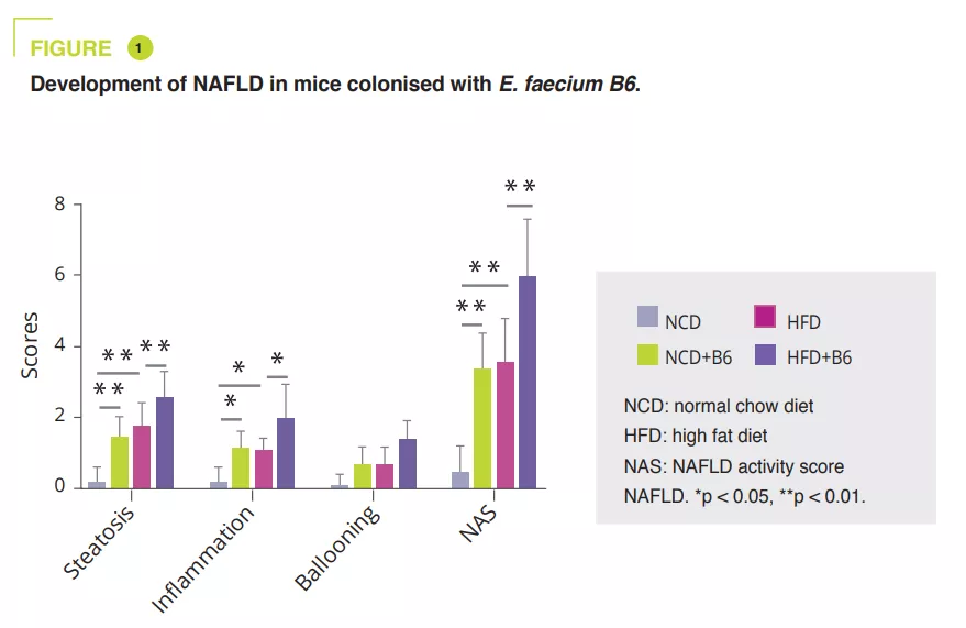
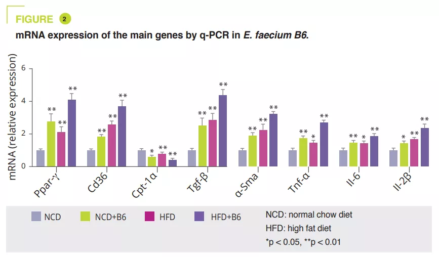
E. faecium B6 in the cell culture obtained after isolating bacterial strains from NAFLD children with obesity showed a lipid-accumulating ability. A murine study compared a normal (NCD) or a fat-enriched (HFD) diet, with or without B6, for 12 weeks. Although E. faecium B6 had no effect on body weight, it aggravated overload-related liver damage, both biologically and histologically (figure 1). Transcriptomic analysis revealed that E. faecium B6 modified gene expression in lipid metabolism and inflammation and fibrosis, such as PPAP, chemokine, NF-kB and TGF-β signalling pathways, in linoleic acid metabolism. Regarding lipid metabolism, mRNA and protein expression of PPAR and CD36 were increased, while that of CPT-1a decreased. The authors reported increased expression of inflammatory cytokines (TNF-α, IL-6, IL-1β) and proteins involved in fibrosis (TGF-β and α-SMA) (figure 2).

Image

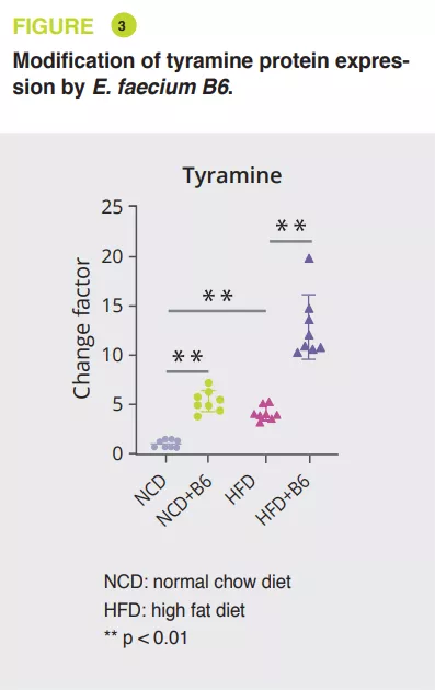
Using mice serum, a broad, non-targeted analysis via tandem mass spectrometry showed that E. faecium B6 increased or decreased metabolites, when on NCD (30 and 85) or HFD (18 and 45) diets. The most notable change was the increase in tyramine (figure 3). Further analyses have suggested that E. faecium B6 is able to produce tyramine. In addition, treating mice on the NCD or HFD diet with tyramine reproduced the development of NAFLD, without effect on weight, in a similar fashion as that observed with E. faecium B6, and similarly altered the expression of genes encoding PPAR , CD36, CPT-1a, TNF-α, IL-6, IL-1β, TGF-β and α-SMA.

Finally, the authors corroborated these findings in 123 NAFLD children with obesity and in 123 controls. Levels of E. faecium B6 and the gene encoding tyramine (mfnA) were higher in the NAFLD group. These levels were correlated with biological markers of NAFLD (AST, ALAT, triglycerides, total cholesterol and LDL) and inflammatory cytokines (TNF-α, IL-6, IL-1β).

Image

What are the consequences in practice?

This study confirms the importance of the gut microbiota in the development of NAFLD and paves the way for new therapeutic strategies. In addition to Enterococcus faecium B6 and tyramine, PPAR y could play a central role in linking lipid accumulation, inflammation and fibrosis.

Key points
  • E. faecium B6, a bacterium isolated from NAFLD children with obesity, exacerbates the disease by producing a bioactive metabolite, tyramine
  • E. faecium B6 and tyramine reproduce NAFLD in a similar way in mice on normal and high-fat diets

CONCLUSION

This study identified Enterococcus faecium B6 as a strain that promotes the development of NAFLD in children with obesity. This bacterium produces a bioactive metabolite, tyramine, which mediates these effects by activating the PPARy signalling pathway

Summary
Off
Sidebar
Off
Migrated content
Désactivé
Updated content
Désactivé
Hide image
Off
Commented article

The maternal microbiome promotes placental development in mice

COMMENTED ARTICLE - Adults’ section

By Pr. Harry Sokol
Gastroenterology and Nutrition Department, Saint-Antoine Hospital, Paris, France

Comments on the article by Pronovost et al. (Science Advances 2023) [1]

The maternal microbiome is an important regulator of gestational health but how it affects the placenta as an interface between mother and foetus has not yet been researched. The authors of this article show that maternal gut microbiota promotes placental development in mice. Depletion of the maternal gut microbiota limits placental growth and impairs foeto-placental vascularisation. The maternal gut microbiota modulates metabolites in maternal and foetal circulation. Short-chain fatty acids (SCFAs) stimulate tube formation in cultured endothelial cells and prevent placental vascularisation abnormalities in microbiota-deficient mice. In addition, in a maternal-malnutrition model, gestational supplementation with SCFAs prevents placental growth restriction and vascular insufficiency. These findings highlight the important role of host-microbial symbioses during pregnancy and reveal that the maternal gut microbiome promotes placental growth and vascularisation in mice.

What do we already know about this subject?

Recent studies have highlighted the significant influences of the maternal microbiome on the development of the offspring during the prenatal period 2, but we still do not understand exactly how the maternal microbiome influences maternal/ foetal health during pregnancy. The highly vascularised placenta, which facilitates the maternal/foetal exchange of nutrients and gas that sustain foetal development, lies at the intersection of the mother and foetus 3. The authors examined the effects of the maternal gut microbiome on placental development in mice, since it is a critical organ that shapes long-term health pathways.

What are the main insights from this study?

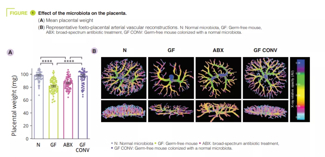
To determine effects of the maternal gut microbiome on placental development, the authors first reared pregnant mice as germ-free (GF) or having depleted the maternal gut microbiome by treating it with broad-spectrum antibiotics (ABX). Absence or depletion of the maternal gut microbiome resulted in reduced placental weight compared to GF controls colonised with a conventional microbiota (CONV) (figure 1). Consistent with the reductions in placental weight, maternal microbiome deficiency led to reductions in total placental volume, along with reduced volume and tissue density in the placental labyrinth - the primary site for maternal-foetal exchange. In addition to maternal ABX-induced placental pathophysiology, the authors observed corresponding decreases in foetal weight and volume. Reduced vascular volume and surface area was observed in the foeto-placental vascularisation of microbiota-deficient mothers, with a visible decrease in vascular branches, as compared to controls (figure 1).This suggests that the maternal microbiome instructs vascular development at critical gestational time points. Given that the maternal microbiome regulates many circulating metabolites, the authors postulated that foeto-placental vascularisation insufficiencies could be linked to the microbiota and could result from key metabolite alterations in foetal circulation. The authors looked specifically at the role of short-chain fatty acids (SCFAs). SCFAs are produced by bacterial carbohydrate fermentation and were observed to be significantly decreased in the maternal and foetal serum from microbiota-deficient mothers.

Image

On the basis of previous research demonstrating that maternal supplementation with SCFAs leads to the direct transfer of SCFAs from maternal circulation to foetal circulation, the authors treated ABX mothers with SCFA-supplemented water or control water. The supplementation strategy significantly increased butyrate and propionate concentrations in foetal whole blood. Treating mothers with SCFAs increased placental weight and corrected alterations in placental growth in ABX mothers to levels comparable to those of controls, with corresponding increases in total placental and labyrinth volumes. Human umbilical vein endothelial cells (HUVECs) were then treated with SCFAs at physiological concentrations. The SCFAs acetate and propionate significantly increased HUVEC branching length compared to vehicle controls, whereas the signal with butyrate was less clear. This effect was dependent on free fatty acid receptors 2 and 3 (FFAR2 and FFAR3). In the context of maternal malnutrition induced by protein restriction, maternal supplementation with SCFAs was sufficient to restore total placental weight and volume and increase foeto-placental vascularisation.

What are the consequences in practice?

This study demonstrates the role of maternal intestinal microbiota in physiology, particularly in placental vascularisation. Placental vascular deficiencies are associated with reduced foetal weight, pre-eclampsia and, in adulthood, an increased risk of a number of diseases. Actions targeting the microbiota to promote SCFA production, primarily through nutrition, could play a protective role.

Key points
  • Overall, the data reveals the key role of the maternal gut microbiome in promoting placental growth and development
  • The maternal microbiome is necessary for the optimal development of foeto-placental vascularisation
  • SCFAs promote placental growth and vascular development, even in conditions of maternal malnutrition

CONCLUSION

This study reveals that the metabolic functions provided by the maternal gut microbiome during pregnancy play an integral part in supporting placental growth and vascularisation in mice. Gaining a better understanding of how the maternal gut microbiome affects the structure and function of the placenta could lead to the development of new approaches to promote maternal and foetal health and reduce the risk of chronic diseases.

Summary
Off
Sidebar
Off
Migrated content
Désactivé
Updated content
Désactivé
Hide image
Off
Commented article

Helicobacter pylori and gastrointestinal microbiota

By Pr. Juozas Kupcinskas
Department of Gastroenterology and Institute for Digestive Research, Lithuanian University of Health Sciences, Kaunas, Lithuania

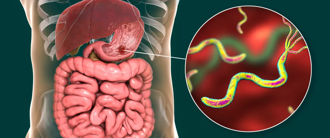
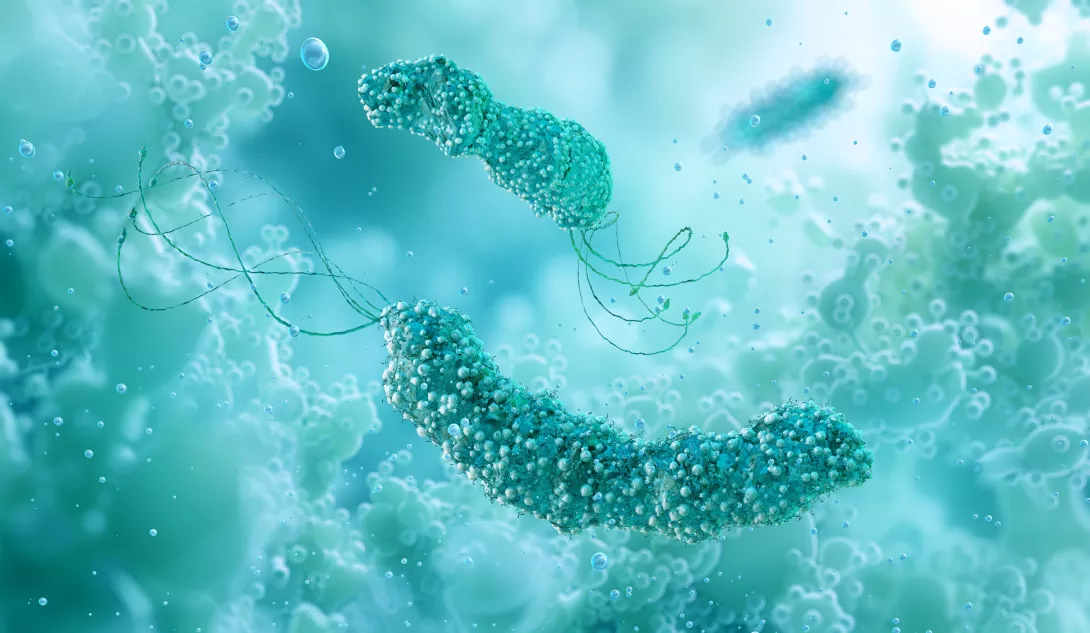
Helicobacter pylori (H. pylori) affects around 50% of the global population and is the most common bacterial infection worldwide. According to the World Health Organization (WHO), H. pylori is classified as a Group 1 carcinogen, which can cause precancerous and cancerous stomach conditions, including gastric adenocarcinoma. It was previously believed that H. pylori was the sole microorganism living in the human stomach, but recent advances in research technologies have provided a better understanding of the gastric microbiome. H. pylori plays a pivotal role in shaping the microbial environment of the stomach. However, the “non-Helicobacter” microbiome of the stomach has also been described and is a hot topic in current research. It is clear that H. pylori is a major modifier of the gastric microbiome, but other species may also contribute to carcinogenic pathways. The effects of proton-pump inhibitors (PPIs) and H. pylori eradication therapies on microbiome alterations have also been studied. In this review article, we aim to summarize recent findings on the gastric microbiome and the role of H. pylori in shaping it, as well as the impact of H. pylori eradication and PPIs on the human microbiome.

H. pylori is the major bacterium shaping gastric microbiome composition

The gastric microbiome is receiving increasing attention, with growing interest in its determining factors. Vilchez-Vargas et al. investigated microbial composition in several gastrointestinal (GI) compartments. Their study involved a cohort of 108 pairs of twins, and gastric microbiome biopsies from the stomach mucosae were analyzed. Microbiome diversity was assessed using V1-V2 regions of the 16S rRNA gene through amplification and sequencing. The results aligned with previous findings, highlighting H. pylori as a key factor in stomach microbiota composition 1.

Hua et al. conducted a study with a cohort of 193 patients to examine H. pylori’s impact on the richness and diversity of gastric microbiota in individuals with chronic gastritis. They profiled the V3- V4 region of the 16S rRNA gene and found significant alterations in gastric microbiota due to H. pylori infection. Specifically, H. pylori suppressed dominant gastric microbiota at the genus level, including Aliidiomarina, Reyranella, Halomonas, Pseudomonas, and Acidovorax. Their findings indicated that virulent strains of H. pylori were significantly associated with chronic atrophic gastritis and decreased gastric microbiota richness 2.

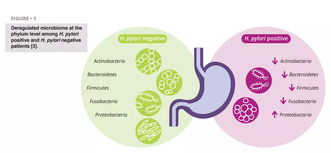
Schulz et al. analyzed differences in microbiota composition between H. pylori-infected and H. pylori-negative patients. They observed a significant difference in the relative abundance of Proteobacteria, which were more prevalent in the aspirates of H. pylori-infected patients. Other phyla showed lower relative abundance (figure 1) 3.

Miftahussurur et al. investigated gastric microbiota variability between H. pylori-positive and H. pylori-negative patients in a cohort of 137 individuals from the Indonesian population. They found that microbial β-diversity and richness were significantly higher in H. pylori-positive samples compared to H. pylori-negative samples. Additionally, their findings suggested that H. pylori plays a leading role in shaping the gastric microbial community in this ethnic group 4.

These studies collectively highlight H. pylori as a crucial factor in influencing gastric microbiome diversity and richness in the harsh stomach environment.

What is a true gastric microbiome?

The stomach presents extreme conditions for living microorganisms. A few decades ago, H. pylori was identified as a bacterium that could withstand these harsh stomach conditions. This discovery sparked curiosity and led to further research into the gastric microbiome. It remains uncertain whether non-Helicobacter bacteria in the stomach represent transient contaminants or part of a persistent microbiota. Spiegelhauer et al. conducted a study involving 22 patients with dyspepsia and 12 with gastric adenocarcinoma 5. They took biopsies from the stomach mucosa and analyzed the V3-V4 region of the 16S rRNA gene, in addition to culturing microorganisms. The authors hypothesized that H. pylori is the only true resident bacterium of the stomach and would persist in washed biopsies. Their findings indicated that bacterial load decreased in washed biopsies, suggesting transient contamination from the oral cavity. However, the diversity of microorganisms did not differ between unwashed and washed biopsies.

Image

It remains unclear whether non-H. pylori microorganisms in the stomach are transient contaminants or genuine residents.

It is possible that continuously swallowed saliva, containing living organisms, may survive acidic conditions for a time. Contamination from the upper oropharyngeal region during gastroscopy and sampling should also be considered 6. These findings highlight the need for further investigation into the true gastric microbiome.

Effect of H. pylori eradication on gastric and gut microbiome

H. pylori is one of the most widespread infections globally, affecting more than half of the human population. Most treatment regimens for H. pylori involve two or more antibiotics, which can impact the gastrointestinal microbiome. Liou et al. investigated long-term changes in gut microbiota following H. pylori eradication. Their multicenter, randomized trial included 1,620 participants who were randomly assigned to three treatment groups. The authors assessed bacterial diversity at various times after eradication by analyzing fecal samples. Results showed that both alpha and beta diversity decreased within two weeks after eradication but returned to baseline levels by week 8 and one year later. These findings indicate only a shortterm perturbation of the gastrointestinal microbiome and suggest that H. pylori eradication therapy is generally safe in the long term 7.

He et al. reported on alterations in the gastrointestinal microbiome following H. pylori eradication, using 16S rRNA gene analysis of gastric mucosal and fecal samples. They found that alpha diversity of the gastric microbiome increased, and the beta diversity of the gut microbiome significantly differed from pre-treatment levels but resembled that of healthy controls 24 weeks after eradication 8.

Image

Guo et al. summarized findings on changes in the gastric microbiome after successful H. pylori treatment. Their systematic review and meta-analysis included nine studies with 546 patients. This meta-analysis is the first to detail changes in alpha diversity after H. pylori eradication. Results indicated no significant differences in microbiota diversity between treatment options, whether quadruple or triple therapy was used. The authors observed increased alpha diversity in the short term, which persisted in long-term follow-up, with a depletion of H. pylori-related taxa and enrichment of common gastric commensals 9. To evaluate the effect of H. pylori eradication on the gut microbiome, Yap et al. conducted a study with 17 young adults. They sequenced the V3-V4 region of the 16S rRNA gene and analyzed the gut microbiome before and 18 months after H. pylori eradication using clarithromycin and metronidazole. No significant changes in microbial diversity were observed between baseline and 18 months post-eradication 10.

Image

PPIs are major modifiers of the gut microbiome

Global consumption of proton pump inhibitors (PPIs) is increasing, making them one of the top 10 most widely used drugs in the world 11. They are a first-line treatment for conditions such as gastroesophageal reflux disease, peptic ulcer disease, dyspepsia, and, when combined with antibiotics, for the treatment of H. pylori 12. PPIs are often used without evidence-based indications or for longer periods than prescribed, and their use has been associated with an increased risk of infections such as Clostridium difficile, Salmonella spp., Shigella spp., Campylobacter spp., and other enteric pathogens 11.

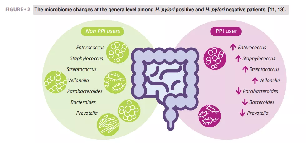
Imhann et al. analyzed the microbiome of 211 subjects using PPIs by sequencing the V4 region of the 16S rRNA gene. They observed a significant decrease in alpha diversity among PPI users and an increase in the abundance of bacteria from the genera Enterococcus, Streptococcus, Staphylococcus, and Veillonella (figure 2). The genera Enterococcus and Veillonella have been linked to a higher susceptibility to Clostridium difficile infections 11. While PPIs are generally considered safe with relatively rare side effects, evidence suggests they negatively impact the gut microbiome.

Zhang et al. conducted a meta-analysis of the effects of PPIs on human gut microbiota, analyzing data from four studies with 16S rRNA gene amplicon sequencing. Their results demonstrated a significant impact of PPI use on microbial diversity, with lower alpha diversity observed in PPI users compared to controls. They found decreases in the genera Parabacteroides, Veillonella, Bacteroides, and Prevotella, as well as from the families Ruminococcaceae and Lachnospiraceae (figure 2) 13.

Weitsman et al. performed a study with 177 subjects, matching PPI users 1:2 with controls. They analyzed stool samples and, for the first time, duodenal microbiomes. No significant differences in alpha or beta diversity were found between PPI users and controls. However, at the family level, they observed a higher relative abundance of Campylobacteraceae (phylum Proteobacteria) and a lower relative abundance of Clostridiaceae (phylum Firmicutes) in PPI users. Stool analysis similarly revealed a reduction in Clostridiaceae and an increase in Streptococcaceae 14.

Image

Overall, these studies indicate that PPIs affect the human microbiome. The clinical significance of these findings warrants further investigation in future studies.

Image

Conclusion

The human stomach presents a harsh environment for living microorganisms, and recent research continues to explore whether the stomach microbiota is persistent or merely reflects transient microorganisms. Despite its clinical significance, H. pylori plays a notable role in shaping the stomach’s microbiome. The eradication of H. pylori appears to have only a temporary and reversible impact on the composition of the gastric and gut microbiomes. Proton pump inhibitors (PPIs) are among the most commonly consumed medications. Scientific studies indicate that PPIs alter the structure of the gut microbiome; however, the clinical significance of these changes requires further investigation.

Sources

1. Vilchez-Vargas R, Skieceviciene J, Lehr K, et al. Gut microbial similarity in twins is driven by shared environment and aging. eBioMedicine 2022; 79: 104011.

2. Hua Z, Xu L, Zhu J, et al. Helicobacter pylori infection altered gastric microbiota in patients with chronic gastritis. Front Cell Infect Microbiol 2023; 13: 1221433.

3. Schulz C, Schütte K, Koch N, et al. The active bacterial assemblages of the upper GI tract in individuals with and without Helicobacter infection. Gut 2018; 67: 216-25.

4. Miftahussurur M, Waskito LA, El‐Serag HB, et al. Gastric microbiota and Helicobacter pylori in Indonesian population. Helicobacter 2020; 25: e12695.

5. Spiegelhauer MR, Kupcinskas J, Johannesen TB, et al. Transient and persistent gastric microbiome: adherence of bacteria in gastric cancer and dyspeptic patient biopsies after washing. J Clin Med 2020; 9: 1882.

6. Klymiuk I, Bilgilier C, Stadlmann A, et al. The human gastric microbiome is predicated upon infection with Helicobacter pylori. Front Microbiol 2017; 8: 2508.

7. Liou JM, Chen CC, Chang CM, et al. Long-term changes of gut microbiota, antibiotic resistance, and metabolic parameters after Helicobacter pylori eradication: a multicentre, open-label, randomised trial. Lancet Infect Dis 2019; 19: 1109-20.

8. He C, Peng C, Wang H, et al. The eradication of Helicobacter pylori restores rather than disturbs the gastrointestinal microbiota in asymptomatic young adults. Helicobacter 2019; 24: e12590.

9. Guo Y, Cao XS, Guo GY, Zhou MG, Yu B. Effect of Helicobacter pylori eradication on human gastric microbiota: a systematic review and meta-analysis. Front Cell Infect Microbiol 2022; 12: 899248.

10. Yap TWC, Gan HM, Lee YP, et al. Helicobacter pylori eradication causes perturbation of the human gut microbiome in young adults. Plos One 2016; 11: e0151893.

11. Imhann F, Bonder MJ, Vich Vila A, et al. Proton pump inhibitors affect the gut microbiome. Gut 2016; 65: 740-8.

12. Kiecka A, Szczepanik M. Proton pump inhibitor-induced gut dysbiosis and immunomodulation: current knowledge and potential restoration by probiotics. Pharmacol Rep 2023; 75: 791-804.

13. Zhang J, Zhang C, Zhang Q, et al. Meta-analysis of the effects of proton pump inhibitors on the human gut microbiota. BMC Microbiol 2023; 23: 171.

14. Weitsman S, Celly S, Leite G, et al. Effects of proton pump inhibitors on the small bowel and stool microbiomes. Dig Dis Sci 2022; 67: 224-32.

Summary
Off
Sidebar
Off
Migrated content
Désactivé
Updated content
Désactivé
Hide image
Off
Article Gastrointestinal

Diversity or function: what defines a healthy microbiota

Within us lies a complex microbial community, the gut microbiome, which greatly impacts our health. It's a site of essential metabolic processes influenced by diet, age and environment. The focus isn’t solely on the variety of microbes, but the essential functions they perform together. Since a healthy state is a dynamic balance between us and these microbes, what defines a healthy microbiome?

We find ourselves in an era of unprecedented exploration into the microscopic world within us, the gut microbiome. A recent, meticulous scientific review 1 has shed light on this complex ecosystem, revealing both the marvels and the mysteries of our internal microbial communities.

This paper challenges the simplistic view of "dysbiosis", highlighting that this term, often used to describe an imbalanced gut, lacks the precision needed for proper understanding.

It poses also a fundamental question: what truly constitutes a ‘healthy’ microbiome?

What defines a healthy microbiome?

Defining a "healthy" gut microbiome is complex, moving beyond simple absence of disease.

It involves assessing gut structure, function, and microbial composition. While a high diversity of microbial species was once thought to be the primary marker of a healthy gut, it's now understood that (sidenote: Functional Diversity This describes the range of metabolic activities performed by the gut microbiota. It is a more important indicator of gut health than simple taxonomic diversity because different microbial compositions can perform similar metabolic functions. Assessing functional diversity can provide a more accurate prediction of physiological states than compositional diversity alone. ) , the range of metabolic activities the microbiome performs, is more important. This means different microbial compositions can perform similar metabolic functions.

  • (sidenote: Strain Specificity This highlights that different strains within the same bacterial species can have vastly different effects on the host. For example, some E. coli strains are pathogenic, while others, like E. coli Nissle 1917, are beneficial. Understanding strain-specific effects is essential for developing targeted therapies and for interpreting research on the gut microbiome. ) is also vital: Different strains within the same species can have vastly different effects. For example, some E. coli strains are pathogenic, while others, like E. coli Nissle 1917, are beneficial.

  • Metabolites like (sidenote: Short chain fatty acids (SCFA) Short chain fatty acids (SCFA) are a source of energy (fuel) for an individual’s cells. They interact with the immune system and are involved in communication between the intestine and the brain. Silva YP, Bernardi A, Frozza RL. The Role of Short-Chain Fatty Acids From Gut Microbiota in Gut-Brain Communication. Front Endocrinol (Lausanne). 2020;11:25. ) , bile acids (BAs), and tryptophan metabolites are key indicators of a functional microbiome. SCFAs, such as butyrate, are essential for colonocyte energy and immune modulation.

  • BAs, produced in the liver and modified by gut bacteria, are crucial for fat digestion, signaling and antimicrobial action.

  • Other indicators include gas production (hydrogen, methane, hydrogen sulfide), gut pH, and inflammatory markers (calprotectin, lactoferrin).

  • Finally, (sidenote: Resilience This refers to the ability of the gut microbiota to maintain a stable composition over time and resist disturbances such as antibiotics or dietary changes. A resilient microbiota can recover quickly from disruptions, reducing the risk of long-term health issues. ) , or the ability of the gut microbiota to resist disturbances, is a crucial marker of a healthy gut.

The mucus layer is also a key component of a healthy gut.

This layer, primarily composed of water, electrolytes, lipids, and mucins, acts as a physical barrier, preventing bacteria from directly contacting the intestinal epithelial cells.

A healthy gut is characterized by an adequate mucus thickness that is not easily penetrable by bacteria. The turnover of the (sidenote: Mucus Layer This is a complex, dynamic barrier lining the gut, primarily composed of water, electrolytes, lipids, and mucins. It physically separates bacteria from the intestinal epithelium, preventing direct contact and maintaining gut barrier integrity. The thickness and turnover of the mucus layer are crucial for a healthy gut. ) which involves synthesis, secretion, and degradation, is a finely tuned process crucial for maintaining proper barrier function.

Factors like prebiotics, such as fructo-oligosaccharides (FOS) and 2′-fucosyllactose (2′FL), can influence mucus production, composition and degradation, enhancing gut barrier integrity and contributing to protection against metabolic disorders.

Disruption of the mucus layer, as seen with some food emulsifiers, can lead to increased intestinal permeability and inflammation.

90-95% Mucus primarily consists of various components, including 90-95% water, electrolytes, lipids (1-2%), proteins and others substances. ¹

45% Approximately 45% of bacterial species are similar between two individuals, their microbiota share 82% common metabolic pathways. ¹

The gut-liver axis: a two-way street

The gut and liver interact closely via the bidirectional gut-liver axis. The liver, as a primary site for detoxification and metabolic regulation, processes and neutralises a variety of environmental toxins, drugs, and metabolic byproducts that are derived from the gut.

The liver produces BAs which are essential for fat digestion and also influence the gut microbiome composition and function. Gut bacteria further metabolise primary BAs into secondary BAs, which have different functions, and some are even associated with longevity.

While the liver is exposed to gut-derived bacterial antigens, it typically does not produce pro-inflammatory cytokines. However, healthy livers produce anti-inflammatory molecules such as IL-1 receptor antagonist (IL-1Ra) to dampen inflammation, as well as specific immunosuppressive macrophages, which are dependent on gut microbiota, to control excessive inflammation.

Factors shaping the microbial ecosystem

The gut microbiome is dynamic, influenced by age, delivery mode at birth, diet and lifestyle.

  • Age and delivery mode at birth are critical. Vaginally born infants tend to have a more diverse and balanced gut microbiota, as do breastfed infants, who have a greater presence of beneficial bacteria such as Bifidobacterium spp

  • Diet is paramount. Dietary fibres, prebiotics, polyphenols, and human milk oligosaccharides (HMOs) promote beneficial microbes. In contrast, saturated fats, artificial sweeteners, and emulsifiers can disrupt microbial balance and impair gut barrier function.

  • Common emulsifiers, like carboxymethylcellulose (CMC) and polysorbate 80 (P80), disrupt the gut's protective mucus layer, leading to increased gut permeability and inflammation.

  • Bioactive lipids also have a crucial, bidirectional role. The host's lipids influence the gut microbiota, and gut microbes produce lipids (e.g., SCFAs, secondary BAs, and other signaling molecules) that impact immune regulation and metabolic health.

A healthy gut microbiome: more than just bacteria

The challenges in defining a universally accepted 'healthy' gut microbiome become increasingly clear. The immense individual variability of the gut microbiome, influenced by genetics, diet, environment, and lifestyle, as well as its dynamic nature, complicates the establishment of universal standards.

The interplay of the gut microbiota, the immune system, and metabolic processes presents a multifaceted challenge. Longitudinal studies are essential to fully understand the dynamic changes within the gut microbiome and their long-term health impacts. This complex area of research calls for a multidisciplinary approach, integrating microbiology, genomics, bioinformatics, clinical research, and personalised medicine. 

Summary
Off
Sidebar
On
Migrated content
Désactivé
Updated content
Désactivé
Hide image
Off
News Gastroenterology

The surprising effect of coffee on the microbiota

Can a single food cause a radical change in microbiota composition? Coffee sure can! According to a new study, coffee strongly stimulates the proliferation of a previously little-known gut bacterium. 1

The gut microbiota Diet

The health benefits of coffee are well known. But how exactly does it affect the microbiota?

To answer these questions, researchers from Harvard University (USA) and the University of Trento (Italy) analyzed the gut microbiota and coffee consumption of more than 22,000 volunteers taking part in an Anglo-American research program. 

They divided the participants into three groups:

  • “Non-drinkers”, who consumed less than three cups of coffee per month;
  • “Moderate drinkers”, who drank between three cups per month and three cups per day.
  • “Heavy drinkers”, who drank more than three cups per day;

Non-drinkers vs. addicts, not the same effects

They found that the microbiota of coffee drinkers differs markedly from that of non-drinkers. The analysis showed that 115 bacterial species react positively to the beverage. 

How many coffees a day? ²

  • 1 cup of filtered coffee (200 ml) = 90 mg of caffeine
  • 1 espresso (60 ml) = 80 mg of caffeine
  • 1 cup of black tea (220 ml) = 50 mg of caffeine

Excessive consumption of caffeine (present in coffee and tea) is associated with cardiovascular problems, sleep disorders and delayed fetal development. What are safe doses?

  • Coffee is safe for all healthy adults up to 200 mg of caffeine per day (up to 400 mg if consumption is spread throughout the day, except for pregnant women).
  • From 100 mg of caffeine per day, sleep may be affected.

Surprisingly, Lawsonibacter asaccharolyticus, a bacterial strain in the microbiota little studied until now, is the microorganism most strongly linked to coffee consumption. The scientists calculated its level to be 4.5 to 8 times higher in the microbiota of “heavy drinkers” than in that of “non-drinkers” and 3.4 to 6.4 times higher in “moderate drinkers” than in “non-drinkers”.

By analyzing another dataset on several thousand individuals across 25 different countries the researchers confirmed that the presence of L. asaccharolyticus is indeed associated with coffee consumption and that the association exists regardless of country or lifestyle.

Virtues of coffee mediated by microbiota?

If there is one food whose beneficial effects have been proven, it is coffee. Studies show regular coffee consumption to be associated with a lower risk of:

  • diabetes 
  • cancer 
  • fatty liver disease 
  • cardiovascular disease
  • all-cause mortality 

The secret? Its polyphenol content, particularly that of chlorogenic acid, an antioxidant present in large quantities in the beverage. This molecule can be broken down and transformed by gut bacteria into multiple potentially beneficial metabolites. The microbiota may therefore be a mediator of the beneficial effects of coffee on health.

In a previous study on 1,000 individuals, the same researchers found that, of 150 foods, coffee had by far the greatest impact on gut microbiota composition.

Caffeine not to blame

To confirm that the exceptional growth of L. asaccharolyticus is directly linked to coffee, the scientists then cultivated the bacterium in vitro in a liquid medium either supplemented or not supplemented with coffee. They found that bacteria grow more quickly in the presence of coffee, even decaffeinated coffee, which removes caffeine from the equation. 

The stimulation of L. asaccharolyticus may be the work of chlorogenic acid, a polyphenol in coffee thought to contribute to its beneficial effects. Chlorogenic acid is metabolized by microbiota bacteria into various molecules, notably quinic acid. The researchers found more quinic acid in the blood of those with higher levels of L. asaccharolyticus.

The next step for researchers is to determine whether foods other than coffee specifically stimulate known beneficial bacteria. Thanks to tests that reveal the presence or absence of certain bacteria associated with a food, it may be possible to design personalized diets. 3

Summary
Off
Sidebar
On
Migrated content
Désactivé
Updated content
Désactivé
Hide image
Off
News

What is this liquid I can see in my panties, is it normal?

Is bacterial vaginosis a disease?

Here's how Prof. Graziottin answers this question from her patients:

Image
Professor Alessandra Graziottin's tips for understanding women's health and addressing patient needs.

First point, the liquid is provoked by the mixture of normal microscopic vaginal transudate secretions through the vaginal wall tissues and the microbiota and microbiome, say the microflora, living in our vagina. Normally it is very mild in quantity, has no scent or color and does not usually stain the underwear (but beware not to consider the urinary stains as vaginal).

When it does, the first causes to be considered are:

  • Gut dysbiosis, particularly when you suffer from diarrhea, constipation, irritable bowel syndrome. Why that? Because this translates into changes of the gut microbiome and because of the close dialogue between the gut microbiome and the vagina, you may have symptoms in the vagina, including leakage, including staining your pants.
  • Second, you can have ovulatory secretions, very limpid and clear at ovulation, at midcycle.
  • Third, we could have a very special condition that is called cervical ectopia (from the Greek word that means “out of place”). What does it mean? Normally, we have two types of epithelia within the uterus and the neck of the uterus, the cervix and the vagina
    • Those within the uterus are a simple layer, red colour.
    • Those in the vagina are multiple layers and gentle pink.

When we have this movement, this migration, we say, of these cells, red colour, covering the cervix, when the gynecologist was looking at it, in the past we used to think, there was a little wound. It's not a wound. Simply, we have this movement of the inner cells towards the external part. It is very frequent. 30-40% of women have, particularly young women. But the point is that this epithelium is much more vulnerable to the presence of different germs like the one we have in the vagina that are different from the cervix and the endometrium. This may translate into increased vagina secretion.

So, read the symptoms and then a competent gynecologist will tell you which situation is this: from the bowel? Is it just ovulation? Or is it a cervical ectopia that could be addressed?

Related content

BMI-24.55

Summary
Off
Sidebar
On
Migrated content
Désactivé
Updated content
Désactivé
Hide image
Off
Dossier detail Gynecology

How do I deal with menopause?

Here's how Prof. Graziottin answers this question from her patients:

Image
Professor Alessandra Graziottin's tips for understanding women's health and addressing patient needs.

First, improve your lifestyles: the more appropriate and consistent they are, the stronger the investment you will personally make in your longevity in smart health!

Physicians, you should:

  • Always take a very accurate family and personal history, to evaluate leading symptoms and signs the menopause is causing, the leading vulnerabilities and the few major contraindications (to hormone replacement therapy, say, hormone replacement cancer breast, endometrial and ovarian cancer, thrombophlebitis/thrombosis).
  • Second, physicians should carry out a comprehensive physical examination, as I do with my patients, including thyroid, breast, abdomen, gynecological and pelvic floor examination, plus blood pressure, body weight, height and abdominal circumference.
  • Third, important to carry out pelvic ultrasonography, pap-smear and/or HPV test, or ask for mammography and bone mineral density (BMD), if the woman is older than 50, or earlier if there is a clinical indication for that. For example, bone mineral density could be appropriate in individual cases, in case of premature ovarian insufficiency, persisting binge eating disorders of restrictive type up to frank anorexia, or cancer treatment with chemotherapy and/or pelvic or total body radiotherapy in also in case of NON-hormone dependent cancers.
  • If a hormone replacement therapy is indicated, then it can be tailored according to the woman’s needs in terms of:
    • types of hormones
    • dosage
    • route of administration
    • regimen: cyclic combined or continuous combined, 

A well-tailored menopausal hormone therapy (MHT) improves the level of health in every organ and tissue: from brain to cardiovascular system, from bones to muscles and joints, from skin to nails and hair, from genitals to bladder. 

MHT improves the gut, the vulva, and vaginal microbiota and microbiome as well, the best secret directors of our health, which have major receptors for sexual hormones. A positive dynamic eubiosis is back, for the best of our systemic health, general and intimate health.

New solid data indicated that very positive health advantages are there also for women who continue far after 65 years of age, always in synergy with healthy lifestyles.

Key point: menopausal hormone therapy replaces the lost hormones, but they work at their best if you take full responsibility for your own health. No alibi, no excuses, please!

Lifestyles are key! 

Related content

BMI-24.55

Summary
Off
Sidebar
On
Migrated content
Désactivé
Updated content
Désactivé
Hide image
Off
Dossier detail Gynecology

“I'm 45, why do I have all these symptoms now (hot flashes, insomnia, dryness, swollen belly)? Is it menopause?”

Here's how Prof. Graziottin answers this question from her patients:

Image
Professor Alessandra Graziottin's tips for understanding women's health and addressing patient needs.

Your body is telling you the truth: your ovaries are losing their last oocytes, say the reproductive female cells, and are losing their follicles, say the cells that nourish the oocytes and produce estradiol, estrogens, and progesterone.

These symptoms tell you that the three major consequences are impending:

  • First, loss of menses = menopause
  • Second, loss of fertility (unless oocytes preservation, with cryo-conservation, has been carried out in the earlier fertile years).
  • And third, loss of health: because these symptoms (hot flashes, insomnia, swollen belly, pain in your joints…) are a real “cry for help”. Your body is asking for help because it wants to have back the hormones lost with ovarian exhaustion. For example, the belly is getting “swollen” because the loss of sexual hormones causes a major dysbiosis in the gut, with reduced biodiversity and an increasing number of gas-producing bacteria.

In parallel, it causes vulvar and vaginal dysbiosis, with the loss of our friend Lactobacilli, increased biodiversity in the vaginal microbiota and microbiome and an increasing number of bacteria like the Gardnerella Vaginalis causing bad smell. Also, this increases the vulnerability to contaminants from the bowel like Escherichia coli

Remember: women are never too young to get menopausal. Therefore, listen carefully to those symptoms even in adolescent girls! A woman can be young and very beautiful, and yet have a very early premature ovarian insufficiency, for a number of reasons (genetic first, autoimmune if she has celiac disease or other autoimmune diseases, after removal of an endometriotic cyst in the ovary or mono-lateral ovariectomy for endometriosis or chemo or radiotherapy.

Related content

BMI-24.55

Summary
Off
Sidebar
On
Migrated content
Désactivé
Updated content
Désactivé
Hide image
Off
Dossier detail Gynecology