A prospective cohort study involving 937 patients suggests that the microbiota present within colorectal tumor tissues is associated with prognosis, independently of established clinical and molecular factors.
Colorectal cancer is the second leading cause of cancer-related death worldwide.
9,3 %
Representing 9.3% of the total cancer mortality in 2022 ¹.
47 582
In France, 47,582 new cases were diagnosed in 2023 – 95% of which occurred in people over the age of 50, with men accounting for 55% of cases and women 45% ².
Benchmark methodology: Next-Generation Sequencing (NGS) on 937 patients
Shi et al. 3 analyzed whole-genome sequencing data from 1,412 tissue samples:
937 tumors and 475 normal adjacent tissues (NAT)
and 462 blood samples from 937 treatment-naive patients recruited from the prospective U-CAN cohort (Uppsala-Umeå, Sweden).
Validation was performed using an independent external cohort (AC-ICAM, n = 246). To address recent methodological controversies in this field, a rigorous bioinformatics pipeline was developed to detect and quantify bacteria present in tumor tissues, prioritizing specificity over sensitivity.
Molecular subtypes of colorectal cancer (CMS1–4)
Colorectal cancer is not a homogeneous group. The international CMS (Consensus Molecular Subtypes) classification distinguishes four subtypes based on the tumor’s molecular profile 4. This classification is central to the interpretation of the results by Shi et al.
CMS1
Characteristics:
hypermutated tumors,
strong immune response,
ascending colon.
Microbial correlation in the study by Shi et al., 2025: relative abundance: enrichment in Fusobacterium (Fn. animalis C2, Fn. nucleatum, Fn. vincentii and Fn. polymorphum), and oral bacteria (Parvimonas, Peptostreptococcus, Treponema) compared to other CMSs. No pks+ prognostic correlation specific to this subtype.
CMS2
Characteristics:
most common (37%),
activation of the WNT and MYC pathways.
Microbial correlation in the study by Shi et al., 2025: specific pks+/Enterobacteriaceae prognostic correlation→ poor prognosis.
CMS3
Characteristics:
metabolic abnormalities
frequent KRAS mutation.
Microbial correlation in the study by Shi et al., 2025: no specific pks+ prognostic correlation identified in this study.
CMS4
Characteristics:
mesenchymal transformation
high risk of metastasis.
Microbial correlation in the study by Shi et al., 2025: prognostic correlation with an enrichment in Fn. animalis C2, Fn. nucleatum, and Fn. polymorphum, linked to reduced survival via specific immunometabolic pathways.
While the prognostic correlation of pks+/Enterobacteriaceae with patient outcome is specific to CMS2, CMS1 and CMS4 exhibit distinct microbial signatures with no such pks+ prognostic association.
361 species identified and a validated microbial risk score
The authors identified 249 genera and 361 bacterial species common to both tumors and NATs, including:
Fusobacterium nucleatum,
various Enterobacteriaceae (E. coli),
Akkermansia muciniphila,
Firmicutes, and species belonging to the genus Treponema.
Microbial signatures varied according to anatomical location, tumor stage, and consensus molecular subtypes (CMS1–4). 17.4% of tumors tested positive for the pks genomic island, which is carried by certain genotoxicE. coli strains that affect colorectal cancer cells via the toxin colibactin. The presence of pks+ and a high abundance of Enterobacteriaceae were specifically associated with a poor prognosis in the CMS2 subtype.
Two microbial risk scores – MRS-T (tumor) and MRS-N (NAT) – were developed and validated across the three cohorts. These scores were able to predict overall survival independently of conventional clinicopathological factors, thereby improving the discriminatory power of the prognostic model compared with models based on host factors alone.
The unfavorable taxa were associated with the activation of pro-inflammatory pathways (hypoxia, MAPK1/3, IL-6) and the modulation of the tumor immune system.
Clinical implications and limitations
The intratissue microbiota of colorectal cancer constitutes an independent source of prognostic information, that could complement current molecular markers. The specificity of the correlation between the presence of pks/Enterobacteriaceae and poor prognosis in the CMS2 subtype suggests that molecular subtyping must be incorporated when interpreting the role of the microbiome.
Limitations to note include:
observational study design,
the inclusion of treatment-naive patients only,
and the fact that WGS is not yet feasible for routine clinical use at this stage.
Mechanistic and prospective studies remain necessary before these findings can have any clinical application.
Bread, yogurt, cheese, vegetables, cured meats... fermented foods play a major role in our diets and culinary traditions. Originally produced to extend the shelf life of seasonal foods, they’re currently all the rage thanks to their health benefits and rich flavors.
They may be in fashion, but fermented foods didn’t need influencers to make it onto the menu, with the earliest being produced in the Mediterranean region as far back as 14,000 B.C. 1
Back then, trial and error revealed fermentation to be an excellent way to preserve food. It wasn’t until 1857 that Louis Pasteur discovered the mechanisms underlying the fermentation process and the role of living organisms (yeasts, bacteria and other microscopic organisms).
In the absence of oxygen, enzymes produced by these microorganisms break down components (often sugars) of the food, resulting in the creation of acids (lactic acid in yogurt, cheese, sauerkraut, kimchi, etc.) or alcohol (alcoholic fermentation in wine) in particular.
The acid lowers the pH and repels pathogens, as does alcohol, an antimicrobial agent. 2
"It is difficult to definitively establish the number of fermented foods produced globally; most estimates suggest that there are in excess of 5000 different kinds." 3
Overview of the most common food fermentations
Source: Valentino, 2024 2
Lactic fermentation
Main taxa involved:
Lactobacillaceae
Leuconostocaceae
Streptococcaceae
Main products:
lactic acid (homolactic)
CO2
ethanol (heterolactic)
Main fermented foods produced:
dairy (yogurt, cheeses, kefir)
sauerkraut
kimchi
pickles
tempeh
fermented meats
Alcoholic fermentation
Main taxa involved:
Saccharomyces spp.
Kloeckera spp.
Main products:
ethanol
CO2
Main fermented foods produced:
wine
beer
kefir
Acetic fermentation
Main taxa involved:
Acetobacter spp.
Gluconacetobacter
Gluconobacter
Main products:
acetate
exopolysaccharides
Main fermented foods produced:
chocolate
coffee
vinegar
specialty beers
water kefir
Propionic fermentation
Main taxa involved:
Propionibacterium spp.
Main products:
propionate
acetate
CO2
succinate (Wood-Werkman pathway)
Main fermented foods produced:
swiss-type cheeses
Rich in microorganisms with probiotic effects
Some fermented foods, such as bread, wine, coffee, or chocolate, no longer contain live microorganisms, since cooking or food processing has eliminated them. 1
However, many foods are still teeming with live bacteria and yeast when consumed.
This is the case with:
yogurt
kefir
most cheeses
miso
nattō
tempeh
unheated fermented vegetables such as pickles,
as well as salami and other unheated sausages. 1
These live microorganisms prevent the growth of pathogens such as the infamous Listeria or Salmonella, which cause food-borne illnesses.
But the story doesn’t end at the cupboard door. When we consume fermented products containing live bacteria and yeast, we ingest these microorganisms, which pass through our digestive system.
Many perish in the acidic environment of our stomach, but some appear to reach our intestines and alter our microbiota, either temporarily, or long term when the food is consumed regularly. 1,3,4,5
Fermented foods aren’t just a source of bacteria; they also feed these microorganisms.
For example, fruits, vegetables, and bread (especially whole-grain bread) contain dietary fiber, i.e., carbohydrates that our bodies can’t digest. 6 These fibers eventually reach our large intestine, where they’re consumed by our bacteria.
Fermentable dietary fibers from fruits, vegetables, and legumes, as well as non-fermentable fibers (such as resistant starch) from grains, all serve as substrates that nourish our gut microbiota 6 and function as prebiotics.
Thanks to them, our gut flora stays in top form.
Fermented foods are also known to be easier to digest. Certain microorganisms, thanks to their own enzymes, do the hard work of digestion for us, breaking down the large molecules of carbohydrates, proteins, or fats into smaller, more digestible molecules.1,3
Fermentation can also improve the bioavailability of certain nutrients. For example, in milk-containing products such as yogurt, calcium and potassium are easier to absorb. 2
Fermentation can also make certain foods “tolerable” to those with intolerances.
The most classic example is lactase, an enzyme which digests lactose during childhood, but which is sometimes lost later in life. Its absence leads to lactose intolerance, which results in bloating at the slightest sip of milk.
Conversely, yogurt and cheese are often well tolerated. Why? Because lactic acid bacteria, thanks to their own lactase, have already broken down a large portion of the lactose, significantly reducing its concentration and the risk of intolerance.1,2,3
Fermentation is also thought to reduce FODMAPs and gluten, somewhat improving tolerability in sensitive individuals. 8
Lastly, fermentation can also lower the concentration of antinutritional factors in foods.
In soy, it reduces the level of trypsin inhibitors, which block a pancreatic enzyme necessary for the digestion of proteins. 1,3
Similarly, the fermentation of grains and legumes lowers the level of phytates, which hinder the absorption of minerals such as zinc, iron, or calcium. 2
Fermentation releases numerous compounds that radically transform the organoleptic qualities of fermented foods in terms of: 3
Flavor: the conversion of sugars into organic acids (lactic, acetic, and propionic acids) alters the aromatic profile of foods. For example, propionic acid gives Swiss cheeses (Gruyère, etc.) their characteristic flavor. The fermentation of citrate by certain lactic acid bacteria imparts nutty and buttery hints to products like cottage cheese or Gouda.
Smell: fermentation releases unique aromatic compounds (esters, alcohols) that give cheeses, breads, and wine their characteristic smells.
Texture and appearance: fermentation can alter physical structure, making yogurt creamy, bread fluffy, and Emmental full of holes thanks to the release of CO2.
Lastly, fermentation is the art of preservation... without preservatives.
It requires little heavy equipment, eliminates the need for additives (since the acids or alcohol produced by microorganisms keep pathogens at bay), and allows food to be stored at room temperature, avoiding the energy consumption needed for months of refrigeration.
In sum, there’s no shortage of reasons to put fermented foods on the menu!
Check out our series of articles on microbiota and fermented foods:
Sarcopenia is often framed as a failure of exercise and nutrition. A new study now links one gut bacterium, Roseburia inulinivorans, to muscle strength in humans and shows a causal effect in micevia amino acid shifts.
The gut microbiota regulates host metabolism, inflammation, and tissue function, yet its role in muscle health has remained a blind spot. While the concept of a gut-muscle axis has gained traction, no specific bacterial species had been causally linked to muscle strength.
A new study 1 published in Gut by Martinez-Tellez et al. from the Leiden University Medical Center and the University of Granada set out to fill that gap.
Gut-muscle axis
A bidirectional communication pathway through which gut microbes, their metabolites, and immune signals influence muscle mass and function. This study provides direct evidence that a single bacterial species within this axis can causally modulate muscle strength. 1
Sarcopenia
The progressive, age-related loss of skeletal muscle mass, strength, and function, contributing to frailty and poor clinical outcomes. The observed decline of R. inulinivorans with age suggests a potential microbiome-based therapeutic avenue for this condition. 1
From human association to causal proof in mice
The team conducted metagenomic analyses in two human cohorts:
90 young adults (18–25 years)
and 33 older adults (65–75 years), extensively phenotyped for muscle strength.
Among all bacterial taxa, the relative abundance of Roseburia inulinivorans, but not other Roseburia species, was positively associated with handgrip strength in both age groups. Older adults with detectable R. inulinivorans exhibited 29% higher handgrip strength (p<0.01). In young adults, higher abundance also correlated with leg press and bench press performance (r≥0.26, p<0.05).
Roseburia inulinivorans
A butyrate-producing anaerobic bacterium of the Lachnospiraceae family, identified here as the only Roseburia species specifically and causally linked to enhanced muscle strength in both human cohorts and a murine model. 1
To test causality, 32 antibiotic-treated mice received oral gavage of :
R. inulinivorans,
R. faecis,
R. intestinalis, or vehicle three times weekly for eight weeks.
Only R. inulinivorans induced a remarkable ~30% increase in forelimb grip strength (p<0.001). Muscle fibre analysis confirmed increased cross-sectional area, a higher frequency of large fibres (>5000 µm²), and a shift from type I to type II fibres, the fast-twitch fibres critical for force generation.
Type II muscle fibres
Fast-twitch skeletal muscle fibres characterised by high glycolytic capacity and rapid force generation. R. inulinivorans supplementation shifted fibre composition toward type II, coinciding with increased muscle fibre size and grip strength in mice. 1
An unexpected mechanism: amino acids, not butyrate
As Roseburia species are established butyrate producers, short-chain fatty acids were the expected driver. Yet caecal SCFA levels remained unchanged.
The key metabolic signature of R. inulinivorans lay elsewhere: a marked decrease in caecal amino acids including :
methionine,
leucine,
isoleucine,
valine,
alanine,
and lysine.
Genomic data indicate that R. inulinivorans depends on a unique succinylation-dependent lysine biosynthesis pathway and cannot use urea as a nitrogen source, likely increasing its luminal amino acid uptake. Downstream, untargeted metabolomics in skeletal muscle revealed activation of the purine and pentose phosphate pathways, central routes for nucleotide biosynthesis, NADPH production, and redox balance. Purine metabolism can be stimulated by mTORC1, a master regulator of anabolic signalling and muscle growth, suggesting a mechanistic link between microbial amino acid consumption in the gut and muscle fibre hypertrophy.
29%
Among older adults, the detectable presence of R. inulinivorans is associated with a 29% increase in grip strength. ¹
A probiotic candidate for aging muscles
R. inulinivorans abundance was significantly lower in older versus young adults, a trend confirmed across 3,512 publicly available metagenomes (p=0.016).
The Roseburia genus is also depleted in:
Notably, strength training has been shown to selectively increase Roseburia abundance, hinting at a bidirectional gut-muscle dialogue.
Taken together, these findings position R. inulinivorans as a species-specific modulator of the gut-muscle axis and a compelling probiotic candidate for nutraceutical strategies against age-related muscle wasting, though rigorous human intervention trials remain the critical next step.
You exercise, you eat your protein… and still, your muscles weaken with age. What if the missing piece isn’t on your plate but in your gut? A new bacterium may just hold the answer.
Picture this: you’re getting older, and no matter how hard you try:
your grip loosens,
your legs tire faster,
your body feels less your own.
Doctors call it sarcopenia, and it affects up to a third of adults over 60.
Diet and exercise help, of course, but a team of scientists 1 in Spain and the Netherlands just found something no one saw coming: a microscopic ally living in your intestines that may be quietly shaping your strength.
They call this route: the gut-muscle axis.
Sarcopenia
It is a syndrome characterised by a decline in muscle strength, often associated with a loss of muscle mass and/or physical performance. When this loss of muscle strength and mass becomes clinically significant, doctors call it sarcopenia. 1
Gut-muscle axis
A recently discovered communication channel between your intestinal bacteria and your skeletal muscles. Think of it as a hidden dialogue where signals from your gut can influence how strong, or weak, your muscles become. 1
A new probiotic for your muscle?
The researchers studied 124 adults, young and old, and discovered that people carrying more of a gut bacterium called Roseburia inulinivorans were measurably stronger. Among older adults, those with detectable levels had 29% greater grip strength than those without.
To test whether this could be reproduced in the lab, they gave this bacterium to mice by mouth, three times a week. Within weeks, the animals’ muscle strength jumped by roughly 30%, without a single minute on a treadmill.
Their muscle fibres even grew larger and shifted toward the type II variety, the powerful, fast-twitch fibres that let you grip, sprint, and lift. Other closely related bacteriadid not show the same effect on grip strength in mice.
This was R. inulinivorans, and it alone.
Type II muscle fibres
The fast, powerful muscle fibres you recruit when you sprint, jump, or grip something tight. In this study, R. inulinivorans shifted muscle composition toward these fibres in mice, helping explain the remarkable strength gains observed. 1
Roseburia inulinivorans
A species of gut bacterium normally found in healthy intestines. Among all the Roseburia family members tested, it was the only one capable of boosting muscle strength, a remarkably specific effect that sets it apart as a probiotic candidate. 1
A bacterium built for performance
Here’s where it gets truly surprising. R. inulinivorans belongs to a family of bacteria celebrated for producing butyrate, a well-known gut metabolite. You’d expect that to be the mechanism. But butyrate levels didn’t budge. Instead, the mice receiving R. inulinivorans showed lower levels of several amino acids in the gut and blood.
Amino acids
The molecular building blocks your body uses to build and repair muscle. In this study, R. inulinivorans consumed amino acids in the gut, which paradoxically seemed to redirect the body’s remaining supply toward muscle tissue. 1
It was as if this bacterium was consuming amino acids, the building blocks of protein, right there in the gut. Think of it as your microbes running their own quiet nutrition strategy.
Paradoxically, by taking amino acids for themselves, they seemed to push the body to reroute its remaining supply toward the muscles.
Once there, they fuelled energy pathways critical for muscle repair, growth, and performance.
Here’s the unsettling part. R. inulinivorans declines as you age, exactly when your muscles need it most. A similar trend appeared in public datasets from over 3,500 people, suggests that age-related muscle loss isn’t just about moving less or eating differently. Your gut ecosystem is changing too, and a silent partner in your strength is quietly fading.
The good news: scientists now see R. inulinivorans as a promising probiotic candidate. Human clinical trials are still needed, but the vision is tangible, a future where maintaining your muscle strength might start not at the gym, but with what lives inside your gut.
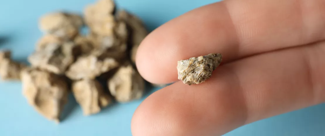
Are kidney stones more than just mineral deposits? A study has revealed that bacteria are part of the internal structure of calcium oxalate kidney stones. This suggests that bacteria play a role in the formation of these stones in the kidneys.
Kidney stones, or nephrolithiasis, are “stones” that form in the kidneys from minerals present in the urine.
Most are so-called calcium stones, which are composed of calcium, particularly in the form of calcium oxalate.
Until now, the formation of these stones was primarily explained by a chemical process: when urine is too rich in calcium and oxalate, crystals form, grow in size, and then clump together in the urinary tract.
70%
Calcium-based stones account for over 70% of stones, making them by far the most prevalent type globally. ¹
1/11
Kidney stone disease prevalence is on the rise globally in recent decades, with a lifetime incidence rate of 1 in 11 people. ¹
80%
A recurrence rate of up to 80% has been observed in certain compositions. ¹
However, this view may be mistaken, just like the belief that urine is sterile, since it harbors a urinary microbiota, important for health.
3
The main types of kidney stones are:
calcium stones, the most common type of stone
struvite stones, usually caused by an infection, like a urine infection
uric acid stones, usually caused by a large amount of acid in your urine. 1
Bacteria in calcium stones
A study 1published in the journal PNAS in 2026 suggests that a biological factor—bacteria—also plays an important role.
What led the researchers down this road was a surprising discovery: the presence in calcium oxalate kidney stones of bacteria and “biofilm” structures (a sort of protective matrix harboring bacterial communities).
Symptoms of kidney stones
You may not notice if you have small kidney stones. You'll usually pee them out without any discomfort.
Larger kidney stones can cause several symptoms, including:
pain in the side of your tummy (abdomen)
severe pain that comes and goes
feeling sick or vomiting. 2
Role of bacteria in stone formation
Upon closer inspection, the researchers discovered that the stones have a non-uniform structure: they are made up of alternating layers composed either of minerals or of organic structures rich in bacteria.
On a microbiological level, bacteria were found in nearly half of the stones studied, even in those of patients with no signs of urinary tract infection.
The most common species were :
Escherichia coli
Enterococcus faecalis
and Proteus mirabilis.
More than one-third of the stones contained multiple bacterial species, forming true microbial communities.
What causes kidney stones ?
Waste products in the blood can occasionally form crystals that collect inside the kidneys. Over time, the crystals may build up to form a hard stone-like lump.
This is more likely to happen if you:
do not drink enough fluids
are taking some types of medication
have a medical condition that raises the levels of certain substances in your urine
After a kidney stone has formed, your body will try to pass it out when you pee. 2
The researchers believe bacteria play an active role in stone formation. They produce biofilms rich in extracellular DNA, capable of capturing calcium ions and concentrating them locally.
These structures create “starting points” conducive to the formation of calcium oxalate crystals.
The crystals then develop around and through these biofilms, which gradually become integrated into the kidney stone in successive layers.
Are kidney stones a biocomposite?
Thus, kidney stones may be more than simple mineral deposits that result from a chemical imbalance. Instead, they may comprise hybrid structures that combine mineral and biological components, i.e., true “biocomposites.”
If confirmed, this model could transform our understanding of the condition and open up new avenues for prevention and treatment that target not only the crystals but also the bacterial biofilms involved in their formation.
The discovery of bacteria and biofilms in calcium oxalate kidney stones suggests that microorganisms may play a role in urinary health through the pathophysiology of this type of kidney stone.
Kidney stones are polycrystalline aggregates which are classified according to their main component.
More than two-thirds of kidney stones are primarily composed of calcium. The formation of calcium oxalate (CaOx) stones was traditionally explained by the nucleation, growth, and aggregation of crystals in urine supersaturated with calcium and oxalate.
However, an additional mechanism involving bacteria from the urinary microbiota may also be at work.
An analysis 1 of patients’ kidney stones revealed the presence of structures typical of bacteria :
in terms of morphology: cocci shape, diameter, etc.;
and components: DNA, membrane-specific lipids, etc
and biofilms within calcium oxalate stones.
In contrast, a calcium oxalate crystal of non-biological, purely mineral origin from the mineral collection at the Natural History Museum of Los Angeles County, contained no bacterial structures or biofilms, confirming the specificity of the human stones studied.
70%
Calcium-based stones account for over 70% of stones, making them by far the most prevalent type globally. ¹
2-6%
Struvite stones have been reported to have a low incidence rate, roughly 2 to 6%. ¹
An analysis of the internal structure of the stones revealed alternating layers either rich in organic matter (bacterial biofilms) or composed primarily of minerals, resulting in an organic–inorganic biocomposite architecture.
3
The main types of kidney stones are:
calcium stones, the most common type of stone
struvite stones, usually caused by an infection, like a urine infection
uric acid stones, usually caused by a large amount of acid in your urine. 1
Which bacteria?
Bacteria were found in nearly half of the kidney stones studied (24 out of 54), including in those of patients with no apparent urinary tract infection at the time of analysis.
Live microorganisms were isolated from 17 of the 22 stones analyzed, including from the calcium oxalate stones.
The most common bacteria were :
Escherichia coli
Proteus mirabilis
and Enterococcus faecalis.
More than 30% of the stones harbored multiple bacterial species, suggesting the existence of complex microbiota within kidney stones.
1/11
Kidney stone disease prevalence is on the rise globally in recent decades, with a lifetime incidence rate of 1 in 11 people. 1
Proposed mechanism
The presence of bacteria both on the surface and inside the stones suggests that bacterial biofilms may serve as a matrix that directly contributes to stone formation and growth. According to this hypothesis, bacteria in urine form biofilms rich in extracellular DNA, which locally trap calcium ions and create “anchoring points” that promote the nucleation of calcium oxalate crystals.
80%
A recurrence rate of up to 80% has been observed in certain compositions. 1
The crystals develop around and through these biofilms, which gradually become integrated into the structure of the kidney stones.
Kidney stone formation is thus not merely a passive chemical process resulting from urinary supersaturation, but also a biological process in which bacterial biofilms act as matrices for nucleation and growth.
In addition to the gut microbiota and the urinary microbiota, a kidney stone microbiota has now been implicated in the condition.
If confirmed, this model may have clinical applications in the treatment and prevention of kidney stones.
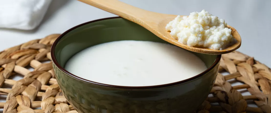
You've seen it on every supermarket shelf and TikTok feed. Kefir, the 3,000-year-old fermented drink, has become a wellness icon, displayed as a healthy food and a nutrition trend. But while marketing races ahead, science is quietly trying to catch up, and what it has to say about its effect on health may surprise you.
The kefir market is racing toward 2.78 billion dollars by 2030. Influencers swear by it. Supermarkets devote whole shelves to it.
Yet a new scientific review tells a humbler story: when researchers asked what kefir actually does to your microbiome, gut health and metabolic balance, the honest answer was, we still don't really know.
Long before lab coats existed, shepherds in the Caucasus mountains were already fermenting milk into a fizzy, tangy drink using mysterious 'grains', clusters of bacteria and yeasts living in a jelly-like matrix. Three millennia later, scientists finally peered inside those grains and found a bustling micro-city:
lactic acid bacteria,
acetic acid bacteria,
and yeasts working together through fermentation.
Among the residents are friendly names like Lentilactobacillus kefiri and Saccharomyces cerevisiae, microbial species already linked to fermented milk ecosystem.
The hope is that drinking this living community could enrich your own, your gut, your mouth, your inner ecosystem, and perhaps influence your immune, metabolic and inflammatory responses.
Microbiome
The vast invisible community of microbes living inside and on your body.
Each region, gut, mouth, skin, has its own unique cast, working quietly to shape your digestion, your immunity, your metabolic health and even your mood.1
Lactic acid bacteria
The friendly fermenters that give kefir its tangy taste.
They turn milk sugar into lactic acid through fermentation, and along the way produce natural compounds that can crowd out less welcome microbial neighbors.1
Your mouth: where kefir scores its clearest win
Of all the body's microbiomes, your mouth is where kefir's effects look most consistent.
Across four studies 1, in adults, in children getting braces, in kids recovering from cavities, kefir drinkers showed lower levels of Streptococcus mutans, the main culprit behind tooth decay, suggesting a possible effect on oral health.
Streptococcus mutans
The main bacterium behind tooth decay. It feeds on sugars in your mouth and produces acid that wears down enamel.
Reducing its numbers is a top goal in modern oral health, and one place where kefir seems to genuinely help, although its clinical effect on cavities still needs stronger evidence.1
Think of it as gentle competition: by flooding your mouth with friendly microbes from fermented food, kefir leaves less room for the troublemakers. But here's the catch, these studies only measured saliva counts, not actual cavities. Whether that translates to fewer trips to the dentist is still an open question for oral and nutrition research.
Fermentation
An ancient kitchen-meets-biology process where microbes transform food, turning milk into kefir, cabbage into sauerkraut, grapes into wine.
The microbes pre-digest the food and leave behind compounds your body can use, that’s why fermentation remains central to nutrition, healthy diet patterns and modern microbiota research.1
Down in the gut, the picture gets murkier.
Some studies hint at small wins: women with polyendocrine metabolic ovarian syndrome (previously known as polycystic ovary syndrome) saw shifts in their microbiota profile; people with inflammatory bowel disease reported feeling better; critically ill patients showed early signs of recovery.
But other studies found almost nothing. Why so messy? Because no two kefirs are alike.
The grains, the milk, the fermentation time, every variable changes the final brew and, in the end, the species composition and the potential effects.
So, if you enjoy kefir, drink it as part of a balanced diet. Just don't expect it to be a miracle food. Science is still figuring out what your microbiome thinks of it.
Numerous studies have implicated Helicobacter pylori in gastric cancer. In recent years, other bacteria in the gut microbiota shave also come under scrutiny, such as Streptococcus anginosus, which is thought to promote gastric carcinogenesis in mice and may serve as a screening biomarker.
To find out more, a research team 1 carried out both in vitro and in vivo studies on the cancer-promoting effect of S. anginosus through its metabolic interactions with the host.
Top 5
The incidence rate and mortality of gastric cancer rank among the top five malignancies worldwide. ¹
Top 3
Gastric cancer was one of the top three causes of cancer-related deaths in China over 2005–2020. ¹
4th and 5th
Gastric cancer is the 4th leading cause of cancer-related deaths among men and the 5th among women. ²
Role of S. anginosus in gastric tumors
A metagenomic analysis conducted on a large clinical cohort of 106 gastric cancer patients and 106 controls with chronic gastritis revealed an enrichment of S. anginosus (and, to a lesser extent, S. constellatus) and methionine in the gastrointestinal tract mucosa and feces of the gastric cancer patients.
Additional in vitro and in vivo experiments highlighted that S. anginosus facilitates the development of gastric tumors via methionine, one of its main metabolic byproducts.
68%
The Asian continent is the major contributor to the expected burden, with more than 10.6 million cases (68% of all cases), followed by the Americas (2.0 million; 13%), Africa (1.7 million; 11%), Europe (1.2 million; 8%) and Oceania (0.07 million; 0.4%). ³
The bacterium thus appears to play a role in the disruptions of methionine metabolism involved in the development of gastric cancer, providing non-dietary sources of essential amino acids required by cancer cells.
968 784
There were 968,784 new cases of stomach cancer in 2022. ⁴
9.2 per 100,000
The global incidence of stomach cancer was 9.2 cases per 100,000 people in 2022, but reached 27.0 per 100,000 in South Korea, 27.6 per 100,000 in Japan, and 35.5 per 100,000 in Mongolia. ⁴
660 175
660 175 personnes sont décédées d’un cancer de l’estomac en 2022. ⁴
Key role of the metE gene
Clinical strains of S. anginosus were isolated and cultured from tumor tissues to verify their ability to promote gastric cancer through methionine production. This also lays the foundation for future work on the subtyping of S. anginosus subspecies.
The authors identified the metE gene as a key player in methionine biosynthesis from S. anginosus. This gene was more abundant in patients with gastric cancer and was strongly associated with the presence of the bacterium.
By creating a ΔmetE mutant strain in which the metE gene was inactivated, they showed that inactivating this gene:
reduced methionine production,
slowed the proliferation of cancer cells,
and limited tumor formation in mice.
This confirms the central role of metE in the carcinogenic effect of S. anginosus.
6,1
The overall mortality rate from stomach cancer worldwide was 6.1 per 100,000 people in 2022, but reached 15.4 per 100,000 in Iran, 10.9 per 100,000 in Vietnam, and 9.4 per 100,000 in China. ⁴
15,6
Assuming no change in future incidence rates, a lifetime estimate of 15.6 million new gastric cancer cases is expected among all men and women born between 2008 and 2017 globally. ³
50
Gastric cancer incidence rates at younger ages (<50 years) are increasing in both low- and high-incidence populations. ³
Future therapeutic target?
A better understanding of the mechanisms by which the pathogenic bacterium S. anginosus promotes gastric cancer could contribute to the identification of new therapeutic or preventive targets. Could inhibiting S. anginosus colonization or growth reduce methionine production and thus slow the growth of gastric tumors?
Could it also improve the effectiveness of immunotherapy or chemotherapy in patients with gastric cancer ? These findings open up many avenues for future research.
The vaginal microbiota, dominated by lactobacilli, seems to play a crucial protective role against HPV and cervical cancer. Could new therapeutic approaches through probiotics help prevent cervical cancer and improve gynecological health?
The vaginal microbiota is receiving growing attention because of its implications for women’s health.
In January 2026, a Mexican research team 1published, a review of the vaginal microbiota and its role in various conditions, particularly cervical cancer and the risk of HPV-associated cervical lesions.
10¹⁰
The vagina hosts approximately 10¹⁰ to 10¹¹ microorganisms. ¹
9 %
Of the total human microbiota, the urogenital region (which includes the vagina) accounts for approximately 9% of the overall composition. ¹
Protected by lactobacilli
The vaginal microbiota is marked by low diversity. It is generally dominated by lactobacilli, especially L. crispatus, L. jensenii, L. gasseri and L. iners. These lactobacilli help maintain vaginal health, protect against pathogens and regulate inflammation, for example by competing for nutrients, producing bacteriocins, modulating the immune system and reducing vaginal pH.
4
A healthy vaginal microbiota is generally dominated by one of 4 Lactobacillus species:
Cervical cancer is the fourth most common cancer in women globally with around 660,000 new cases and around 350,000 deaths in 2022.
This uterine cancer is a major issue for women. 2
Vaginal microbiota and HPV
In the specific case of HPV, vaginal dysbiosis promotes viral progression by creating a pro-inflammatory environment.
The reduction in lactobacilli and the proliferation of anaerobic bacteria (Gardnerella, Sneathia,Atopobium) inhibit apoptosis, increase inflammatory cytokines and raise pH. These changes all make it easier for HPV to become established in the cervix and adjacent uterine tissue.
Conversely, a microbiota rich in lactobacilli is associated with a less inflammatory microenvironment. According to some studies, it may modulate the expression of the viral oncoproteins E6/E7 in infected cells. This context is associated also with more favorable HPV clearance and may help limit progression to cervical lesions.
In addition, excision of precancerous cervical lesions induces a change in the composition of the vaginal microbiota only in women who clear HPV, with a shift from pathogenic bacteria (Prevotella, Sneathia) to protective lactobacilli (L. iners then L. crispatus). By contrast, in cases of persistent HPV, no change appears.
90-70-90
Countries around the world are accelerating efforts to eliminate cervical cancer, guided by the global 90–70–90 targets :
90% of girls fully vaccinated with HPV vaccine by age 15,
70% of women screened by ages 35 and 45,
and 90% of women with pre-cancer or invasive cancer receiving appropriate treatment.
Illustrating the importance of screening, vaccination and treatment in prevention for women’s health. 2
Finally, probiotics, namely Lactobacillus strains (L. crispatus, L. reuteri, L. casei and/or L. rhamnosus) administered orally or intravaginally, appear to improve HPV clearance and reduce cervical lesions.
Results remain mixed, with effects that vary by strain, duration and population, but some probiotics could still complement treatment and prevention strategies.
8
As of 2025, there are 8 licensed HPV vaccines, five of which have received WHO pre-qualification and are available globally.
All these protect against the high-risk HPV types 16 and 18, which cause ~76% of cervical cancers.
This underscores the importance of vaccination in cervical cancer prevention and risk reduction. 2
Differences from one population to another
The composition of the vaginal microbiota varies across populations, and according to geographic origin, pregnancy, the menstrual cycle and age.
The dominant Lactobacillus species thus differ among Canadian, Chinese, Indian and Mexican women. In cases of HPV, progression to cervical lesions and then cancer is associated with changes in the microbiota: among Mexican women, L. crispatus and L. iners predominate in women without disease, while Sneathia spp. appears in lesions and Fusobacterium in cervical cancer.
According to the authors, current research points to the need for additional studies, such as exploring how cervical cancer cultures respond to different Lactobacillus species.
The aim is to improve therapeutic treatment approaches and reverse dysbiosis to support recovery and even prevent these cervical cancers.
Are you more of an eggs person or an oatmeal person in the morning? Researchers have tested two approaches in overweight adults. Weight loss, appetite, gut microbiota... the findings reveal some unexpected differences.
Breakfast is an essential meal for anyone seeking to lose weight or stay in shape.
But should we prioritize protein (dairy, eggs, ham, etc.), known for its satiating effect, or dietary fiber (oatmeal, whole-grain bread, fruit, etc.) which is beneficial for gut health?
50%
Diet accounts for more than 50% of the variability of the microbiota. ¹
Hearty, balanced, but distinct
To try to find some answers, British researchers 2 put 19 overweight or obese adults on a balanced but low-calorie diet for 28 days. This diet was based on hearty breakfasts (45% of daily calories) and light dinners (a mere 20%).
For 28 days, each participant followed:
a high-fiber diet (30 g per day, with 15% of calories from protein)
then a high-protein diet (30% of calories from protein, with less than 15 g of fiber per day)
or vice versa, with a washout period in between.
The scientists measured changes in weight, satiety, blood markers, and more, and collected stool samples from the participants to analyze their microbiota. The results?
In both groups, weight loss was significant: nearly 5 kg in the fiber group and nearly 4 kg in the protein group. Blood pressure, fasting blood sugar, blood lipid levels, and other markers also decreased significantly, reflecting a marked improvement in the participants’ health.2
However, satiety was more pronounced in the protein group, suggesting that protein may be useful for regulating appetite outside of a diet or for sustaining energy levels during periods of restriction.
And the gut microbiota? That’s a different story...
What is a “healthy” diet (according to the WHO)?
The WHO 3 emphasizes that rather than a single “model,” there are instead four principles: adequacy, balance, moderation, and diversity.
In practice, a healthy diet mainly involves:
unprocessed or minimally processed and varied foods
at least 400 g of fruits and vegetables per day (for those aged 10 and older);
at least 25 g of fiber per day (for those aged 10 and older) from whole grains, legumes, fruits, and vegetables;
less free sugar: ideally <10% of total energy intake (or even <5% for greater health benefits);
higher-quality fats: limit saturated fats (<10%) and trans fats (<1%), and prioritize unsaturated fats (vegetable oils, fish, nuts)
Free sugar
Sugar added to foods/beverages (by the manufacturer, the cook, or you) plus sugar naturally present in honey, syrups, fruit juices, and juice concentrates.
Saturated fats
Fats found mainly in animal products (butter, cream, cheese, fatty meats) and also in certain vegetable fats (e.g., coconut/palm oil). Limit their intake.
Trans fats
Industrially-modified fats (often through “hydrogenation”) found mainly in certain ultra-processed foods. The WHO recommends a very low intake.
Unsaturated fats
Fats found primarily in vegetable oils (olive, rapeseed, sunflower, etc.), nuts/seeds, and fatty fish. These are the fats that should replace saturated or trans fats.
Fiber, the microbiota’s ally
An analysis of bacterial strains showed that, compared to the fiber diet, the protein diet led to a significant loss of diversity and a decrease in the abundance of bacteria known for their beneficial effects: bifidobacteria and butyrate-producing bacteria, such as Faecalibacterium, Roseburia, and Anaerostipes.
According to previous studies, this type of microbiota profile—which results from the fact that high-protein, low-fiber diets lead to a marked reduction in these bacteria and their beneficial metabolic products—is associated with an increased risk of several diseases.
Butyrate is a short-chain fatty acid ( (sidenote:
Short chain fatty acids (SCFA)
Short chain fatty acids (SCFA) are a source of energy (fuel) for an individual’s cells. They interact with the immune system and are involved in communication between the intestine and the brain.
Silva YP, Bernardi A, Frozza RL. The Role of Short-Chain Fatty Acids From Gut Microbiota in Gut-Brain Communication. Front Endocrinol (Lausanne). 2020;11:25.)) that is important for maintaining good gut health. It serves as an energy source for gut cells, strengthens the gut barrier, and is believed to possess anti-inflammatory and anti-carcinogenic properties.
In practice, the high-protein breakfast was more effective at curbing appetite. However, the high-fiber breakfast was associated with a more “beneficial” microbiota, containing more bifidobacteria and butyrate-producing bacteria. So, which of the two is right for you?




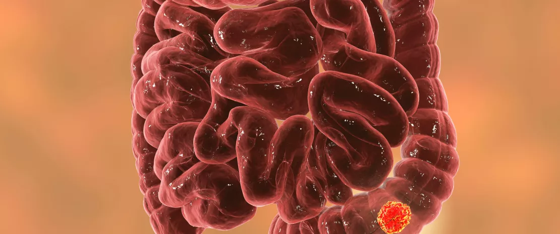
 Aspirin: antibiotic effect in colorectal cancer?
 Commensal clostridiales strains mediate effective anti-cancer immune response against solid tumours