
Endometriosis: the immune pathway opens new treatment avenues

Endometriosis is linked to disruptions of innate and adaptive immunity, worsened by endometrial dysbiosis, which promotes inflammation and lesion progression. Could this be an avenue for new therapeutic strategies?
Photo: Endométriose : la piste de l’immunité ouvre de  nouvelles voies de traitement

In recent years, growing attention has been paid to immune disorders in endometriosis, a condition that affects about 1 in 10 women.

10% Endometriosis is one of the most prevalent gynecological diseases, affecting up to 10% of women of reproductive age worldwide... ²

190 million which represents 190 million women, worldwide. ²

These women show several disruptions in innate immunity (rapid, non-specific, without immune memory) and adaptive immunity (antigen-specific). Hence, the relevance of this narrative review, which draws on 198 publications to summarize current knowledge on the immunopathogenesis of endometriosis. 1

Disruptions in innate and adaptive immunity

The literature confirms that the pathogenesis of endometriosis appears to be driven by disruptions affecting both innate immunity (macrophages, neutrophils, NK cells, complement) and adaptive immunity (T and B lymphocytes). This is true across every stage of development and for all forms of the condition (ovarian, deep infiltrating, extra-genital or adenomyosis).

1st Endometriosis is the leading cause of subfertility. ²

4 - 12 years Currently, the average time to diagnosis is between 4 and 12 years. ²

Innate immune cells create an inflammatory microenvironment that supports the survival of ectopic endometrial cells, while dysfunction in adaptive immunity (reduced cytotoxic T-cell activity, Treg-mediated immune tolerance) allows these cells to escape elimination once implanted.

Together, these two mechanisms promote the implantation of endometriotic lesions, their growth, angiogenesis and progressive tissue damage, maintaining a self-perpetuating inflammatory cycle.

Endometriosis encompasses 2 distinct forms:

  • external endometriosis, characterized by ectopic lesions outside the uterus;
  • internal endometriosis (adenomyosis), in which endometrial tissue penetrates into the myometrium.

What is the role of the endometrial microbiota?

In women with endometriosis, dysbiosis of the endometrial microbiota is observed: an increase in pathogenic bacteria such as Escherichia coli, Shigella and Streptococcus, and a decrease in protective Lactobacillus species.

Vaginal microbiome may predict the severity of endometriosis

Learn more

This dysbiosis is associated with activation of the complement system, which may enhance local inflammation and promote the attachment of endometrial cells to the peritoneal mesothelium.

Due to chronic inflammation, macrophages that should be in “destruction” mode (M1 phenotype) shift to an anti-inflammatory and healing mode (M2 type). Instead of eliminating displaced endometrial cells, they “calm” the immune response, support tissue repair and promote lesion survival. 

Therapeutic avenues

Beyond advancing knowledge, understanding the mechanisms of endometriosis immunopathogenesis paves the way for targeted immunotherapy. Goal: directly modulate dysfunctional immune components. 

Among the strategies under study are immunomodulators, cytokine/chemokine inhibitors, gene therapy, nanotechnology, immune checkpoint targeting (PD-1/PD-L1, CTLA-4) and therapies guided by macrophages and NK cells. Probiotics may also play an important role, as oral administration of Lactobacillus gasseri activates NK cells and reduces lesion size in a mouse model.

Research worth following!

Towards a microbiotic marker for endometriosis?

Learn more
Summary
Off
Sidebar
On
Migrated content
Désactivé
Updated content
Désactivé
Hide image
Off
News Gynecology Off

How gut flora improves or disrupts sleep

So you think sleep is all about the brain? Think again. Your gut flora also has a say. And it has a lot to say.

The gut microbiota
Photo: Comment votre flore intestinale dope ou sabote vos nuits

If you’re not sleeping well, your gut bacteria might be to blame. Your gut microbiota, the billions of microorganisms living in your gut, is in constant communication with your brain, and this conversation directly affects the quality of your sleep. 1

Studies continue to reveal links between gut microbiota, the brain, and sleep. For example:

  • insomniacs and people with sleep apnea have less diverse gut flora;
  • night shifts and jet lag are often associated with increased intestinal permeability and inflammation;
  • and imbalances in the gut microbiota are observed in several disorders frequently linked to poor sleep (depression, Parkinson’s, Alzheimer’s); etc.

Three communication pathways

How do gut bacteria communicate with the brain? Gut bacteria produce compounds such as:

  • serotonin (the “happiness hormone”),
  • melatonin (the “sleep hormone”),
  • and GABA (a calming neurotransmitter),

which enter the bloodstream and travel to the brain. 

But that’s not all: gut bacteria also act on the vagus nerve—a direct connection between the gut and the brain—and influence the brain regions associated with sleep. A stressed microbiota means a frazzled brain and a sleepless night ahead.
The third communication pathway is inflammation. Poor sleep triggers an inflammatory reaction in the gut, which weakens its lining and, in turn, affects the nervous system, including the brain.

Thus, the gut-brain axis enables two-way communication, creating the risk of a vicious circle that can take a toll on your nights.

1 in 10 Insomnia affects 1 in 10 adults. ²

2 in 10 adults experience occasional symptoms of insomnia. ²

Anti-insomnia bacteria?

How can you return to restful sleep? Since traditional sleeping pills come with a list of side effects as long as a sleepless night, researchers are looking to the microbiota for novel treatments. 

Thus, personalized sleep medicine may one day pass through your gut.

In the meantime, eating well, especially fiber-rich foods, means nourishing your gut microbiota and increasing the odds you’ll get a good night’s sleep. Ultimately, it may be true that those who eat well, sleep well.

Summary
Off
Sidebar
On
Migrated content
Désactivé
Updated content
Désactivé
Hide image
Off
News Off

At nursery, babies also build up their gut microbiota

At nursery, babies meet their first peers... and their peers’ gut bacteria, which they’ll actively exchange. This plays just as big a role in the development of their gut flora as the microbiota inherited from their own family.

The gut microbiota

Worried your baby might catch a virus at nursery? You’re not entirely wrong—but the surprising reality is far more upbeat. It’s true that nursery is a true hub for the exchange of microbes among infants. But far from being bad news, it’s actually a boon for their health.

A major Italian scientific study 1, microTOUCH-baby, has recently confirmed this. For a year, researchers followed 43 infants in three public nurseries in Trento, as well as their parents, siblings, pets, and nursery staff—134 subjects in total. They analyzed the composition of each subject’s gut microbiota at regular intervals.

Babies share a lot

The results? Infants start nursery with a gut microbiota partly inherited from their mother and father and enriched by any siblings. Very quickly, babies begin to share their gut bacteria.

After one year, microorganisms received from nursery peers account for a larger portion of their gut flora than those transmitted by their own parents. Over the months, the transmission network becomes increasingly complex: certain bacterial strains spread from parent to baby, then to the baby’s playmate, then to the playmate’s parents... like an invisible game of pass-the-parcel.
 

Greening of daycare centers and microbiota: flower power!

Learn more

Influencing factors

Babies who have siblings at home have a more diverse microbiota and are less likely to acquire new strains on arriving at nursery, since the space is already taken. 

Infants who receive antibiotics see their flora temporarily collapse and then rapidly replenish itself with bacteria from their peers, as if their microbiota, depleted by the treatment, were trying to rebuild itself quickly, much faster than what is observed in adults.

Other findings will reassure moms: the influence on a child’s gut microbiota of the mode of delivery (vaginal or cesarean) or the possible administration of antibiotics shortly before birth appears to have disappeared by 10 months, the median age of entry into nursery in this study.

A catalyst for microbial exchange

In short, social interactions in early childhood actively contribute to the development of the gut microbiota, which plays a key role in long-term health. So the next time your baby comes home from nursery with a runny nose, remember that they also have some fantastic new residents in their gut!

Summary
Off
Sidebar
On
Migrated content
Désactivé
Updated content
Désactivé
Hide image
Off
News Off

Nurseries: microbiota exchange hubs

Microbiota transmission between infants during their first year in nursery is intense, suggesting social interactions during early childhood are key drivers of gut microbiota development in infants.

An infant’s microbiota develops gradually after birth. The mother is the primary source of microorganisms, but she is far from being alone.

To assess the role of early social interactions, particularly in nursery, researchers launched a longitudinal metagenomic study 1, microTOUCH-baby, in three public nursery centers in Trento, Italy. The study involved 43 infants, 7 of their siblings, 39 mothers and 30 fathers, 5 pets living in the home, and 10 nursery staff members.

Microbial colonization as a determinant factor for health during the first 1000 days of life

Learn more

Nursery acts as transmission hub

When they entered nursery (median age 10 months), the infants’ microbiota was heavily influenced by that of their mother and father, but still differed significantly from that of adults. The presence of siblings at home was also a source of enrichment. In contrast, the influence of birth practices (mode of delivery, peripartum antibiotics) on microbial diversity was no longer statistically significant at this age.

As early as the first month of nursery, microbiota transmission between babies began. Eight strains spread very efficiently: initially detected in no more than one infant, after a few months, they were present in more than half of the group.

After three months in nursery, a convergence was observed in the infants’ gut microbiota: diversity between children decreased (-7% during the first quarter), and the proportion of strains acquired from nursery peers (28.4%) ultimately surpassed those acquired from family (20.0%).

In total, the study identified more than 565,000 instances of strain sharing over 15 months, with complex chains of transmission. Pets also shared strains with infants, but to a more limited extent.

Factors influencing acquisition 

Unsurprisingly, antibiotic treatment was the factor that most influenced transmission: it reduced the retention of existing strains but was followed by a massive influx of new strains, more pronounced in the infants than in the adults. Exposure in nursery could contribute to this rapid recovery of microbial diversity after treatment.

Infants with siblings acquire significantly fewer strains at nursery than only children. Early acquisition via siblings could “saturate” the potential for acquiring new strains.

In the study, certain species, such as the beneficial Bifidobacterium longum subsp. infantis, typically found in breastfed children, was present only in the infants. Other highly transmissible species included fiber-degrading bacteria, which suggests these social transmissions may promote healthy microbiota development.

Thus, social interactions such as those in nursery appear to be essential to microbiota development, acting, according to the authors, as a “microbial enrichment hub” beyond the family circle.

Summary
Off
Sidebar
On
Migrated content
Désactivé
Updated content
Désactivé
Hide image
Off
News Pediatrics Off

Your breast milk is feeding your baby's microbiome

Your breast milk is not just food, it is a living ecosystem. Every feed delivers bacteria straight into your baby’s gut. New science now shows exactly which ones arrive, take hold, and shape your child’s health from the inside out.

The gut microbiota

You already know breast milk is remarkable. But scientists have just revealed something that goes deeper than nutrition: your milk is alive with bacteria, and those bacteria travel directly into your baby’s gut, take up residence, and begin shaping their health within the first weeks of life.
A major new study 1 in Nature Communications, following 195 mother-baby pairs across six months, used technology powerful enough to tell individual bacterial “fingerprints” apart, and traced them, one by one, from milk to baby.

The tiny resident that holds everything together

Among all the bacteria scientists found, one stood out: Bifidobacterium longum. It was present in the guts of 98 out of every 100 babies at one month of age.
But what makes it remarkable is not just how common it is, it’s what it does. Babies whose guts were dominated by this species, especially a subspecies called B. longum subsp. infantis, had a gut microbiome that held remarkably steady over the following months.
It settled, rather than swinging. Why? Because this bacterium has evolved a special ability to break down the natural sugars in breast milk, sugars that exist, it now seems, precisely to feed it.

The longer mothers breastfed exclusively, the more this species flourished. Babies who stopped earlier showed less stable communities taking hold. What you feed your baby shapes who moves in, and who stays.

Disclaimer

At the Biocodex Microbiota Institute, we recognize that breastfeeding, including prolonged breastfeeding, is not always possible or suitable for every mother and family. This article is intended to share the latest scientific findings on the subject, while respecting the diversity of feeding journeys and individual circumstances.

From your milk to their gut: a direct line of inheritance

Here is where the research becomes genuinely astonishing. Using a technique that can tell apart bacterial “twins”, strains so similar they look identical to standard tests, scientists found something never confirmed before with this precision: the exact same strain detected in a mother’s breast milk was also found, weeks later, living in her baby’s gut. Not just a similar species. The same genetic identity. Your bacteria know your baby’s address, and they deliver themselves there.

They also discovered something unexpected about direction. Some shared bacteria were species normally found in the mouth, suggesting that when your baby feeds at the breast, microbes from their mouth travel back into the milk. Breastfeeding is not a one-way delivery. It is a conversation between two microbiomes, flowing in both directions with every feed.
Birth mode leaves its mark here too. Babies born vaginally retained their gut bacteria for significantly longer, their microbial community at six months looked more like it had at one month.
Babies born by C-section showed a more fluid picture, with fewer strains persisting. Neither is a verdict. But how a baby enters the world shapes the microbiome they carry through it.

Infant microbiota: could the drawbacks of a C-section be reduced by breastfeeding?

Learn more

The genes your baby was born carrying 

Every baby in the study, including the two-thirds who had never received a single antibiotic, carried genes linked to antibiotic resistance. This sounds alarming. It isn’t. These genes, known collectively as the resistome, are a normal, ancient feature of the human gut, far older than antibiotics themselves. What scientists set out to understand was simply: where does a newborn’s resistome come from?

Resistome

The collection of antibiotic resistance genes living in a microbiome. Ancient and normal, these genes existed long before antibiotics were developed. Babies carry them from birth. What this study shows is that breastfeeding helps keep them in check.

The answer, in significant part, is breast milk. Mothers and their babies shared far more resistance genes with each other than unrelated pairs did, clear evidence that feeding is a transmission route. But here is what matters most: babies whose guts were dominated by Bifidobacterium, exactly the bacteria that breast milk cultivates, carried substantially fewer resistance genes than those with other microbial communities. 

Breastfeeding doesn’t just build a thriving microbiome. It actively displaces a less desirable one. Every feed is, in the most literal biological sense, an act of protection.

Antibiotics: what impact on the microbiota and on our health?

Read more
Summary
Off
Sidebar
On
Migrated content
Désactivé
Updated content
Désactivé
Hide image
Off
News Off

Does breast milk wire the infant resistome?

Breastfeeding is universally recommended. But which live bacteria does milk actually transmit, and can they reshape the infant gut resistome? Strain-level metagenomics now answer both, with direct implications for perinatal counselling.

Breastfeeding guidance has long rested on binary choices: breast vs. formula. A landmark study in Nature Communications by Ferretti, Allert et al. dissolves that simplicity entirely 1.

Using (sidenote: Shotgun Metagenomics This is a high-resolution sequencing method that analyzes all the genetic material from every microbe in a sample. Unlike older techniques that just identify bacterial families, it allows for precise identification down to the species level and reveals the functional genes those bacteria possess. ) on 507 samples from 195 mother–infant pairs, the team tracked bacteria not just at species level but at strain level; the genetic resolution required to prove transmission, not merely infer it. What they found reframes breastfeeding as an active, strain-specific microbial intervention with measurable consequences for the infant (sidenote: Resistome The complete set of antimicrobial resistance genes (ARGs) harbored by a microbiome. In this study, the infant gut resistome was significantly shaped by maternal breast milk, even in antibiotic-naive infants. ) .

When one species holds the microbiome together

Bifidobacterium longum was present in 98.2% of infant stool samples at one month, but prevalence alone understates its role. Infants whose guts were dominated by B. longum, and particularly by its subspecies B. longum subsp. infantis, maintained significantly more stable microbiome composition between one and six months than those dominated by other species.
The mechanism is specific: B. longum subsp. infantis carries the enzymatic machinery to degrade human milk oligosaccharides (HMOs), giving it a decisive competitive advantage in the breastfed gut.
Its mean relative abundance surged from 3.2% at one month to 23.8% at six months. Infants with non-bifidobacteria-dominated guts showed the most volatility.

The clinical read: breastfeeding duration is not just a nutritional variable; it is a determinant of microbiome architecture.

Twelve confirmed transmissions, and what they reveal about the gut–milk axis

Twelve instances of strain-level sharing between maternal milk and infant gut; same species, identical genetic fingerprint; demonstrate that breast milk is a bona fide transmission route.
Shared taxa spanned commensals (B. longum, B. bifidum), gut-associated species (Phocaeicola vulgatus), and typical oral residents such as Streptococcus salivarius and Rothia mucilaginosa, the latter two suggesting retrograde colonisation from the infant’s oral cavity back into the milk during suckling, a biologically plausible bidirectional axis.

Most notable was the detection of Klebsiella pneumoniae as a confirmed shared strain. No infants showed clinical manifestations, consistent with silent commensal carriage; nonetheless, this finding signals that culture-independent, strain-level milk surveillance may add meaningful risk stratification in high-risk neonatal settings beyond what standard culture screens can offer.

Delivery mode compounds this picture: 19.4% of infant gut strains at one month persisted to six months, and vaginally born infants retained significantly more of them than those born by C-section (p = 0.018), evidence that birth mode shapes not just early colonisation, but the durability of the microbial community across the first half-year of life.

Antibiotics and cesarean delivery: how do they affect the newborn microbiota?

Learn more

The resistome is inherited and breastfeeding can modulate it

Every infant carried antibiotic resistance genes (ARGs) at one month, including the 67% who had no recorded pre-, peri-, or postnatal antibiotic exposure. Resistance classes for tetracycline, MLS (macrolide-lincosamide-streptogramin), aminoglycoside, and beta-lactams were all present. This is not a signal of clinical failure; it is the baseline ecology of the human neonatal gut, assembled through mechanisms independent of antibiotic selective pressure and largely invisible to standard clinical workups.

What this study adds is the transmission axis and, critically, a modifiable countermeasure. Mother–infant pairs shared significantly more ARGs than permuted pseudo-pairs (p < 0.016).The dominant shared genes were MACB (MLS class), ACRD (aminoglycoside), and TETQ (tetracycline). Sharing was highest in the two pairs with confirmed strain transmission events, providing a mechanistically coherent explanation. Yet infants with bifidobacteria-dominated guts carried markedly fewer ARGs than those dominated by other species (p = 7.6×10⁻¹⁰).

The implication for practice is direct: supporting Bifidobacterium colonisation through exclusive breastfeeding and, where indicated, B. longum subsp. infantis-containing probiotics does not only enrich the microbiome, it also suppresses the resistome.

Clinicians now have strain-level, resistome-level evidence that the how and how long of breastfeeding counselling carries consequences that extend well beyond nutrition.

Summary
Off
Sidebar
On
Migrated content
Désactivé
Updated content
Désactivé
Hide image
Off
News Pediatrics General Medicine Off

Vitiligo: the gut microbiota at the heart of skin oxidative stress

The gut microbiota may play an important role in the onset and progression of vitiligo. Hippuric acid, a metabolite produced by the microbiota, may be a key mediator of oxidative stress… and a potential biomarker or even a therapeutic target?

Vitiligo, an autoimmune skin disease, is characterized by the destruction of melanocytes, leading to depigmented patches. However, cutaneous oxidative stress—a key factor in the progression of the disease—appears to be influenced by the gut microbiota. Here’s a breakdown of a study that examines the mechanisms at work.1

Oxidative stress linked to the gut microbiota

The first step in the researchers’ work involved analyzing lesions in patients and in a mouse model. This revealed, in the depigmented areas, an overexpression of genes associated with oxidative stress and responses to reactive oxygen species (ROS). This confirms that these processes are crucial in the pathogenesis of the disease.

Next step: the researchers discovered that mice with vitiligo exhibit a significant accumulation of ROS in the skin, as well as mitochondrial abnormalities in their melanocytes. Eliminating the gut microbiota with antibiotics reduced the abnormal accumulation of ROS and the mitochondrial abnormalities in the melanocytes, resulting in a significant improvement in depigmentation. Thus, the gut microbiota appears to directly regulate the skin’s oxidative stress status.

Vitiligo around the world

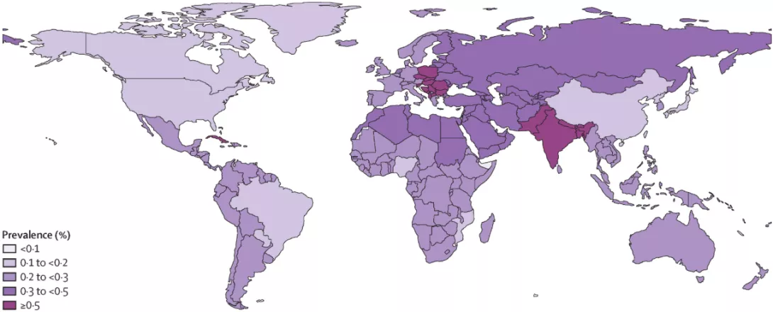
  • The global lifetime prevalence of vitiligo diagnosed by a physician or dermatologist is estimated at 0.36% in the general population, 0.67% in the adult population, and 0.24% in the pediatric population.2
  • Among adults, prevalence estimates range from 0.43% in East Asia to 0.98% in Central Europe.2
  • Among children, the prevalence of vitiligo ranges from 0.16% in East Asia to 0.35% in South Asia and 0.35% in Central Europe.2
Image

From the gut microbiota to the skin

Subsequent research by scientists shows that the microbiota of mice with vitiligo is imbalanced (increased Clostridiales, decreased Verrucomicrobiae). Transferring this dysbiotic microbiota (through cohabitation of animals or fecal transplantation) exacerbates depigmentation. Conversely, administering probiotics slows the progression of the disease, suggesting a potential therapeutic approach.

But how is the gut microbiota linked to the skin? Metabolomic analysis of fecal, blood, and skin tissues from mice identified a key factor: hippuric acid, a metabolite derived from the microbiota, accumulates excessively in the feces, serum, and skin of mice with vitiligo. Injections of hippuric acid are sufficient to reproduce the accumulation of ROS and exacerbate depigmentation in mice. And in humans? Serum levels of hippuric acid are found to be higher in patients with vitiligo.

A mechanistic hypothesis

This leads to the following hypothesis: in mice with vitiligo, the intestinal mucosal barrier is weakened (due to a reduction in goblet cells responsible for mucus production and a decrease in mucosal thickness), which increases intestinal permeability. This increased permeability would facilitate the passage of hippuric acid into the bloodstream and then to the skin. Hippuric acid would induce oxidative stress by directly binding to two proteins (NOS2 and MAPK14); this direct molecular interaction would then promote the production of ROS in skin tissues.

Thus, intestinal dysbiosis would orchestrate cutaneous oxidative stress in vitiligo via hippuric acid. These results also suggest that probiotics may play a role in slowing the progression of the disease.

Gut bacteria turn stress hormones into progestins hormones: a surprising role for hydrogen gas

Learn more
Summary
Off
Sidebar
On
Migrated content
Désactivé
Updated content
Désactivé
Hide image
Off
News Dermatology Off

How the gut microbiome affects sleep quality

The gut microbiota and sleep interact with each other in both directions through metabolic, neural, and immune pathways. Does this open the door to new therapeutic strategies for sleep disorders?

The Gut-Brain axis

What if there is a bidirectional relationship between sleep and the gut? To go beyond simple correlational studies1, this review delves into the literature on the microbiota-gut-brain axis and its role in sleep regulation to summarize the potential mechanisms linking the composition and function of the gut microbiota to sleep disorders.

18% The estimated prevalence of obstructive sleep apnea (OSA) in the adult population is 18%.

10% The estimated prevalence of insomnia in the adult population is 10%.

3% The estimated prevalence of restless legs syndrome (RLS) in the adult population is 3%.

Sleep Disorders and the Gut-Brain Axis

Different sleep disorders are associated with changes in the composition of the gut microbiota and its metabolites:

- Insomniacs show reduced microbial diversity, with (though results vary by study) a decrease in the abundance of certain bacteria (such as Ruminococcaceae) correlating with a decrease in their metabolites (fewer secondary bile acids, produced by bacteria from liver bile acids).

- There is also less diversity in patients suffering from obstructive sleep apnea, and (sidenote: Fecal Microbiota Transplantation (FMT) A therapeutic procedure to restore the gut microbiota by transferring fecal bacteria from a healthy donor to a recipient. Explore https://www.science.org/doi/10.1126/scitranslmed.abo2750 ) from individuals suffering from hypoxia can disrupt the sleep cycles of healthy animals.

- Night work and jet lag also cause significant changes in the microbiota, increasing intestinal permeability and inflammation. 

- Narcolepsy may be associated with an imbalance between immunosuppressive and immunostimulatory microorganisms.

- In the case of restless leg syndrome, the multiplication of bacteria in the small intestine may play a role.

Dysbiosis is also observed in sleep-related disorders: in major depressive disorders, the composition of the gut microbiota is associated with sleep quality; in Parkinson’s disease, (sidenote: Mendelian randomization An approach that exploits natural genetic variation to test causal relationships between a biological factor (e.g., cholesterol, vitamin D, BMI) and a disease (e.g., heart attack). Researchers use genetic variants associated with the biological factor of interest (e.g., patients with alleles that increase LDL cholesterol vs. patients without) to test whether there is a causal relationship between the biological factor (LDL cholesterol) and the disease (heart attack). The random distribution of alleles in the population reduces biases (confounding factors, etc.), mimicking some of the advantages of a randomized trial. ) studies suggest a causal link with gut bacteria; in Alzheimer’s disease, sleep disturbances are directly related to the accumulation of beta-amyloid protein, in parallel with changes in the bacterial flora that could be potential diagnostic and even therapeutic biomarkers.
 

0,03% The estimated prevalence of narcolepsy in the adult population is 0.03%.

>50% More than one in 2 people with dementia (including Alzheimer’s disease) suffer from insomnia.²

Two-way communication

The review also highlights three main bidirectional communication pathways through which the gut-brain-microbiota axis coordinates sleep:

- Metabolic and endocrine pathways: secondary bile acids and (sidenote: Short chain fatty acids (SCFA) Short chain fatty acids (SCFA) are a source of energy (fuel) for an individual’s cells. They interact with the immune system and are involved in communication between the intestine and the brain. Silva YP, Bernardi A, Frozza RL. The Role of Short-Chain Fatty Acids From Gut Microbiota in Gut-Brain Communication. Front Endocrinol (Lausanne). 2020;11:25. ) produced by bacteria affect sleep through systemic circulation. In addition, the microbiota produces or regulates key molecules of the wake-sleep cycle, such as GABA, serotonin, tryptophan and melatonin.

- Neural pathways: gut bacteria and their metabolites interact with the gut nervous system and cooperate with vagal nerve pathways, influencing brain areas and circuits related to sleep. In addition, the microbiota and its metabolites modulate the stress response and the corticosterone rhythm, influencing wakefulness-related hyperactivity.

- Immune pathways: Sleep deprivation triggers systemic and gut inflammation, weakening the gut barrier and affecting the central nervous system.

Did you know?

  • In high-income European countries, obstructive sleep apnea (OSA) is the most costly sleep disorder (€184 billion), followed by insomnia (€158 billion), restless legs syndrome (RLS) (€79 billion), narcolepsy (€905 million) and REM sleep behavior disorder (RBD) (€436 million). Direct and indirect costs account for 48% and 52% of the total, respectively, with no data available on informal care costs.3

  • Sleep disorders are very common in people with Parkinson’s disease, with up to 98% of patients experiencing them at some point. REM sleep behavior disorder (RBD) is seen in about 46% of patients with Parkinson’s disease.

  • Restless legs syndrome (RLS) affects 15-20% of patients with Parkinson’s disease, although early-onset RLS does not predispose to the later development of Parkinson’s disease. However, severe RLS may be an early warning sign of the disease. 

Modifying the Microbiome to Improve Sleep

Since traditional methods of treating sleep disorders often involve side effects, microbiome-targeted therapies represent promising strategies:

Is science heading towards precision medicine in the field of sleep?


The gut microbiota and sleep disorder: a therapeutic approach based on modulation of the microbiota?

Find out more!
Summary
Off
Sidebar
On
Migrated content
Désactivé
Updated content
Désactivé
Hide image
Off
News Gastroenterology Off

A Gut Metabolite May Predict CAR-T Success

We know antibiotics harm CAR-T outcomes. But why? New multicenter data reveals the gut microbiota, specifically butyrate-producing bacteria, drives therapy success in non-Hodgkin lymphoma. Circulating butyrate emerges as both biomarker and potential therapeutic enhancer, reshaping how we prepare patients for cellular therapy.

The gut microbiota has emerged as a critical modulator of cancer immunotherapy, but its precise mechanisms in CAR-T cell therapy have remained elusive. A compelling multicenter study1 published in Clinical Cancer Research now demonstrates that a specific microbial metabolite, (sidenote: Butyrate A short-chain fatty acid produced by gut bacteria through fermentation of dietary fiber. ) , may represent both a prognostic biomarker and a potential therapeutic lever for patients with Non-Hodgkin Lymphoma (NHL) undergoing CD19 CAR-T treatment.

When antibiotics disrupt more than infection

The study enrolled 84 NHL patients across four centers and confirmed what recent American and German cohorts suggested: non-prophylactic antibiotic exposure before CAR-T infusion significantly impairs progression-free survival. Patients receiving two or more lines of non-prophylactic antibiotics showed markedly worse outcomes, with high-risk antibiotics including meropenem, cefazolin, ceftriaxone, and piperacillin-tazobactam demonstrating the strongest negative associations.

Researchers from Hospital 12 de Octubre didn't stop at correlation; they examined the mechanism. Through (sidenote: 16S rRNA sequencing A method that reads a bacterial “barcode” gene to identify and differentiate species. ) of stool samples collected at lymphapheresis, they found that antibiotic-exposed patients had significantly lower (sidenote: Microbiota diversity The variety and evenness of bacterial species within the gut ecosystem, measured by indices such as Shannon and Pielou. ) indices. Critically, lower diversity itself predicted worse survival, creating a mechanistic chain from antibiotic use through dysbiosis to clinical failure.

Short-chain fatty acids emerge as the missing link

Taxonomic analysis revealed a striking pattern: responders to CAR-T therapy harbored significantly higher relative abundances of (sidenote: SCFA-producing bacteria Bacterial taxa that metabolize dietary substrates into short-chain fatty acids including acetate, propionate, and butyrate.  ) . Specifically, taxa including Prevotella, Ruminococcus, Butyricicoccus, and the Clostridiaceae family were enriched in patients achieving complete or partial responses. Non-responders, conversely, showed elevated lactic acid bacteria including Lactobacillales and Enterococcus. The functional consequence became clear when researchers measured serum metabolites. Patients with higher circulating butyrate at baseline demonstrated superior progression-free and overall survival. A multivariate analysis confirmed butyrate as an independent prognostic factor, with low levels conferring a more than six-fold increased hazard of progression.

Butyrate reprograms CAR-T cells for enhanced killing

To validate butyrate's direct role, investigators exposed CAR-T cells to physiologically relevant concentrations in vitro. Butyrate-stimulated CAR-T cells showed increased activation marker expression, higher transduction efficiency, and a shift toward central memory phenotypes, characteristics associated with superior persistence. 

Functionally, these cells generated significantly greater specific lysis of lymphoma targets at multiple effector-to-target ratios. Whole transcriptome sequencing revealed upregulation of 145 genes involved in cytotoxicity, chemokine responsiveness, and T cell proliferation, while senescence-associated genes were downregulated. Pathway enrichment analysis confirmed enhanced inflammatory signaling and cytotoxic function. Remarkably, oral butyrate supplementation in a xenograft mouse model significantly reduced tumor burden and extended survival compared to controls, demonstrating in vivo proof of concept. 

The takeaway for this isn't to immediately supplement all patients, but to recognize that the microbiota-butyrate axis represents a modifiable determinant of CAR-T efficacy worthy of prospective evaluation.

Antibiotics disrupt cancer immunotherapy via gut and immune effects

Find out more!
Source

1. García-Vicente R, et al. The Potential of the Gut Microbiota and Butyrate to Enhance CAR-T Cell Therapy in Non-Hodgkin Lymphoma. Clin Cancer Res. 2025. https://doi.org/10.1158/1078-0432.CCR-25-1676

Summary
Off
Sidebar
On
Migrated content
Désactivé
Updated content
Désactivé
Hide image
Off
News Oncology Off

Gut bacteria linked to liver cancer risk?

We link liver cancer to viral hepatitis, alcohol, and metabolic disease. Yet a 12-cohort study reveals that gut barrier dysfunction, detectable years before diagnosis via bacterial translocation markers, independently predicts liver cancer. This gut-liver axis mechanism reshapes our understanding of hepatocarcinogenesis.

Liver cancer development has been primarily attributed to well-established risk factors including viral hepatitis, excessive alcohol consumption, and metabolic conditions. However, a groundbreaking nested case-control study published in the International Journal of Cancer examined 867 liver cancer cases and 867 matched controls across 12 United States cohorts, revealing that immunological markers of (sidenote: Bacterial translocation The passage of viable bacteria or bacterial products such as lipopolysaccharide and flagellin across the intestinal barrier into systemic circulation. When gut barrier function is compromised, bacterial translocation triggers immune activation and chronic inflammation that may contribute to hepatocarcinogenesis. ) , measured an average of 12 years before diagnosis, are independently associated with liver cancer risk1.

The gut barrier under scrutiny

The gut barrier serves a critical dual function, allowing nutrient absorption while preventing translocation of harmful bacterial products into systemic circulation. Researchers from multiple institutions measured pre-diagnostic serum concentrations of (sidenote: Lipopolysaccharide-binding protein An acute-phase protein primarily synthesized by hepatocytes that binds to lipopolysaccharide from gram-negative bacteria to form an LPS-LBP complex. ) , (sidenote: Soluble CD14 A co-receptor protein that recognizes the LPS-LBP complex and facilitates immune signaling to trigger inflammatory responses. ) , and antibodies against lipopolysaccharide and flagellin. These markers reflect the body's response to gut-derived bacterial products that have breached the intestinal barrier.

LBP, an acute-phase protein synthesized primarily by hepatocytes, binds to lipopolysaccharide from gram-negative bacteria and forms complexes recognized by soluble CD14, triggering inflammatory cascades. The most striking finding was that LBP concentrations showed the strongest association with liver cancer risk, with an odds ratio of 1.48 per doubling in concentration. Anti-flagellin IgA and IgG, anti-lipopolysaccharide IgG, and soluble CD14 were also positively associated with risk.

Hepatocellular carcinoma versus cholangiocarcinoma

When analyses were stratified by liver cancer subtype, a critical distinction emerged. LBP concentrations were positively associated with hepatocellular carcinoma with an odds ratio of 1.77 per doubling, but showed no association with intrahepatic cholangiocarcinoma with an odds ratio of 0.67. This finding suggests potential specificity in the bacterial translocation pathway for hepatocellular carcinoma development.

Animal models have previously demonstrated that lipopolysaccharide accumulation activates (sidenote: Toll-like receptor 4 A pattern recognition receptor that recognizes lipopolysaccharide and initiates inflammatory signaling pathways. Animal studies have shown that TLR4 activation by bacterial lipopolysaccharide promotes hepatic inflammation and accelerates hepatocellular carcinoma development, providing a mechanistic link between bacterial translocation and liver cancer. ) signaling, promoting hepatic inflammation and tumor formation. The prospective nature of this study, with biomarkers measured years before diagnosis, suggests that LBP elevation represents an early etiological factor in hepatocarcinogenesis rather than merely a consequence of underlying liver disease. Associations were generally consistent across subgroups and remained significant even after excluding participants with hepatitis B or C infection.

The findings highlight the (sidenote: Gut-liver axis The bidirectional relationship between the gastrointestinal tract and the liver, mediated by the portal venous system that carries nutrients and bacterial products from the gut to the liver. ) as a modifiable pathway in liver cancer development, necessitating further investigation into interventions that maintain gut barrier integrity.

Bacterial metabolites and immunotherapy effectiveness

Recommended by our community
Summary
Off
Sidebar
On
Migrated content
Désactivé
Updated content
Désactivé
Hide image
Off
News Oncology Off