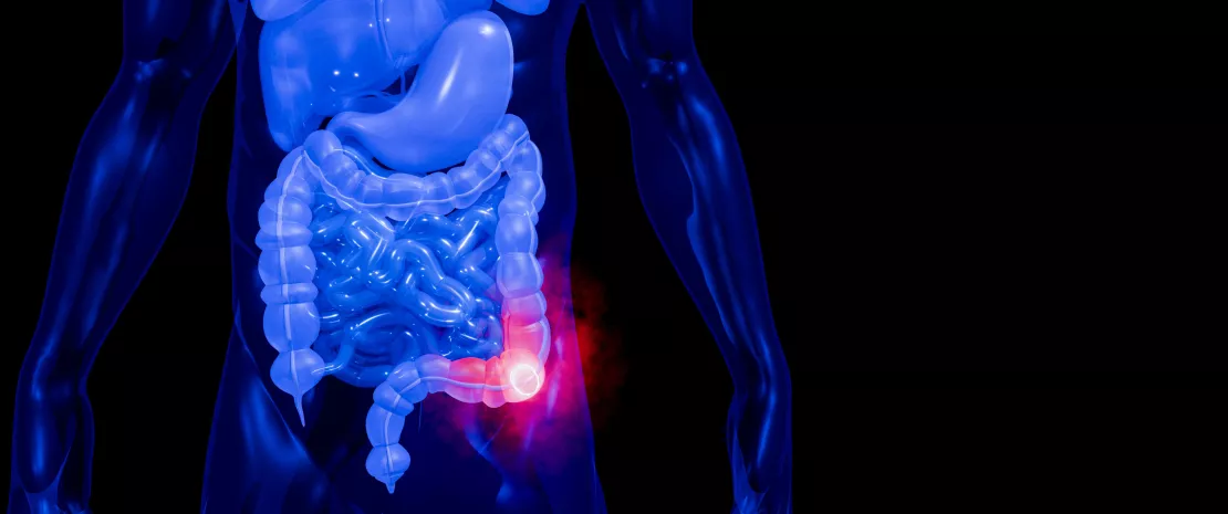

Could rebalancing your gut microbiota help alleviate depression ?

Le microbiote intestinal pourrait-il influencer notre santé mentale en agissant sur notre énergie ? Une étude chez l’humain et la souris explore ce lien entre bactéries intestinales, métabolisme et symptômes dépressifs. 

The gut microbiota

Rééquilibrer son microbiote pour sortir de la dépression ?

What if restoring a healthy gut microbiota could help you alleviate depression by giving your body an energy boost?

Depression doesn’t just affect mood and cognitive function. It frequently goes hand in hand with changes in energy metabolism and gut microbiota, as confirmed by a study¹ involving 100 patients with major depressive disorder and 68 healthy controls. 

Major depressive disorder

Major depressive disorder (MDD) is a highly disabling psychiatric condition with a substantial global burden, exhibiting a lifetime prevalence of 10%–20%.¹

1,5 Depression is about 1.5 times more common among women than among men.²

10% Worldwide, more than 10% of pregnant women and women who have just given birth experience depression (perinatal depression).²

A link between microbiota, metabolism, and depression

The results are clear: in people with depression, several pathways involved in energy production are disrupted, with some metabolites present in excess—such as lactate, glutamate, and arginine—and others in lower-than-normal levels, including glucose and ornithine. These metabolic disturbances are associated with more severe symptoms, including increased anxiety, more pronounced depressive symptoms, and greater cognitive difficulties.

But that’s not all. The authors also found differences in the gut microbiota of people with depression: a dysbiosis is present, involving around 100 bacterial species that are either more or less abundant than in stool samples from healthy controls. 

What if everything were connected? Among these microorganisms that are over- or underrepresented in the gut, several may influence our energy metabolism and, in turn, negatively affect our mood. Or, conversely, they could improve our mental health—provided we give good bacteria in our gut a little boost. But before considering such an approach, researchers first needed to establish a causal relationship. That has now been demonstrated.

4% An estimated 4% of the population experience depression, including 5.7% of adults (4.6% among men and 6.9% among women), and 5.9% of adults aged 70 years and older.²

332 million Approximately 332 million people in the world have depression.³

3rd Suicide is the third leading cause of death in 15–29-year-olds.²

A mouse experiment to test causality

Researchers subjected mice to chronic stress that induced depressive-like behaviors. As observed in patients with depression, the mice developed alterations in both their gut microbiota and energy metabolism.

The study focuses on the microbiota—the gut bacterial community—and its links to metabolism and mental health.
Previous studies had already identified changes in the gut bacteria of patients with major depressive disorder, but the links between these changes and metabolism had remained largely correlational.

The researchers then transplanted the mice’s own fecal microbiota, collected before they were exposed to chronic stress, back into the animals after they had developed depression-like behaviors. Following this procedure, known as an autologous fecal microbiota transplant (each mouse received its own gut microbiota), several metabolic abnormalities were corrected, and the mice showed improvements in their mental health. Mitochondrial function—which provides energy—was also restored in brain regions involved in mood and memory.

"The transplant reintroduced bacteria that had been present in the gut before exposure to the stressor.”

“From a mental-health perspective, these findings add to the growing evidence supporting the importance of the gut–brain axis. However, they do not demonstrate that interventions targeting this axis can treat depression in humans.”

What about humans?

So far, this causal relationship has only been demonstrated in animals. Further studies are needed to determine whether the same mechanism exists in people with major depressive disorder and, above all, whether modulating the gut microbiota could one day have therapeutic potential. In the meantime, to give your mood a boost, you can start by taking care of your gut microbiota. How? Through diet, for example, by choosing citrus fruits and cutting out soda.

Three examples to help you understand

Reducing excess (toxic) lactate

In people with depression, chronic stress can impair mitochondrial function—the mitochondria being the powerhouses of our cells—and cause brain cells to switch to an alternative energy-producing pathway known as anaerobic glycolysis. This can lead to an abnormal and harmful buildup of lactate in the blood and brain. This excess of lactate may contribute to depressive behaviors and memory deficits. However, the gut bacterium Anaerostipes hadrus may alleviate anxiety and depression by reducing brain lactate levels, thus allowing the mitochondria to resume their normal function and reversing this anaerobic glycolytic shift.

Reducing excess (toxic) glutamate

In people with depression, glutamate is thought to accumulate at abnormally high levels in the blood and brain. These elevated glutamate levels are thought to interfere with brain energy metabolism and may exacerbate depressive symptoms. The gut bacterium Anaerostipes hadrus may play a potentially beneficial role in improving depression and anxiety: it seems to have a protective effect by reducing certain key metabolites, including glutamate, as well as lactate and isocitric acid.

Increasing ornithine (a protective agent) when levels are low

In people with depression, ornithine levels are believed to be very low in the blood and brain. This deficiency could contribute to the accumulation of toxic metabolites by impairing ammonia detoxification and increasing glutamate production. It may also deprive the brain of neuroprotective compounds, since ornithine serves as a precursor for their synthesis. Together, these changes could increase vulnerability to depression, fatigue, and cognitive decline. However, several intestinal bacteria, such as Dorea formicigenerans and Eubacterium ramulus , appear to be major sources of ornithine for the host. 

Sources

1. Lei P, Qi Z, Ma Q et al. Gut microbiota reshapes host energy metabolism to modulate depressive behaviors. Gut Microbes. 2026 Dec 31;18(1):2662556.
2. https://www.who.int/news-room/fact-sheets/detail/depression. 
3. 2021 Global Burden of Disease. 

Summary
Off
Sidebar
Off
Migrated content
Désactivé
Updated content
Désactivé
Hide image
Off
News Off

Depression: could the microbiota-metabolism axis be involved?

What if the gut microbiota contributes to depression by disrupting energy metabolism? A multi-omic study in humans, complemented by a mouse model, reveals a potential link between the gut microbiota, metabolites, and depressive phenotypes.

 Dépression : un axe microbiote-métabolisme en cause ?

Major depressive disorder (MDD) is not limited to affective and cognitive symptoms; it is also associated with substantial disturbances in energy metabolism, including blood glucose dysregulation, mitochondrial dysfunction, and alterations in key metabolic pathways such as glycolysis, the Krebs cycle, and amino acid metabolism. At the same time, gut dysbiosis is frequently observed in patients with MDD, with alterations in specific bacterial species, including Faecalibacterium prausnitzii and Escherichia coli, that may indirectly influence host metabolic pathways. This led the researchers1 to ask: could the gut microbiota influence the host’s energy homeostasis and in turn, alleviate depression? 

Major Depressive Disorder (MDD)

Major Depressive Disorder (MDD) is a highly disabling psychiatric condition with a substantial global burden, exhibiting a lifetime prevalence of 10%–20%.1

A microbiota-metabolites-depressive phenotype axis

Analysis of serum and fecal samples from 100 patients with depression and 68 healthy controls revealed significant disruptions in key energy metabolism pathways in individuals with depression: 14 metabolites were significantly altered, including lower serum levels of glucose, ornithine, AMP, and cyclic AMP, and higher levels of lactate, glutamate, arginine, citrulline, and isocitrate. Several of these metabolites were also associated with measures of depression, anxiety, and cognitive function, suggesting a potential link between these metabolic alterations and the severity of depressive symptoms.

Most importantly, the authors identified 92 bacterial species that differed significantly in individuals with depression, including 50 that were overrepresented and 42 that were underrepresented, particularly within the Firmicutes phylum. The researchers also identified 36 bacterial species whose abundance was potentially associated with mitochondrial fatty acid synthesis, ketogenesis, and amino acid metabolism. Among these, 28 species appeared to be involved in the relationship between the gut microbiota, energy metabolites, and mood, cognitive function, and anxiety. These findings thus point to a potential “gut microbiota–metabolites–depressive phenotype” axis, in which certain bacterial metabolites act as mediators.

332 million Approximately 332 million people in the world have depression.²

Preclinical evidence supporting a causal role for the gut microbiota

As is often the case in microbiota research, an important question remained: were these findings simply correlational, or could the gut microbiota play a causal role? To investigate this, the researchers first collected samples of the gut microbiota from mice and then subjected them to 14 days of chronic stress, which induced depressive-like behaviors (including reduced sucrose preference and increased immobility) as well as impaired memory. An autologous fecal microbiota transplantation (FMT) was performed after each episode of stress. FMT reduced depressive-like behaviors and improved memory, while also restoring the composition and function of the gut microbiota. However, the intestinal barrier remained impaired. Most importantly, FMT corrected several metabolic disturbances—including alterations in glucose, lactate, glutamate, and the ornithine cycle—and restored mitochondrial structural integrity in the prefrontal cortex and hippocampus of the mice. According to the authors, these findings highlight the role of the gut microbiota in regulating energy metabolism in depressive disorders. 
Although the experiment has so far been conducted only in mice, they raise an intriguing question: could early autologous FMT one day become a therapeutic approach for depression in humans? This remains an important avenue for future research.

Modulation of the microbiota by FMT: controversial results

Learn more

In the meantime, maintaining a balanced diet that includes some citrus fruits and engaging in regular physical activity such as running, may be beneficial.

Summary
Off
Sidebar
On
Migrated content
Désactivé
Updated content
Désactivé
Hide image
Off
News Off

Can AI identify promising vaginal probiotic strains?

We often consider Lactobacillus crispatus a hallmark of vaginal health. But not all strains share the same functional properties. A new study shows how machine learning could help identify strains with the most promising in vitro probiotic potential.

Can AI identify promising vaginal probiotic strains?

When considering a vaginal probiotic, we may read a species name off the label and assume it carries a defined benefit. A new study 1 challenges that assumption directly: does the functional potential of Lactobacillus crispatus belong to the species as a whole, or to individual strains we've been lumping together far too generously?

One species, forty-seven versions of it

The team collected 1,126 bacterial isolates from vaginal swabs collected from 36 healthy women aged 18 to 30. Of these, 639 turned out to be Lactobacillus crispatus. Phylogenomic analysis grouped these 639 isolates into 47 distinct clades, revealing substantial diversity within the species. Across the 639 isolates, the pangenome contained 4,482 gene families, of which 1,579 belonged to the core genome. This highlights meaningful genomic diversity within L. crispatus, despite an overall conserved genomic structure.

This diversity isn't just academic: strains differed in the presence and copy number of genes encoding pullulanase (PulA), an enzyme involved in glycogen degradation that may help L. crispatus exploit this carbon source in the vaginal environment. Existing general probiotic screening tools may struggle to capture these strain-level differences. When the researchers ran the strains through an existing tool called iProbiotics, nearly every one scored well, averaging 97.58%, which illustrates the challenge of distinguishing strain-level functional potential using existing general probiotic prediction approaches.

Strain-level functional potential: the measurable capacity of one isolate, not one species, to acidify, produce lactic acid and H2O2, and inhibit pathogens. The 67 strains tested varied fivefold in lactic acid output alone.

Pullulanase (PulA): an enzyme involved in glycogen degradation. Its presence may help L. crispatus use vaginal glycogen and adapt to the vaginal environment (supports colonisation and sustained acidification).

The phenotypes that distinguish higher-performing strains

The researchers then tested 67 representative strains on five in vitro traits that may matter clinically and considered relevant to vaginal probiotic potential: how well they grow, how much they acidify their environment, how much lactic acid and hydrogen peroxide they produce, and how well they block vaginal pathogens. The results varied widely. Lactic acid output ranged from 2.02 to 10.29 g/L, and hydrogen peroxide levels from 9.39 to 18.3 µmol/L, even though all the strains belonged to the same species. Final pH ranged from 3.8 to 4.45, so every strain made its environment more acidic, just not to the same degree.

Strains also differed in how well they blocked
Gardnerella vaginalis, a bacterium linked to bacterial vaginosis : the inhibition zone averaged 15.35 mm but varied by several millimeters from one strain to the next. The researchers combined these five measurements into a single score, giving the heaviest weight, 30%, to how well a strain fought off BV-related pathogens. They then split the strains at the median into a higher functional-potential group, labeled Lcris-SFS, and a lower functional-potential group.. The three top-performing strains are now being tested in animals and in early clinical studies.

Lcris-SFS: labels for strains scoring above or below the median on the composite functional score. They rank functional potential and are not a judgement of safety.

Vaginal dysbiosis: a shift away from a Lactobacillus-dominated vaginal microbiota toward a more diverse community enriched in anaerobic bacteria. Bacterial vaginosis is a common dysbiotic state.

A model that reads function from the genome

To avoid months of lab culture work, the researchers built a computer model called VLCPredictor. It reads short DNA snippets from each strain's genome, narrows 349,184 of these snippets down to the 534 most informative ones, and uses that pattern to predict a strain’s functional potential, helping prioritize candidates for further experimental testing. Eight prediction methods were tested, and the best performing model, a random forest, achieved a mean AUC of 0.742 for distinguishing strains with higher versus lower functional potential.. Applied to 103 published genomes, the model flagged about 76% of strains from healthy women as having higher predicted functional potential, compared with only 31% of strains from women with bacterial vaginosis. The model isn't perfect: it was trained on genomes from a single region and ethnic group, and only 16 genomes came from women with bacterial vaginosis, so it still needs more validation. The importance of strain-level selection is also illustrated by previous clinical studies cited by the authors: one L. crispatus-based probiotic reduced bacterial vaginosis recurrence following antibiotic treatment, whereas another Lactobacillus-based probiotic combination did not improve cure rates in a separate trial. 

Vaginal lactobacilli's anti-inflammatory superpowers

Learn more

The clinical message is not that genomic prediction can already select a probiotic for an individual patient. Rather, this study reinforces the importance of strain-level characterization: belonging to a health-associated species such as L. crispatus does not guarantee identical functional properties. VLCPredictor could help researchers prioritize promising candidates, but in vivo and clinical validation remain essential before translating these predictions into practice.

Summary
Off
Sidebar
On
Migrated content
Désactivé
Updated content
Désactivé
Hide image
Off
News Gynecology Obstetrics Off

Oral microbiome dynamics during travel : environmental exposure, dysbiosis, and resilience

The ENT microbiota
Evolución de la microbiota bucal durante los viajes: exposición ambiental, disbiosis y resiliencia

 1. How much do people travel each year ?

According to the European Commission, EU residents aged 15 years and older made nearly 1.2 billion tourism trips with overnight stays in 2024, corresponding to approximately three trips per person per year.1 This frequency of travel is biologically significant, as repeated movement between environments creates repeated opportunities for microbial exposure and short-term ecological disruption. While travel-associated changes in the gut microbiome and the clinical syndrome of travelers’ diarrhea are well documented, the oral microbiome—including saliva-, tongue-, and plaque-associated communities—has received far less attention, despite serving as a primary interface between the host and the external environment. 

Travel repeatedly perturbs the microbiome through exposure to new environments, diets, and water sources, yet the oral microbiome remains far less studied than the gut despite being the first point of contact.

2. Why travel should reshape the oral microbiome

The oral cavity is continuously exposed to the external environment, making it particularly sensitive to rapid lifestyle and environmental changes. Although oral microbial communities are generally stable within individuals, they respond to shifts in diet, oral hygiene practices, circadian rhythms, stress, and immune status. Oral health depends on community balance (homeostasis), and dysbiosis—disruption of community structure or function—is associated with both oral and systemic disease.2
Travel concentrates multiple perturbations simultaneously, including exposure to new environments, water sources, dietary patterns, and social contacts,3-4 all of which can influence resident microbial communities. As the entry point to the gastrointestinal tract, the oral cavity may also influence downstream microbial ecology. Large, multi-country studies have shown that oral microbes can transmit to and colonize the gut more frequently than previously appreciated, even in healthy individuals.5

Travel-induced microbiome disruption is often transient but not always fully reversible, with microbial and resistance-related changes persisting beyond clinical symptom resolution.

3. When travel introduces new microbes: dysbiosis, food poisoning, and diarrhea

A well-recognized risk of travel is acute gastrointestinal infection, often caused by food- or water-borne pathogens, which can coincide with microbial community disruption. Longitudinal studies show that microbiome composition shifts during travel and that diarrheal episodes can destabilize community structure and increase antimicrobial resistance gene abundance for weeks.6-7 Travel is also associated with colonization by multidrug-resistant organisms, particularly among individuals who experience travelers’ diarrhea.6 Clinically, most uncomplicated cases of travelers’ diarrhea resolve within days. However, microbiome studies suggest only partial resilience: microbial composition often trends back toward baseline after return, but recovery may be delayed or incomplete. Notably, travel-associated antimicrobial resistance signatures can persist for weeks following the initial disruption in some cohorts.6, 8-9

Traveling shapes our lives...but also our microbiota and antibiotic resistance

Learn More

4. The underexplored questions: acclimation time, stability, and permanence

Despite growing interest in travel-related microbiome dynamics, direct longitudinal sampling of the oral microbiome during travel remains rare. Time-series studies of the gut microbiome in non-travel settings reveal substantial day-to-day variability even under stable conditions, highlighting the need for dense longitudinal sampling to distinguish normal temporal variation from true travel-induced effects.4

Leveraging a year-long international gap-year program, Dr. Wendy Lin is tracking oral microbiome changes in a student cohort as they travel across seven regions, including the United States, Brazil, France, Turkey, Kenya, India, and China. This study aims to address fundamental questions that remain poorly characterized for the oral microbiome:

  • How long does it take for the oral microbiome to acclimate to a new environment?

  • Is the travel-associated oral microbiome stable or highly dynamic?

  • How quickly does the oral microbiome revert to baseline after returning home?

  • Do newly introduced taxa or strain-level variants persist long-term?

The timing, stability, and long-term persistence of travel-associated changes in the oral microbiome remain largely unknown.

Conclusion

Travel predictably alters environmental exposures and dietary patterns, creating a natural experiment for studying human microbiome ecology. Evidence from gut microbiome studies of travelers demonstrates rapid compositional shifts during travel and additional disruption during diarrheal illness, often followed by delayed or incomplete recovery. The oral microbiome—despite being the first point of contact for food, water, and environmental microbes—remains comparatively undercharacterized in this context. Longitudinal studies of traveling cohorts with dense oral sampling have the potential to answer foundational questions about microbial acclimation, stability, and long-term persistence, and to determine whether travel leaves lasting microbial imprints after individuals return home. 

Summary
Off
Sidebar
On
Migrated content
Désactivé
Updated content
Désactivé
Hide image
Off
News Off

Coffee and decaf: what effects do they have on the microbiota?

According to a new exploratory study, caffeinated coffee is not the only brew that affects the brain. Decaf may have an impact too. And your gut microbiota could be the missing link.

The gut microbiota

It is now well established that coffee can influence cognitive function, affecting attention, alertness, memory, and the body’s response to stress. Yet the underlying mechanisms remain largely unclear. Are they mediated by the microbiota? And perhaps more intriguingly, is caffeine really the key compound behind coffee’s effects on the brain—or is there more to the story?

-27 % The estimated reduction in the risk of Alzheimer's disease among coffee drinkers.²

3rd most popular beverage in the world Coffee is said to be the third most widely consumed beverage in the world, after water and tea.³

2 billion That's the number of cups of coffee consumed worldwide every day.³

Understanding the links between coffee, the gut, and cognition

This is a legitimate question. Several nutritional studies have shown that coffee, through its bioactive compounds (chlorogenic acids, melanoidins, and other polyphenols), can significantly modulate the composition of the gut microbiota. Moreover, the gut microbiota and the brain are in constant communication through the microbiota-gut-brain axis. Beyond caffeine, this axis may help explain some of coffee’s effects on health.

To test this hypothesis, researchers in Ireland recruited 62 healthy adults who had not recently taken probiotics, prebiotics, or antibiotics. Half were habitual coffee drinkers, consuming three to five cups a day, while the other half did not drink coffee. The coffee drinkers first abstained from coffee for two weeks before resuming their usual habit for three weeks. During this period, they consumed either regular or decaffeinated coffee, while keeping their usual diet unchanged. The aim was to disentangle the effects of caffeine from those of coffee’s other compounds.

Throughout the study, the researchers collected stool, urine, and blood samples and assessed participants’ cognitive and behavioral performance at several time points, while also monitoring their dietary intake.

The surprising effect of coffee on the microbiota

Learn more

Positive effects on mood and cognition

Published in Nature Communications,¹ the study found that habitual coffee drinkers exhibited notable differences in their gut microbiota. In particular, they showed a higher abundance of Firmicutes CAG:94, a bacterial strain that has been associated with positive emotional states in women.

Following a period of coffee abstinence and subsequent reintroduction, both caffeinated and decaffeinated coffee were found to affect seven specific bacterial strains in the gut microbiota, without altering overall microbial diversity. 

The effects on the brain were equally intriguing. Reintroducing coffee—regardless of its caffeine content—was associated with several beneficial outcomes, including reductions in:

  • perceived stress,
  • experienced depressive symptoms,
  • impulsivity, defined as the tendency to act quickly without considering the potential consequences.

However, only caffeinated coffee was associated with reduced anxiety, improved cognitive performance, and greater resilience to stress.

Perhaps more surprisingly, the consumption of decaffeinated coffee—but not caffeinated coffee—was associated with improved episodic memory, increased physical activity, and better sleep quality. 

The microbiota: a likely mediator between coffee and the brain

Analysis of stool samples from coffee drinkers revealed that, before the abstinence period and compared with non-drinkers, they had lower levels of indole-3-propionic acid–a metabolite involved in nervous system function–, and GABA, a neurotransmitter that plays an important role in anxiety and cognitive function.

The researchers also found that levels of five coffee-related metabolites (theophylline, hippuric acid, 1,7-dimethylxanthine, and two others) were strongly associated not only with cognitive and behavioral outcomes, but also with numerous species of gut bacteria.

According to the researchers, these fecal metabolite profiles primarily reflect the metabolism of coffee’s various polyphenols, regardless of whether the coffee is caffeinated or decaffeinated. Decaf is therefore clearly more than just 'placebo coffee'.

Overall, the findings of this far-reaching study point to a clear association between coffee consumption, gut bacteria, metabolites, and cognitive and behavioral outcomes. They therefore lend further support to the hypothesis that the gut-microbiota-brain axis may play a role in the effects of coffee. We can therefore conclude that there are several potential health benefits, but this is certainly not a free pass to go overboard with coffee either. As we know, excessive consumption can also have adverse effects on health.

Further research will be needed to move beyond these associations and determine whether there is a true causal relationship between coffee, the gut microbiota, metabolites, and cognitive and behavioral outcomes.

Summary
Off
Sidebar
On
Migrated content
Désactivé
Updated content
Désactivé
Hide image
Off
News Off

When an oral bacterium turns immune cells into killers

We've long blamed Fusobacterium nucleatum for fueling colon cancer. New research shows it can also pull immune cells into the tumor and switch them into killers. Whether that happens depends on a gene your patient carries.

When an oral bacterium turns immune cells into killers

Neutrophils in colorectal tumors have a bad reputation, and so does Fusobacterium nucleatum. A new study1 puts the two together and finds something unexpected: the bacterium draws neutrophils into the tumor and switches them on to kill it. Whether that happens in your patient comes down to one receptor.

Tumor-associated neutrophils (TANs)

Neutrophils recruited into the tumor microenvironment. Long assumed to be immunosuppressive, they are shown here to kill CRC cells once the right bacterial signal reaches them.1

Not every gut bacterium recruits neutrophils the same way

Of the two bacteria most common in colorectal tumors, only F. nucleatum pushed tumor cells to release neutrophil-attracting signals, and drew far more neutrophils toward it in lab tests than Bacteroides fragilis did. In mice, F. nucleatum raised neutrophil levels inside the tumor; B. fragilis did not. Direct contact matters too: within five minutes, F. nucleatum latched onto up to 25% of circulating neutrophils and switched on an activation marker that peaked by three hours.

Links between gut microbiota and colorectal cancer are confirmed

Learn more

One receptor flips the switch, and it isn't TLR4

Neutrophils exposed to F. nucleatum released a cocktail of antimicrobial and tumor-killing compounds. Their conditioned media killed colon cancer cells in the lab and slowed tumor growth in mice when injected directly into it; B. fragilis-conditioned media did nothing. Blocking TLR4 stopped the activation marker from appearing but left the killing intact. Blocking Siglec-5/14 did the opposite: killing stopped. Siglec-14 is the one that binds the bacterium tightly, while its partner Siglec-5 barely does. Unlike most of its receptor family, which calms neutrophils down, Siglec-14 switches them on instead.

Siglec-14

A sialic-acid-binding immunoglobulin-like lectin on neutrophils. It binds F. nucleatum LPS and is required for cytotoxicity; blocking it abolishes killing without preventing activation.1

A common gene variant may explain the conflicting outcomes data

Patients carrying two copies of a common SIGLEC14 gene variant have neutrophils that still respond to F. nucleatum but can't kill: the more copies of the variant, the weaker the response. That variant is present in under 10% of Europeans but up to 70% of East Asians, which may explain the split literature: European and US cohorts link neutrophil infiltration to better survival, Chinese cohorts to worse. Mice lack the activating receptor entirely, which is likely why animal studies have painted F. nucleatum as purely harmful. In one 444-patient sample, high neutrophil markers predicted longer survival, but only in tumors that also carried F. nucleatum. In a large public gene-expression database (The Cancer Genome Atlas), the 31 patients whose tumors lacked SIGLEC14 did worse than the 351 who had it.

This is a biomarker question before a prescribing one. The next time the evidence on neutrophil infiltration in CRC looks irreconcilable, the missing variables may be which bacteria occupy the tumor and which Siglec-14 allele the patient inherited.

Summary
Off
Sidebar
On
Migrated content
Désactivé
Updated content
Désactivé
Hide image
Off
News Off

Menstrual cup: reassuring findings regarding infection risk

Among women who use menstrual cups during their periods, current evidence does not suggest an increased risk of genital infections. On the contrary, available findings point to a possible reduction in bacterial vaginosis and STIs, while also indicating that a healthy vaginal microbiota is maintained.

Coupe menstruelle : résultats rassurants sur les infections

A menstrual cup is an intravaginal device that comes in different sizes, depending on the model. Unlike an IUD, it is not inserted into the uterus. Instead, it sits in the vagina and collects menstrual blood. As a reusable menstrual product, the menstrual cup has progressively become more popular alongside conventional products such as tampons, as well as newer alternatives such as period underwear. However, despite the growing use of menstrual cups for menstrual hygiene, their potential effects on the vaginal microbiota, infections, and reproductive health have not yet been extensively studied.
This underlines the importance of this systematic review and meta-analysis1 that investigated the relationship between menstrual cup use for menstrual protection and reproductive tract infections (RTIs), sexually transmitted infections (STIs), and the vaginal microbiota.

1.8 billion Every month, almost 1.8 billion people worldwide menstruate.¹

65 days On average, menstruation accounts for a total of 65 days per year.¹

18 % In the United States, 18% of women reported using a menstrual cup.¹

No increased risk of infection 

A total of 11 studies were included in the review, 8 of which were published after 2019. Together, they involved 10,268 adult and adolescent women from Africa (Kenya and Tanzania), the Americas (Brazil and the United States), and Europe (France, Belgium, and Denmark). One important limitation is that only three of the studies were considered to be of high quality. This limits both the overall scope of the review and the robustness of the results.Despite this limitation, the findings are broadly reassuring—and even encouraging—with regard to the use of menstrual cups: no increased risk of infection was observed among menstrual cup users for the infections evaluated. 
The findings actually point in the opposite direction. Observational studies and randomized trials conducted across three continents found a lower prevalence of bacterial vaginosis among menstrual cup users (around -20%).
Menstrual cup use also appears to be associated with a lower prevalence of STIs (- 26 %) than other menstrual products and protection methods. This finding comes from two randomized controlled trials conducted among young women in Kenya. One trial also suggested a possible reduction in the risk of HSV-2 (herpes simplex virus type 2) infection, although the difference did not quite reach statistical significance.

Bacterial vaginosis: sexual transmission & genomic insights

Learn more

A beneficial effect on the vaginal flora?

Regarding the vaginal microbiota, menstrual cup users appear more likely to have a community-state type I (CST-I) microbiota, characterized by a predominance of Lactobacillus crispatus. This was observed in populations from the three European countries and in Kenya. This potential benefit may be partly explained by how the cup works. By collecting menstrual blood in the upper part of the vagina, the cup may reduce the exposure of the vaginal microbiota to iron-rich blood, which can promote the growth of Gardnerella vaginalis and L. iners. Menstrual cups may therefore help preserve a healthy vaginal microbiota dominated by L. crispatus, a profile associated with greater protection against vaginal dysbiosis and certain STIs. No cases of toxic shock syndrome were reported among menstrual cup users in the two studies that provided this information.
Overall, the authors conclude that menstrual cups represent an affordable, easy-to-implement, and sustainable option that could help reduce the burden of preventable gynecological conditions while supporting menstrual and intimate hygiene.

Summary
Off
Sidebar
On
Migrated content
Désactivé
Updated content
Désactivé
Hide image
Off
News Off

Menstrual cup: a potential benefit for vaginal health ?

Contrary to some concerns, using a menstrual cup does not appear to increase the risk of genital infections. On the contrary, its use seems to be associated with a lower prevalence of bacterial vaginosis and STIs, as well as a vaginal microbiota that is more often in optimal condition.

The vaginal microbiota
Coupe menstruelle : un atout pour la santé vaginale ?

In recent years, menstrual cups have become an increasingly popular menstrual product. Reusable, affordable, and environmentally friendly, they are available in different sizes depending on the model. Unlike an IUD, a menstrual cup does not need to be inserted into the uterus. Instead, it sits in the vagina, where it simply collects menstrual blood. But how does using a menstrual cup affect menstrual hygiene, the vaginal microbiota, and the risk of infection when it remains in the vagina for up to 12 hours?
A comprehensive review of the literature,1 compiling the findings from 11 studies involving more than 10,000 participants across Africa, Europe, Brazil, and the United States, offers some reassuring answers.

1.8 billion Every month, almost 1.8 billion people worldwide menstruate.¹

65 days On average, menstruation accounts for a total of 65 days per year.¹

Not more infections… in fact, fewer

First finding: no increased risk of infection was observed among menstrual cup users. In fact, several findings point to a potential protective effect. For instance, some studies found that women using a menstrual cup had 21% lower incidence of bacterial vaginosis than those using other menstrual products, particularly tampons or pads.
A similar finding was observed for sexually transmitted infections (STIs). Two trials conducted among young women in Kenya found a lower risk of STIs
among menstrual cup users. The risk of HSV-2 (herpes simplex virus type 2) infection may also be reduced, although this finding has not yet been confirmed.

18 % In the United States, 18% of women reported using a menstrual cup.¹

12 heures Menstrual cup can be used for up to 12 h prior to being emptied, carefully cleaned and reinserted.¹

An ally of the vaginal microbiota

These potential benefits may be related to the menstrual cup’s protective effect on the vaginal microbiota. Menstrual cup users are more likely to have a microbiota dominated by Lactobacillus crispatus, a bacterial species considered a marker of good vaginal health and associated with protection against vaginal dysbiosis, bacterial vaginosis, and certain STIs. No cases of toxic shock syndrome were reported among menstrual cup users in the two studies that provided this information.
Why does this happen? The authors put forward a simple hypothesis: unlike tampons or pads, the menstrual cup collects menstrual blood in the upper part of the vagina. As a result, the vaginal microbiota may have less exposure to iron-rich blood, an environment that can favor the growth of bacteria such as Gardnerella vaginalis and Lactobacillus iners, which are often associated with an imbalance in the vaginal flora.

Female anatomy, microbiotas and intimate hygiene

Learn more

Promising findings… pending further confirmation

The authors remain cautious: the studies are still scare, involve comparatively small numbers of women, and are not always of high quality. Based on the current evidence, the review provides a reassuring message: menstrual cups do not appear to pose a risk to your vaginal microbiota and may, in fact, have a beneficial effect.

Summary
Off
Sidebar
On
Migrated content
Désactivé
Updated content
Désactivé
Hide image
Off
News Off

Your vulvovaginal dryness checklist

How many women do you see who may experience vulvovaginal dryness, discomfort, irritation, pain during sex or urinary symptoms? How often is the topic spontaneously discussed during consultation or at the pharmacy counter?

Vulvovaginal dryness is common (1 in 5 women affected)1, 2, but often under-recognized and rarely discussed. It can occur across life stages, not only after menopause, and may affect comfort, intimacy and quality of life. It may be linked to low-estrogen states, but also to postpartum and breastfeeding, hormonal contraception, some medications, cancer therapies, dermatologic, neurologic or autoimmune conditions, and other situations.

≥1 in 5 women Vulvovaginal dryness can occur across life stages, not only after menopause.

In the context of menopause-related symptoms, the communication gap is striking: in the European REVIVE Survey, 65% of symptomatic women expected their healthcare professional to initiate the conversation, yet healthcare professionals initiated the discussion about vulvovaginal atrophy symptoms in only 10% of cases where the topic was discussed.3

65% vs 10% a communication gap

This gap is precisely why the Vulvovaginal Dryness Checklist was developed:

to help healthcare professionals ask, recognize, explain and guide care with confidence.

Dr Clarence de Belilovsky, Dr Isabelle Dehaene, Dr Brigitte Letombe, Prof. Rossella E. Nappi and the Biocodex Microbiota Institute have created a practical checklist dedicated to vulvovaginal dryness.

This tool supports healthcare professionals in:
  • identifying vulvovaginal dryness and associated symptoms; 
  • distinguishing vulvar and vaginal symptoms; 
  • exploring sexual comfort and intimacy with sensitive wording; 
  • recognizing warning signs and differential diagnoses; 
  • understanding the role of estrogens, tissue changes and vaginal microbiota; 
  • guiding general management and follow-up; 
  • helping pharmacists open the conversation and refer when needed.

Download the vulvovaginal dryness checklist

Summary
Off
Sidebar
Off
Migrated content
Désactivé
Updated content
Désactivé
Hide image
Off
Article Gynecology Off

Peanut anaphylaxis: microbiota modulates severity

A mechanistic study combining mouse models with two clinical cohorts (n=19, then n=120) shows that oral and gut microbiota break down major peanut allergens and influence the severity of IgE-mediated anaphylaxis. Peanut allergy persists in more than 70% of patients and affects health and daily life, particularly in children.

Peanut anaphylaxis: microbiota modulates severity

Microbial metabolism of allergens modulates anaphylaxis in mice

In mouse models with controlled, minimal or complex microbiota, gut microbiota composition directly determines the ability to break down Ara h 1 and Ara h 2, the major peanut allergens. 

Three sensitization protocols were compared1 to establish this link in the development of the allergic response. The first exposed the allergen orally, which is a route that involves gut microbiota from the sensitization phase onward. The second injected the allergen directly into the peritoneum, bypassing the digestive tract and therefore the microbiota. The third passively transferred serum already containing anti-peanut IgE, without an active sensitization step. 

During oral sensitization, the group of mice with minimal microbiota, which break down Ara h 1 and Ara h 2 less effectively, developed both more specific IgE and a more severe allergic reaction than mice with complex microbiota. In the case of intraperitoneal sensitization, which bypasses the gut microbiota, mice with minimal microbiota and mice with complex microbiota produced similar IgE levels. However, during the subsequent oral challenge, the allergic reaction in mice with complex microbiota was less severe, demonstrating the direct involvement of the gut microbiota in the allergic reaction.


To confirm the results, serum rich in peanut-specific IgE was injected directly into naïve mice, thus eliminating any active sensitization step. Even in this case, the group of mice with minimal flora reacted more strongly to the oral challenge than mice with complex flora, confirming that the microbiota also influences severity at the time of allergen exposure. Microbiota therefore influences peanut allergy, both through IgE production during oral exposure and, independently, through the number of Ara h 1 and Ara h 2 allergens that actually reach the bloodstream at the time of the challenge.

Can microbiota prevent peanut allergy?

Learn more

Rothia and Staphylococcus: strain-dependent degradation

In humans, the bacterial genus Rothia accounted for up to 43% of the salivary microbiota in the healthy donors studied. All Rothia strains tested in the study consistently degraded Ara h 1 and Ara h 2 by cleaving the proteins, particularly at epitopes recognized by the IgE of patients with peanut allergy, thereby reducing mast cell activation in functional tests. 
Genome sequencing identified candidate proteases, including a subtilisin-like family in Rothia, already known to degrade gluten. The proposed mechanism has not yet been directly demonstrated, as researchers have not deleted the gene to establish its role.

Severe allergic reaction (IgE-mediated anaphylaxis)

Acute, generalized reaction that can be fatal within minutes, triggered when an allergen binds to IgE attached to mast cells, causing the rapid release of inflammatory mediators. 

Food allergy

Immune system reaction directed against a normally tolerated food protein, in this case the major peanut allergens Ara h 1 and Ara h 2. 

In Staphylococcus, the effect depended on the strain studied, as two different tested strains of S. aureus showed opposite behaviors: one partially broke down Ara h 1, while the other had no detectable effect on either allergen. Serum allergen levels in mice colonized by the latter were higher, and an ex vivo Ussing chamber test confirmed increased allergen passage through the intestinal mucosa.
The authors suggest two possible explanations: partial breakdown that may facilitate the passage of allergen fragments, or impaired intestinal barrier permeability caused by certain
Staphylococcus strains, independently of their ability to break down the allergen.

A clinical association to be prospectively confirmed

In 19 patients about to start oral immunotherapy, the peanut allergy tolerance threshold, determined through a controlled food challenge, is associated with a higher abundance of Micrococcales—an order that notably includes Rothia—regardless of specific IgE levels. This association was found in an external cohort of 120 children who underwent double-blind, placebo-controlled food challenges, with the Rothia aeria species being more abundant in non-allergic children and in allergic children with a high peanut tolerance threshold.

Allergic tolerance (eliciting dose threshold assessed through a controlled challenge)

Amount of allergen a patient can consume without developing a clinical reaction, measured during an allergy assessment using a double-blind, placebo-controlled oral food challenge. 

This data remains observational and cross-sectional. The authors call for longitudinal monitoring of the oral microbiota during immunotherapy treatment. From a clinical perspective, this research points to the potential for functional characterization of the oral microbiota in food allergy assessment, alongside IgE testing, without replacing established diagnostic tools at this stage.

Degradation capacity and clinical association according to genus / strain

Rothia (R1- R.aeria, R2- R.dentocariosa, R3- R.mucilaginosa): 

  • Degradation capacity (Ara h 1 / Ara h 2) : effective degradation of both allergens.
  • Clinical association observed in this study : abundance associated with a higher tolerance threshold, clinical cohorts n=19 and n=120.

Micrococcus (related genus):

  • Degradation capacity (Ara h 1 / Ara h 2) : effective degradation.
  • Clinical association observed in this study : no clinical association tested in this study.

Staphylococcus epidermidis S1:

  • Degradation capacity (Ara h 1 / Ara h 2) : effective degradation of both allergens.
  • Clinical association observed in this study : no clinical association tested in this study.

S. aureus strain S3: 

  • Degradation capacity (Ara h 1 / Ara h 2) : partial degradation, Ara h 1 only.
  • Clinical association observed in this study : no clinical association tested in this study.

S. aureus strain S2:

  • Degradation capacity (Ara h 1 / Ara h 2) : no detectable degradation
  • Clinical association observed in this study : associated with increased transmucosal passage of the allergen, mouse model.

Streptococcus, Gemella:

  • Degradation capacity (Ara h 1 / Ara h 2) : limited or no degradation capacity for most strains tested.
  • Clinical association observed in this study : no clinical association tested in this study.

Microbial metabolism of allergens

Certain microbiotal bacteria* can directly transform the molecular structure of a food allergen, thereby modifying its recognition by the immune system**, regardless of the level of IgE sensitization.

*Microbiotal bacteria (enzymatic breakdown of food proteins) : microorganisms in the oral cavity and intestine that use proteolytic enzymes to break down food proteins resistant to human digestion, including peanut allergens. 

**Immune system : all the cells and molecules that protect the body against infections, abnormal cells and foreign substances. In an allergy, IgE and mast cells are involved in recognizing the allergen and triggering the reaction
. 

Summary
Off
Sidebar
On
Migrated content
Désactivé
Updated content
Désactivé
Hide image
Off
News Pediatrics Off